
Diagnosis and Treatment of Achilles Tendon Injury
Rongbin Wang
Guangzhou Foreign Language School, Guangzhou, China
Keywords: Achilles Tendon Injury, Diagnostic Imaging, Surgical Treatment.
Abstract: The Achilles tendon is the thickest and strongest tendon in the human body, and due to its unique anatomical
location and physiological function, it is also one of the most prone tendons to rupture. With the development
of biomechanics research in recent years, more biomechanical concepts and research methods have been
incorporated into the etiology analysis of Achilles tendon injuries, especially Achilles tendon ruptures. This
study suggests that Achilles tendon injury has become a potential threat for athletes in track and field as well
as ball sports during running and jumping. The probability of Achilles tendon injury varies depending on the
sports they participate in and their grade level. Senior college students engaged in jumping and competitive
sports have a higher probability of developing Achilles tendon injury. Achilles tendon injury is one of the
most common symptoms in orthopedic clinical practice, and there are many causes of Achilles tendon injury,
with the highest number of cases of accidental injury to the Achilles tendon due to improper exercise. The
treatment of chronic Achilles tendinitis in Achilles tendon injury is relatively simple, and the treatment
methods are also relatively single. The treatment of Achilles tendon rupture is mainly divided into
conservative and surgical methods, including surgical treatment, conservative treatment, rehabilitation
treatment, and cytokine action.
1 INTRODUCTION
The Achilles tendon is the most powerful and thickest
tendon in a human body, located behind the ankle
joint. Its starting point is one-third of the lower back
of the calf, and its endpoint is the calcaneal
tuberosity. Therefore, it can transfer strength to the
feet by pulling the muscles of the posterior calf
muscle group, and its main physiological function is
to help the plantar flexion of the foot and ankle joint.
Common Achilles tendon injuries are mainly divided
into acute and chronic injuries, and diseases mainly
include Achilles tendon rupture and Achilles
tendinitis. The Achilles tendon is one of the most
easily breakable tendons in the human body, mainly
due to its unique physiological function and
anatomical location. Once Achilles tendon injury
occurs, the quality of life of patients will be greatly
affected.
The blood supply of Achilles tendon is mainly
provided by branches of the posterior tibial artery,
followed by branches of the fibular artery. The
vascular distribution is relatively sparse 2-6 cm above
the insertion point, which is prone to ischemic
degeneration of the tendon structure. Tearing and
rupture of the Achilles tendon often occur in this
segment. The microvessels and capillaries in the
upper, middle, and lower segments of the Achilles
tendon are not evenly distributed, with significantly
fewer in the middle segment than in the upper and
lower segments. This may be the pathological
anatomical basis for the frequent occurrence of
Achilles tendon rupture in the middle segment.
According to the epidemiology, in recent years, with
the progress of people's living standards and
lifestyles, the awareness of sports has gradually
increased, and the incidence rate of acute has also
gradually increased. The incidence rate of male
patients is higher than that of female patients, mainly
aged 35-39 years (Lantto et al.,2015).
2 DIAGNOSIS
The known risk factors for Achilles tendon injury
include previous intratendinopathy, history of
fluoroquinolone use, history of steroid injection, and
history of inflammatory arthritis (Haapasalo et al.,
2018). With the development of biomechanics
research in recent years, more biomechanical
concepts and research methods have been
incorporated into the etiology analysis of Achilles
Wang, R.
Diagnosis and Treatment of Achilles Tendon Injury.
DOI: 10.5220/0014494900004933
Paper published under CC license (CC BY-NC-ND 4.0)
In Proceedings of the 1st International Conference on Biomedical Engineering and Food Science (BEFS 2025), pages 439-443
ISBN: 978-989-758-789-4
Proceedings Copyright © 2026 by SCITEPRESS – Science and Technology Publications, Lda.
439
tendon injury, especially Achilles tendon rupture.
Which is subjected to dynamic loads at or near failure
during movement, and fatigue induced damage may
be a factor leading to its ultimate fatigue and failure
(Noback et al., 2018).
The diagnostic method for Achilles tendon injury
is supported by clinical manifestations and imaging
and ultrasound examination results. Imaging mainly
relies on X-rays and MRI. In recent years, progress in
the diagnosis of Achilles tendon injury has mainly
been made through the update of imaging and
ultrasound equipment, as well as new research and
exploration by doctors based on new diagnostic
equipment. A clinical trial has shown that high-
frequency ultrasound has high diagnostic sensitivity
and specificity for Achilles tendon. Elastic ultrasound
imaging technology can accurately reflect muscle
tension and assess muscle strength in patients with
acute Achilles tendon injury. At the same time, there
is a linear correlation between the longitudinal tensile
force borne by the tendon and the elastic modulus
value measured by elastic ultrasound imaging
technology, which can reflect the elasticity of the
Achilles tendon and achieve effective diagnosis of the
patient's disease.
MRI has the characteristics of multi plane
imaging and high soft tissue resolution, which can
well display the morphology and signal changes of
Achilles tendon, determine the location and degree of
tear, and also display the changes in surrounding soft
tissue structure. It can comprehensively and
accurately evaluate Achilles tendon tear and is the
preferred imaging examination method for acute
Achilles tendon tear. The normal Achilles tendon
shows low signal shadows on both T1WI and T2WI
sequences, and there may be strip like high signal
shadows inside. The strip like high signal shadows
are parallel to Achilles tendon and have a width of
less than 1.5mm. When the magic angle phenomenon
occurs (the main magnetic field forms a 55 ° angle
with the normal tendon diameter), the phenomenon in
the magic angle rarely occurs in the conventional
position. If the ruptured tendon bends, the effect of
magic angle may occur, which needs to be analyzed
comprehensively (Peh and Chan, 1998).
The MRI manifestations of complete Achilles
tendon tear include retraction of the rupture end, and
the width of the rupture crack reflects the degree of
retraction of both ends. The ruptured fibrous bundles
interlock and overlap with each other in a "brush"
shape, and the ruptured fibrous bundles retract in a
"oak" shape. When the torn tendon retracts
significantly, it can appear wavy. MRI manifestations
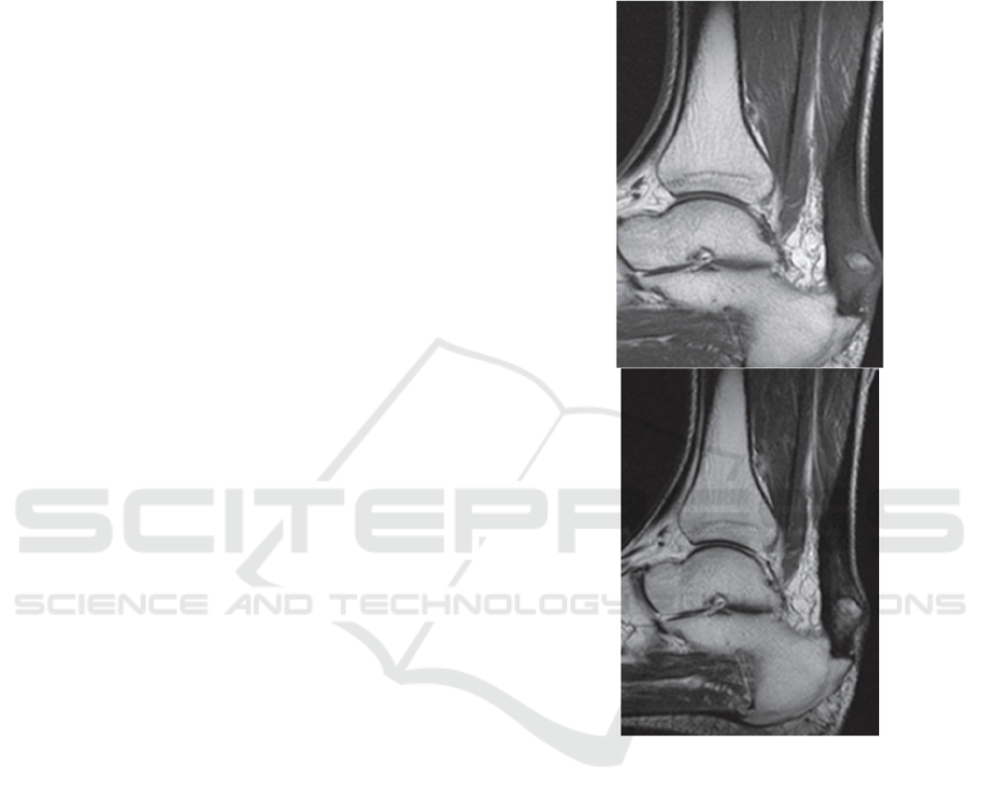
of incomplete Achilles tendon tear: Partial Achilles
tendon tear, with a stripe like, patchy, and focal high
signal shadow at the tear site compared to normal
Achilles tendon. Incomplete Achilles tendon tear
shows continuity on at least one level. Partial
incomplete tearing occurs on the basis of certain
lesions, as shown in Figures 1 (Xiao et al., 2014).
Figure 1: MRI manifestations of incomplete Achilles
tendon tear.
3 ACHILLES TENDON INJURY
AND MOTOR FUNCTION
Achilles tendon injury has become a potential threat
for athletes in athletics and ball sports during running
and jumping. Previous literature studies have
reported that runners should be aware of the overuse
injuries which is on the tendon, and athletes engaged
in athletics and ball sports should pay attention to
Achilles tendinopathy. Epidemiological data shows
that in the past few decades, the incidence that about
the Achilles tendon rupture has increased from
11/100000 to 37/100000, and the incidence is still on
BEFS 2025 - International Conference on Biomedical Engineering and Food Science
440

the rise. More than 60% of tendon ruptures are related
to participating during in the sports. Therefore, in the
field of sports medicine, sports researchers should
pay special attention to and attach importance to the
issue of Achilles tendon injuries in athletes engaged
in athletics and ball sports during training and
competition.
The probability of Achilles tendon injury varies
depending on the sports program and grade level.
Senior college students who engage in jumping and
competitive sports have a higher probability of
Achilles tendon injury. In relevant research literature,
Leppilahti et al.(1996) believe that continuous jump
movements, strides, and single foot landing
movements in basketball, as well as forward strides,
large forward strides, light jump forward strides, and
light jump backward movements in badminton, are
more likely to cause Achilles tendon injury, even
tearing (or rupture), compared to other sports.
Numerous studies have shown that there are
significant differences between conservative
treatment and surgical treatment in terms of recovery
time, treatment efficacy, and recurrence rate. It can be
seen that conservative treatment can achieve good
therapeutic effects in patients with chronic Achilles
tendon injury and partial tear injury under certain
circumstances. However, compared with surgical
treatment, there are still disadvantages such as
difficult healing and high recurrence rate in some
patients.
The Achilles tendon in the human body shows the
largest tendon group, and its importance to the body
is self-evident. Its proximal end refers to the muscle
belly of the soleus muscles, and its distal end extends
to the calcaneal tuberosity. Its main function is to
maintain the stability of the ankle joint when
standing, prevent the body from tilting forward, and
assist in completing movements such as walking,
jumping, and running, playing a key role in the body's
load-bearing capacity. Under normal circumstances,
the force on the tendon bone complex in the human
body is uniform and consistent. However, during
intense exercise, the position of force on the entire
foot changes significantly, resulting in a decrease in
the contraction and coordination ability of the active
muscles, which in turn leads to an imbalance in the
weight-bearing capacity of the tendon bone complex,
causing damage to the weak area of the Achilles
tendon or increasing its risk of injury. The injury is
one of the most common symptoms in orthopedic
clinical practice, and there are many causes of
Achilles tendon injury, with the highest number of
cases of accidental injury to the Achilles tendon due
to improper exercise.
In real life, there are two mechanisms that may
cause injury to patients. One is due to long-term long-
distance jumping exercise, which can lead to
degenerative changes in the tendon and ultimately
trigger Achilles tendon injury. The injury caused by
this reason is closed and the surface skin of the
affected area is not affected; Another type of injury is
caused by sharp cutting or direct impact, and in
severe cases, it can even lead to Achilles tendon
rupture, which belongs to open type. Patients with
closed Achilles tendon injuries often suffer from
injuries caused by the inability to bear weight at the
moment when the limb suddenly jumps and the toe is
close to the ground. It is necessary to treat the affected
limb with congestion, swelling elimination, and pain
management to prevent infection. At the same time,
patients should be guided to undergo proper
rehabilitation training to ensure a speedy recovery.
4 TREATMENT
The treatment in Achilles tendon injury is relatively
simple, and the treatment methods are also relatively
single. The treatment is mainly divided into two
treatment methods: conservative and surgical. There
is no unified understanding in medicine regarding the
choice of the two treatment methods, but surgical
treatment is still the main means for orthopedic
surgeons to treat Achilles tendon rupture or defect. In
recent years, the development of surgical methods
has mainly focused on minimally invasive techniques
and the improvement and research of existing
technologies. This also requires clinicians and family
members to consider comprehensively and make
accurate judgments when choosing treatment
methods. The basis for accurate judgment is not only
the patient's symptoms and examination results, but
also the patient's prognosis and the acceptance of the
prognosis by the patient and family members.
Therefore, patients should be fully informed of the
advantages and disadvantages of the two treatment
methods before treatment, and the final decision
between surgical or non-surgical treatment should be
based on the joint decision-making and specific
factors from the patient.
4.1 Surgical Treatment
The surgical treatment in the injury about Achilles
tendon mainly involves the treatment in the rupture
or defect. Currently, there are two main methods:
open and minimally invasive surgery. Minimally
invasive treatment is currently the preferred choice to
Diagnosis and Treatment of Achilles Tendon Injury
441

avoid postoperative complications as much as
possible. For example, arthroscopic percutaneous
anastomosis technology and Achilles tendon
anastomosis devices are used in the treatment, but
they have a clear range of applications and are more
suitable for patients with clear preoperative
examination results and small Achilles tendon
rupture or defect area. In the study of postoperative
infection incidence, a meta-analysis including 5
RCTs and 4 cohort studies showed that the deep
infection rate of minimally invasive treatment was
significantly lower than that of open treatment (Yang
et al., 2017).
Open surgery is a relatively traditional surgical
method, including V-Y tendon reconstruction,
gastrocnemius fascia flap, tendon transplantation,
allogeneic transplantation reconstruction, autograft
reconstruction, artificial transplantation
enhancement, and biomaterial enhancement. In
recent years, there have been many studies on clinical
doctors using open surgery for Achilles tendon repair.
V-Y tendon reconstruction is an effective and
economical method in classic surgery, suitable for
large and medium-sized (2 cm or more) defects. A
mid - to long-term follow-up study on the
reconstruction using V-Y tenoplasty showed that it
can produce satisfactory functional outcomes and
lower incidence of complications, without the need
for expensive synthetic implants (Lin et al., 2019).
4.2 Conservative Treatment
In terms of conservative treatment, traditional
conservative treatment requires 6-8 weeks of plaster
fixation. Within the first 4 weeks, place the ankle in a
cast which in a plantar flexion position, and then
place it in a neutral position for the next 2-4 weeks.
The cast provides protection for the tendon during the
maximum treatment in healing, but fixation may
increase the risk of calf muscle atrophy, gait
abnormalities, and thrombosis (Healy et al., 2010). In
recent years, functional braces have been involved in
conservative treatment of Achilles tendon injuries.
The calf in a patient is placed in a removable walking
boot that includes wedges for lifting the heel.
Compared with surgical treatment, a long-term
research showed that patients who use functional
braces have longer underground time and healing
cycle than those who use surgical sutures, making
them more suitable for patients with severe
comorbidities and less lifestyle exercise.
4.3 Rehabilitation Treatment
After long-term follow-up studies at multiple centers,
it has been found that rehabilitation therapy, whether
surgical or conservative, has a significant impact on
the prognosis of patients by helping to strengthen
muscle strength and improve ankle joint mobility
(Westin et al., 2018). The first is to prevent
complications. The highest incidence rate of
complications after Achilles tendon surgery is deep
vein thrombosis. The latest research found that deep
vein thrombosis will affect the prognosis of patients
with Achilles tendon injury through their subjective
and functional factors (Svedman et al., 2020).
Intermittent pneumatic compression during the
rehabilitation period of Achilles tendon injury and leg
fixation has been shown to reduce the risk. At the
same time, studies have shown that intermittent
initiation of compression may promote the growth of
Achilles tendon ends by upregulating the synthesis of
type I collagen. Secondly, it can promote functional
recovery. A prospective cohort study found that
immediate weight-bearing and early functional
activity of the ankle joint during the early healing
period after surgery can better restore patients'
function and muscle strength compared to fixation in
a plaster model for the first 2 weeks (Aufwerber et al.,
2020).
4.4 Cytokine Effects
There are two mechanisms of healing after Achilles
tendon injury, one is endogenous healing and the
other is exogenous healing. The Exogenous healing
mainly relies on fibrous connective’s growth, which
is tissued into the Achilles tendon, accompanied by
fibrous adhesions. Endogenous healing refers to the
division and proliferation of fibroblasts, including in
the tendon itself, blood vessels during the healing
process, or outer membrane of the tendon. The
normal tendon collagen fibers are formed through
self-cell proliferation and participate in the repair
process. Therefore, the increasing of the endogenous
healing can reduce the occurrence of adhesions
(Boyer et al., 2005). How to increase endogenous
healing of tendons, reduce exogenous healing of
tendons, and thereby reduce tendon adhesion and the
regulatory mechanism of endogenous healing of
tendons has been a hot topic in scientific research in
recent years.
BEFS 2025 - International Conference on Biomedical Engineering and Food Science
442

5 CONCLUSION
As the thickest and most powerful tendon in the
human body, the Achilles tendon located behind the
ankle joint is one of the most prone tendons to
rupture. Once Achilles tendon injury occurs, it will
have a huge impact on the patient's quality of life. In
the analysis of the etiology of Achilles tendon injury,
especially Achilles tendon rupture, it is necessary to
incorporate the concept and research methods of
biomechanics. The conclusion of this study is that the
diagnosis of Achilles tendon injury is mainly based
on the update of imaging and ultrasound equipment,
as well as new research and exploration conducted by
doctors on the basis of new diagnostic equipment.
The probability of Achilles tendon injury is related to
the sports they engage in and different grades. Senior
college students who engage in jumping and
competitive sports have a higher probability of
Achilles tendon injury. There are many reasons that
can cause Achilles tendon injury, and improper
exercise has the highest number of cases of accidental
injury to the Achilles tendon. In real life, there are
two mechanisms that can cause injury to patients: one
is due to long-term long-distance running and
jumping exercise, which leads to degenerative
changes in the Achilles tendon, and the other is due
to injury caused by sharp cutting or direct impact. The
treatment of Achilles tendon rupture is mainly
divided into two methods: conservative and surgical.
Before treatment, patients should be fully informed
of the advantages and disadvantages of both
treatment methods, and the final decision should be
based on the patient's specific factors and joint
decision-making. In short, in the field of sports
medicine, special attention and importance should be
paid to the issue of Achilles tendon injuries in athletes
engaged in athletics and ball sports during training
and competition, and continuous research and
improvement of treatment methods and effects for
Achilles tendon injuries should be carried out.
REFERENCES
Aufwerber, S., Edman, G., Gr vare Silbernagel, K., et al.
2020. Changes in tendon elongation and muscle
atrophy over time after Achilles tendon rupture repair:
A prospective cohort study on the effects of early
functional mobilization. Am J Sports Med 48(13):
3296-3305.
Boyer, M.I., Goldfarb, C.A., Gelberman, R.H., 2005. The
modulation of tendon healing with rehabilitation
variables. J Hand Ther 18(2): 80-85.
Haapasalo, H., Peltoniemi, U., & Laine, H.J., et al. 2018.
Treatment of acute Achilles tendon rupture with a
standardised protocol. Arch Orthop Trauma Surg
138(8): 1089-1096.
Healy, B., Beasley, R., & Weatherall, M. 2010. Venous
thromboembolism following prolonged cast
immobilisation for injury to the tendo Achillis. J Bone
Joint Surg Br 92(5): 646-650.
Lantto, I., Heikkinen, J., & Flinkkil, T., et al. 2015.
Epidemiology of Achilles tendon ruptures: increasing
incidence over a 33 year period. Scand J Med Sci Sports
25(1): 133-138.
Leppilahti, J., Puranen, J., & Orava, S. 1996. Incidence of
Achilles tendon rupture. Acta Orthop 67(3): 277-279.
Lin, Y.J., Duan, X.J., & Yang, L., 2019. V-Y tendon
plasty for reconstruction of chronic achilles tendon
rupture: A medium term and long term follow up.
Orthop Surg 11(1):109-116.
Noback, P.C., Freibott, C.E., & Tantigate, D., et al. 2018.
Prevalence of asymptomatic Achilles tendinosis. Foot
Ankle Int 39(10): 1205-1209.
Peh, W.C., & Chan, J.H., 1998. The magic angle
phenomenon in tendons: effect of varying the MR echo
time. The British Journal of Radiology 71(841): 31-36.
Svedman, S., Edman, G., & Ackermann, P.W., 2020. Deep
venous thrombosis after Achilles tendon rupture is
associated with poor patientlorreported outcome. Knee
Surg Sports Traumatol Arthrosc 28(10): 3309-3317.
Westin, O., Svensson, M., & Nilsson, Helander, K., et al.
2018. Cost effectiveness analysis of surgical versus
nonlorsurgical management of acute Achilles tendon
ruptures. Knee Surg Sports Traumatol Arthrosc 26(10):
3074-3082.
Xiao, Meng-qiang., Liu, Jin-feng., & Shen, Zi-xuan., et al.
2014. MRI Appearances of Achilles Tendon Rupture,
Chinese Journal of CT And MRI 12(7): 99-102.
Yang, B., Liu, Y., & Kan, S., et al. 2017. Outcomes and
complications of percutaneous versus open repair of
acute Achilles tendon rupture: A meta analysis. Int J
Surg 40: 178-186.
Diagnosis and Treatment of Achilles Tendon Injury
443
