
Robotic-Assisted Surgery: State-of-the-Art Development, Clinical
Challenges, and Future Directions
Ruiwen Zhu
College of Mechanical and Vehicle Engineering, Chongqing University, Chongqing, 400030, China
Keywords: Surgical Robotics, Minimally Invasive Surgery, Precision Medicine.
Abstract: Surgical robots, as an advanced integration of artificial intelligence, precision engineering, and biomedical
technology, have gained widespread adoption in modern clinical practice. This paper provides a
comprehensive review of the current advancements, challenges, and future trends in surgical robotics across
multiple specialties, including laparoscopic, orthopedic, neurosurgical, cardiovascular interventional, and
puncture robots. While robotic systems have demonstrated superior precision, minimal invasiveness, and
improved clinical outcomes compared to traditional methods, significant challenges remain, such as high costs,
limited haptic feedback, and technical complexities in certain procedures. For instance, the Da Vinci system
has revolutionized minimally invasive surgery but faces economic sustainability issues. Future directions
emphasize AI-enhanced preoperative planning, multi-modal imaging fusion, miniaturization, and 5G-enabled
remote surgery, which promise to further refine robotic precision, expand accessibility, and optimize surgical
workflows. By analyzing these developments, this paper aims to offer valuable insights for researchers and
industry stakeholders, facilitating the evolution of surgical robotics toward greater intelligence, affordability,
and clinical efficacy.
1 INTRODUCTION
With the continuous advancement of clinical
medicine, surgical robots have become increasingly
mature in multidisciplinary clinical applications.
They are now widely utilized in procedures such as
hysterectomy, prostatectomy, lobectomy, and spinal
pedicle screw implantation, demonstrating clinical
outcomes comparable to or even superior to
traditional surgery, particularly in complex
operations where they exhibit higher precision and
safety (Neis et al., 2024, Ping et al., 2024, Xue et al.,
2024). Undoubtedly, as a product of the deep
integration of artificial intelligence, precision
engineering, and biomedical technology, surgical
robots are driving modern surgical practices into a
new era of intelligence and precision. Recent
breakthroughs in 5G remote control, augmented
reality (AR) navigation, and autonomous decision-
making algorithms have further expanded their
application scope, covering specialized fields such as
general surgery, orthopedics, neurosurgery, and even
vascular interventions. However, these surgical
robots also faces numerous challenges, including high
costs leading to poor economic accessibility, low
cost-effectiveness, and unsustainable economic
models. Investigating the current status and trends in
this field, as well as analyzing key bottlenecks in
clinical translation, will contribute to enhancing the
efficacy of surgical robots and better serving public
healthcare.
This paper aims to provide a comprehensive
analysis and overview of the current developments,
challenges, and future trends in laparoscopic surgical
robots, orthopedic surgical robots, neurosurgical
robots, cardiovascular interventional robots, and
puncture robots, with the goal of offering
multidimensional insights for scientific research and
industrial advancement in the field of medical
robotics.
2 LAPAROSCOPIC SURGICAL
ROBOTS
Laparoscopic surgical robots represent an advanced
medical device that integrates traditional laparoscopic
Zhu, R.
Robotic-Assisted Surgery: State-of-the-Art Development, Clinical Challenges, and Future Directions.
DOI: 10.5220/0014362900004718
Paper published under CC license (CC BY-NC-ND 4.0)
In Proceedings of the 2nd International Conference on Engineering Management, Information Technology and Intelligence (EMITI 2025), pages 565-573
ISBN: 978-989-758-792-4
Proceedings Copyright © 2025 by SCITEPRESS – Science and Technology Publications, Lda.
565

techniques with robotic technology, aiming to
enhance surgical precision, flexibility, and safety.
The fundamental principle involves performing
minimally invasive surgery through remotely
operated robotic arms, reducing surgeon hand fatigue
and surgical trauma while providing clearer three-
dimensional visualization and more stable instrument
control. Typically, a laparoscopic surgical robot
system consists of a master console, slave devices,
and surgical instruments. The master console allows
surgeons to remotely control the robotic arms via
joysticks or foot pedals, while the slave devices
include multi-degree-of-freedom robotic arms and an
endoscope for precise instrument manipulation and
real-time imaging.
2.1 Thoracic Laparoscopic Robot
In thoracic surgery, robot-assisted thoracoscopic
surgery (RATS) is increasingly becoming
mainstream in lung cancer treatment. The PORTaL
study (n=6,646 cases) demonstrated that robotic
lobectomy outperformed conventional thoracoscopic
surgery in terms of intraoperative blood loss, number
of lymph nodes dissected, and postoperative
complication rates, highlighting its feasibility and
safety (Lee et al., 2024). Single-port robotic
lobectomy (SP-RATS) has shown low postoperative
complication rates (e.g., median hospital stay of 3
days and a conversion-to-thoracotomy rate of only
0.8%) in treating large tumors (such as NSCLC with
diameters >5 cm), making it a viable alternative in
select cases and for experienced surgeons (Lee et al.,
2024). The RAVAL trial further confirmed that the
robot-assisted group exhibited lower postoperative
pain scores and shorter hospital stays (average
reduction of 1.5 days) in early-stage lung cancer
treatment (Lee et al., 2024).
In mediastinal tumor resection, particularly for
complex anterior and posterior mediastinal lesions,
robotic systems demonstrate significant advantages.
Single-port robotic technology reduces postoperative
trauma through high-precision maneuvers, leading to
shorter recovery times and decreased analgesic
requirements. This approach enables bilateral
visualization, particularly in identifying the left
phrenic nerve, while the flexibility of robotic wristed
instruments and adjustable cameras enhances surgical
safety and efficiency (Manolache et al., 2023). The
robotic 3D visualization and articulating instruments
facilitate systematic lymph node dissection (e.g.,
mediastinal lymph nodes), significantly increasing
the average number of lymph nodes retrieved (≥10
stations) and reducing the risk of missed lesions
(Cerfolio et al., 2017).
Regarding improvements in robotic arm precision
and flexibility, the da Vinci Xi system exemplifies
advancements. Compared to the Si system, all Xi
instruments feature extended arm lengths, while the
inter-arm distance can be reduced from 6 cm (Si) to 8
cm (Xi). An additional joint (patient clearance joint)
allows the arms to rotate away from the patient’s body
or other arms, minimizing collisions and enhancing
maneuverability. Furthermore, as shown in Fig. 1, the
Xi platform incorporates a laser alignment system and
an integrated stapler, further improving surgical
accuracy and safety (Ricciardi et al., 2017).
Figure 1: Xi surgical cart positioning: laser crossair
(Ricciardi et al., 2017).
2.2 Gynecological Laparoscopic Robot
Compared to traditional gynecological surgery,
robot-assisted surgery demonstrates superior
precision and safety in gynecologic oncology. The
latest Xi Da Vinci surgical system incorporates a
four-arm architecture, Firefly™ fluorescence
imaging (for real-time tissue perfusion assessment),
and an optimized port placement strategy,
significantly improving accessibility in deep pelvic
dissection (Matsuura et al., 2024). Its enhanced
targeting system minimizes arm collision risks,
facilitating complex procedures such as
lymphadenectomy in ovarian cancer with greater
efficiency (Settnes & Topsoee, 2015).
In benign gynecological surgeries, including
ovarian cystectomy, the Senhance® robotic system
has gained widespread adoption. By integrating
haptic feedback and eye-tracking technology, it
achieves comparable efficacy to conventional
laparoscopy while reducing surgeon fatigue. Unlike
the Da Vinci® system, Senhance® employs reusable
EMITI 2025 - International Conference on Engineering Management, Information Technology and Intelligence
566

instruments, allowing surgeons to leverage their
laparoscopic expertise more effectively and
addressing some limitations of the Da Vinci®
platform (Šiaulys, 2019).
China’s domestically developed "MicroHand S"
robotic system has also seen preliminary applications
in gynecology. Featuring 7-degree-of-freedom
instruments and 3D visualization, it has been
successfully used in single-port laparoscopic ovarian
cystectomy, with costs over 30% lower than imported
systems (Šiaulys, 2019).
Additionally, Medtronic’s HUGO™ RAS system,
a newly launched robotic-assisted surgery platform,
has demonstrated its capability in cadaveric
gynecological studies. The system efficiently
performed various surgical tasks—including
retraction, cutting, coagulation, and dissection—
across different anatomical regions without technical
complications. Its customizable docking
configuration allows adaptation to complex pelvic
surgeries, such as radical ovarian cancer resection
(Alletti et al., 2022).
3 UROLOGICAL
LAPAROSCOPIC ROBOT
Robot-assisted surgery has been widely adopted in
various urological procedures, including
pyeloureterectomy, adrenal tumor resection, lymph
node dissection, and radical laparoscopic
lymphadenectomy for nonseminomatous testicular
cancer. These robotic techniques enhance surgical
precision and safety while minimizing trauma and
shortening recovery time (Autorino & Porpiglia,
2020).
Recent advancements in intraoperative imaging
have achieved significant breakthroughs.
Indocyanine green (ICG) labeling of vascular and
lymphatic systems substantially improves
intraoperative visualization of anatomical structures.
In robot-assisted partial nephrectomy (RPN), near-
infrared fluorescence (NIRF) imaging enables precise
identification of renal artery branches, reducing
intraoperative bleeding risks. The combination of
ICG and NIRF enhances landmark recognition,
facilitates complex reconstruction, and improves
oncological outcomes (Cacciamani et al., 2020).
Further integration of intraoperative ultrasound
and 3D modeling enables real-time surgical
navigation. For instance, the NeuroSAFE technique
(nerve-sparing frozen section analysis during robotic
prostatectomy) combines frozen-section pathology
and augmented reality (AR) guidance to optimize
nerve-sparing surgery (NSS) in radical prostatectomy
(RP). Studies demonstrate that NeuroSAFE increases
the number of patients eligible for NSS without
compromising surgical margin status or biochemical
recurrence (BCR) rates (van der Slot et al., 2022).
Artificial intelligence (AI) integration in robotic
surgery demonstrates significant advancements
across the surgical workflow. For preoperative
planning, deep learning techniques enable automated
analysis of CT and MRI images to identify and
segment critical anatomical structures including
tumors, blood vessels, and organs. During
intraoperative navigation, the combination of robotic
systems with deep learning algorithms facilitates real-
time tracking of surgical instruments and internal
organs, ensuring surgical precision. Machine learning
models further analyze intraoperative data to provide
real-time decision support regarding optimal
resection paths and avoidance of critical structures.
Postoperatively, deep learning technology automates
image analysis to evaluate surgical outcomes,
assessing tumor resection completeness and residual
lesions (Bellos et al., 2024).
4 ORTHOPEDIC SURGICAL
ROBOTS
Orthopedic surgical robots represent a technological
platform that integrates robotic arms, navigation
systems, and artificial intelligence assistance to
achieve precise bone positioning, implant placement,
and minimally invasive procedures. The core value
lies in surpassing the limitations of manual operations
while enhancing surgical standardization and
reproducibility. Through advancements in navigation
accuracy, AI integration, and robotic arm flexibility,
these systems have significantly improved clinical
outcomes in hip/knee arthroplasty and spinal
surgeries.
The Stryker Mako system utilizes CT scans to
generate patient-specific 3D bone models, combined
with haptic feedback-enabled robotic arms, reducing
acetabular cup positioning errors to within 1°. By
integrating 3D preoperative planning with
intraoperative robotic assistance, surgeons benefit
from real-time feedback to ensure accurate acetabular
cup placement and leg length restoration. The system
Robotic-Assisted Surgery: State-of-the-Art Development, Clinical Challenges, and Future Directions
567
merges preoperative planning with robotic execution,
allowing surgeons to prepare the acetabulum and
precisely position the cup using a handheld robotic
arm, minimizing complications (Ram et al., 2023).
A flexible drilling system developed by the
University of Hamburg enables curved femoral
milling in total hip arthroplasty (THA). The
experimental team tested the integrated system—
comprising mechanical assembly, embedded position
sensing, optical tracking, and navigation—on
sawbone models. Results demonstrated milling
boundary accuracy of 75.232% within ±1SD and
93.924% within ±2SD, confirming its capability to
perform curved-path milling in femurs, addressing
challenges posed by complex anatomy inaccessible to
rigid tools (Fujad et al., 2018).
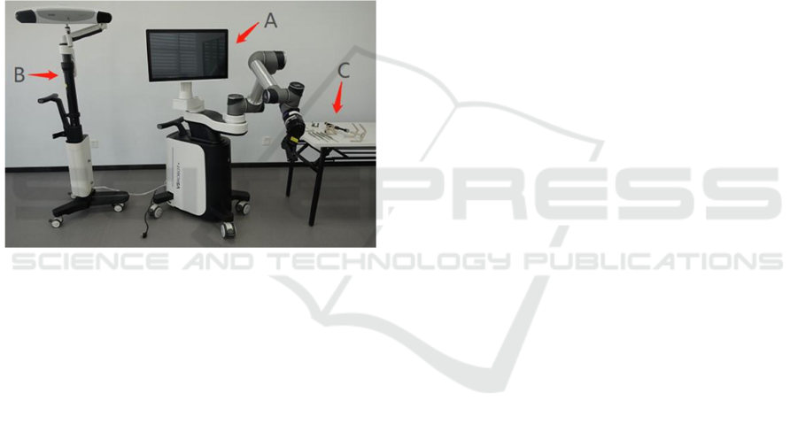
Figure 2: “Tuoshou” Robotic base station (A), optical
tracking system (B), and toolset (C) (Chang, J., et al., 2022).
Nanjing Tuoshou Medical’s high-precision
surgical robot, "Tuoshou," employs deep learning
algorithms to intraoperatively identify bone
landmarks in real time, reducing registration duration.
As shown in Fig.2, The robot consists of a robotic
base station, an optical tracking system (OTS), and a
toolset for navigation and positioning, together with
surgical navigation and positioning software. In a
multicenter randomized controlled trial comparing
thoracolumbar pedicle screw fixation with the
TiRobot system, safety assessments revealed no
significant differences in operative time, instrument
success rate, technical success rate, or procedural
success rate. However, the Tuoshou group exhibited
smaller K-wire placement deviations and higher
pedicle screw accuracy than the TiRobot group,
demonstrating superior precision and reduced
invasiveness for spinal applications (Chang et al.
2022, Gandhi, 2023).
5 NEUROSURGICAL ROBOTS
Neurosurgical robotic systems represent an advanced
integration of robotic arms, image-guided navigation,
and artificial intelligence, designed to enhance
procedural safety through precise positioning and
stabilized operation. Recent breakthroughs in this
field focus on three key areas: frameless high-
precision localization, flexible miniaturized designs,
and multimodal image fusion.
Kim et al. developed an MRI-compatible
continuum robot capable of navigating narrow
anatomical pathways to access deep brain regions
while minimizing collateral tissue damage. Utilizing
smart materials (e.g., McKibben pneumatic artificial
muscles), these robots achieve high-precision
bending and extension, adapting to intricate
intracranial environments (Gandhi, 2023). Advances
in soft robotics have further expanded neurosurgical
applications, with researchers implementing sliding
mode control for stretchable soft robotic modules to
improve motion accuracy and response speed,
thereby enhancing stability in dynamic brain tissue
(Gandhi, 2023).
For multimodal image fusion, the ROSA One
system employs 3D structured light technology to
achieve submillimeter registration accuracy. By
integrating real-time imaging updates to compensate
for brain shift caused by cerebrospinal fluid loss, it
addresses localization inaccuracies inherent in
conventional navigation (Zhou et al., 2023). Modern
deep learning models can predict tumor margins and
vascular distribution, enabling robotic systems to
optimize surgical trajectories. The CyberKnife
system, leveraging AI-driven respiratory
synchronization, maintains 0.5 mm targeting
precision in spinal radiosurgery (Eljamel, 2008).
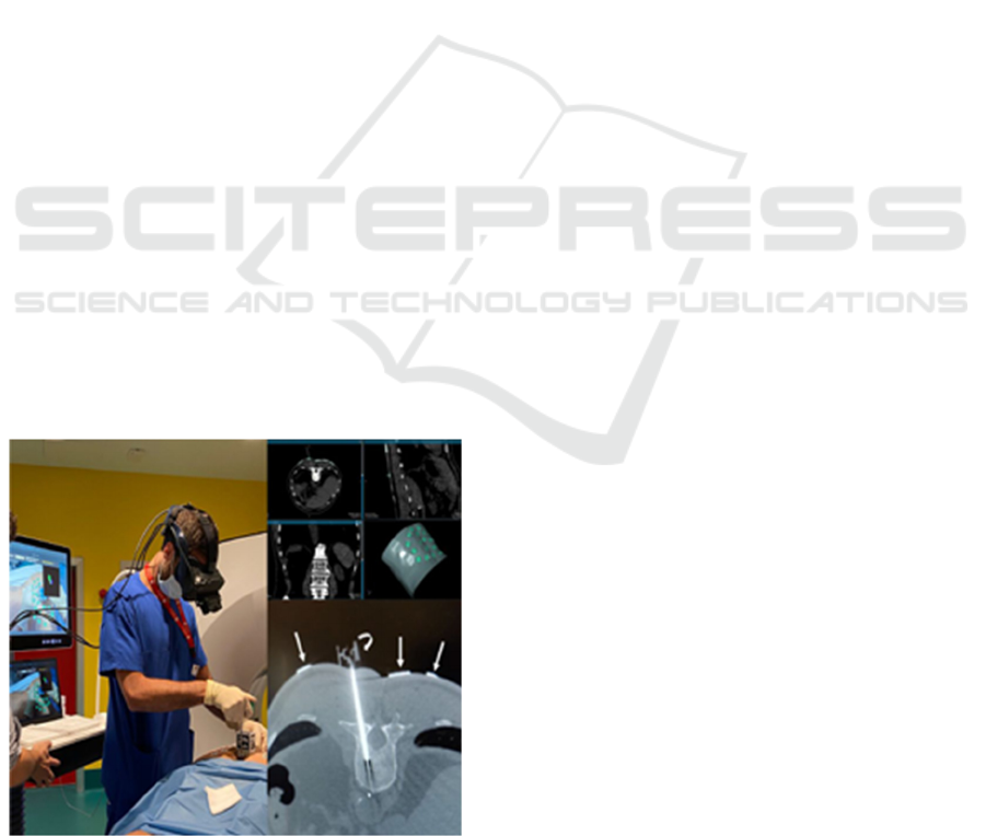
Additionally, neurosurgical robots are being
integrated with virtual reality (VR) simulation
platforms. As shown in Fig.3, the Lindbergh surgical
rehearsal system allows surgeons to train for complex
procedures in a virtual environment, shortening the
learning curve. A collaborative effort between
Mexico’s National Autonomous University and the
University of Tokyo introduced an interactive VR
simulator for transsphenoidal tumor resection. Its
dynamic motion scaling (DMS) feature refines fine
motor control near target areas, reducing healthy
tissue damage. Although this approach increases
operative time, it significantly improves safety
(Heredia-Pérez et al., 2019).
EMITI 2025 - International Conference on Engineering Management, Information Technology and Intelligence
568

(a)
(b)
Figure 3: Interactive VR simulator for transsphenoidal
tumor resection (Heredia-Pérez et al., 2019). (a) User
interacting with the simulator through two haptic interfaces
and a stereo‐monitor; (b) screenshot of the simulation
indicating virtual components
6 CARDIOVASCULAR
INTERVENTIONAL ROBOTS
Cardiovascular surgical robotics is a technology that
utilizes robotic systems to assist surgeons in
performing minimally invasive procedures on the
heart and blood vessels. Its core lies in integrating
three-dimensional high-definition imaging, multi-
degree-of-freedom robotic arms, and remote control
technologies to overcome the physical limitations of
traditional surgery. In recent years, key technological
innovations have been achieved in structural design
and material advancements, AI-assisted instrument
tracking, as well as force feedback and operational
precision.
The third-generation robotic system, co-developed
by Beijing Institute of Technology and Kagawa
University, employs dual linear sliding mechanisms
to enable simultaneous delivery of catheters and
guidewires. Equipped with advanced force-sensing
capabilities, the system supports coordinated
manipulation of catheters and guidewires, surpassing
human surgeons in performance and enabling more
intricate and complex surgical procedures (Zhao et
al., 2022).
A convolutional neural network (CNN)-based
cross-frame real-time recognition model has
demonstrated high accuracy and stability in tracking
and localizing surgical instruments. Specifically, the
model exhibits a low RMSE value, indicating
minimal positioning error, while its high AUC value
confirms superior accuracy in distinguishing different
instrument states, thereby replacing traditional
manual assessment (Zhang et al., 2024).
Meta’s Segment Anything Model (SAM), trained
on over 110 million medical images, achieves zero-
shot transfer learning for segmentation tasks,
adapting to novel image distributions while matching
the performance of fully supervised models. Users
can guide the model via various prompts—such as
clicks, bounding boxes, or text descriptions—to
facilitate target segmentation. The optimized SAM
operates efficiently in real-time environments,
making it suitable for time-sensitive applications
(Zhang et al., 2024).
Researchers from the Medical Robotics and
Micro-Nano Devices Research Center at the
Shenzhen Institute of Advanced Technology, Chinese
Academy of Sciences, have designed and developed
a compact 2-degree-of-freedom (2-DOF) robotic
catheter system. By employing long short-term
memory (LSTM) and gated recurrent unit (GRU)
networks, the system predicts the slave robot’s
position and computes appropriate compensation
values. Simulation studies in CoppeliaSim and
physical experiments validate the effectiveness of the
neural network controller. Results demonstrate that
the controller significantly enhances master-slave
position tracking while minimizing positional errors,
contributing to autonomous navigation and improved
patient safety (Ricciardi et al., 2017).
7 PUNCTURE ROBOTS
Puncture robots are automated medical devices
typically composed of robotic arms, sensors, control
systems, and image-guidance modules. They are
designed to perform precise and navigated puncture
procedures on targeted tissues or organs within the
patient's body. The robotic arm executes the puncture
operation, while integrated sensors provide real-time
monitoring of needle position and orientation to
ensure procedural accuracy. The control system
utilizes image-processing algorithms to determine
Robotic-Assisted Surgery: State-of-the-Art Development, Clinical Challenges, and Future Directions
569
optimal puncture trajectories and guides the robot in
performing highly precise maneuvers. Due to their
ability to enhance surgical precision, reduce
physician radiation exposure, minimize patient
discomfort, and alleviate clinician workload, these
robotic systems are widely adopted in interventional
radiology, oncology, and ultrasonography.
7.1 Interventional Radiology Puncture
Robot
Interventional radiology puncture robots integrate
robotic arms and control systems with advanced
imaging modalities such as CT, MRI, and ultrasound
to perform a variety of clinical functions, including
biopsy, ablation therapy, injection therapy, and
neurointerventions. These robotic systems have been
widely adopted in vascular interventions, particularly
in coronary, peripheral, and neurovascular
procedures. Commercial platforms such as the
CorPath GRX and Magellan Robotic System leverage
remote-control technology to achieve precise
manipulation of guidewires and catheters,
significantly reducing operator fatigue while
enhancing procedural stability. Studies have
demonstrated that these robotic systems achieve
comparable—if not superior—precision compared to
manual techniques (Zhang et al., 2024).
In musculoskeletal interventions, puncture robots
are extensively utilized for needle biopsies, deep
brain stimulation electrode placement, and skull-base
biopsies. For instance, as shown in Fig.4, in bone
biopsy procedures, augmented reality (AR)-guided
navigation systems enable real-time overlay of digital
Figure 4: CT-Guided Lumbar Biopsy with AR Navigation
system (Albano et al., 2023).
content onto the surgical field, substantially reducing
the number of required CT scans and radiation
exposure. This advancement not only minimizes
patient radiation dose but also shortens procedural
duration while maintaining safety and efficacy
(Albano et al., 2023).
7.2 Oncology Puncture Robot
Oncology puncture robots represent an advanced
integration of medical imaging navigation (including
CT, MRI, and ultrasound) with robotic manipulator
systems, designed to assist physicians in performing
percutaneous biopsies, ablations, and other minimally
invasive procedures with high-precision targeting and
trajectory planning. By leveraging CT or MRI-based
image guidance, these robotic systems achieve
accurate tumor localization. Equipped with multi-
degree-of-freedom robotic arms, they enable flexible
needle insertion at various angles while maintaining
operational stability.
During the puncture procedure, the system
provides real-time visualization of needle tip position
and insertion trajectory to ensure targeting accuracy.
Furthermore, the robotic platform can autonomously
optimize puncture paths based on patient-specific
anatomical considerations, thereby avoiding critical
tissues and organs. These technological
advancements not only improve procedural success
rates and reduce complications but also significantly
decrease physicians' radiation exposure, enhancing
overall operational safety (Kissler & Settmacher,
2016).
7.3 Ultrasound-Guided Puncture
Robot
Ultrasound-guided puncture robots represent an
advanced medical technology that integrates real-
time ultrasonic imaging with robotic control systems
to achieve highly accurate percutaneous procedures.
These systems employ three core technological
components: (1) multi-degree-of-freedom robotic
positioning, (2) dynamic ultrasound image feedback,
and (3) intelligent control algorithms.
In prostate cancer diagnostics, Stoianovici et al.
developed an MRI-compatible robotic system for
transrectal ultrasound (TRUS)-guided prostate
biopsies. The system's modular design achieved
submillimeter needle positioning accuracy (<1 mm),
with clinical validation leading to FDA clearance. For
breast interventions, Navarro-Alarcon et al. created a
EMITI 2025 - International Conference on Engineering Management, Information Technology and Intelligence
570

compact robotic system featuring ultrasonic motor
actuation and Bowden cable transmission, enabling
MRI-compatible remote operation with 1.29 mm
targeting precision. This system's open-control
architecture significantly enhanced trajectory
planning flexibility for complex breast biopsy
procedures (Palep, 2009).
8 DISCUSSION
Despite remarkable advancements and demonstrated
innovation potential across multiple medical
specialties, the widespread adoption of surgical
robotic systems continues to confront significant
challenges. The most prominent barrier remains the
prohibitively high total cost of ownership. Taking the
da Vinci Surgical System as a representative
example, the initial capital expenditure encompasses
not only the robotic console and associated
peripherals (including specialized instruments and
stereoscopic camera systems) but also installation and
comprehensive training programs, with complete
system configurations frequently exceeding several
million dollars (Longmore et al., 2020). Furthermore,
recurring operational expenses - comprising single-
use disposable instruments, periodic maintenance
contracts, and mandatory recalibration of reusable
robotic arms - substantially increase the lifetime cost
of ownership.
Within the domain of laparoscopic robotic
systems, current technological limitations manifest
primarily as prolonged operative durations
(particularly evident in complex procedures such as
pancreaticoduodenectomies) and the absence of
sophisticated haptic feedback mechanisms (Kissler &
Settmacher, 2016). This tactile deficiency forces
surgeons to rely exclusively on visual compensation
for precision control, potentially elevating the risk of
iatrogenic tissue trauma (Sebastian, 2017). Emerging
solutions focus on the integration of multimodal
imaging fusion technologies, combining indocyanine
green fluorescence imaging (e.g., Firefly technology),
real-time intraoperative ultrasonography, and
preoperative CT/MRI datasets to enhance surgical
navigation capabilities (Li et al., 2024).
Gynecological robotic platforms face distinct
technical obstacles including restricted visual fields
during large myomectomy procedures and
insufficient end-effector articulation degrees-of-
freedom (Park et al., 2023). Next-generation systems
are evolving toward precision personalized medicine
through artificial intelligence-enhanced preoperative
planning algorithms. Recent investigations
demonstrate that AI-generated three-dimensional
navigation models can automatically delineate tumor
margins and vascular architecture with 92% accuracy
in endometrial cancer surgeries, establishing reliable
"no-fly zones" that significantly reduce ureteral
injury rates (Knigin et al., 2024).
Orthopedic robotic assistance is currently
constrained by substantial physical footprints (with
systems like the da Vinci occupying approximately
100 cubic feet of operating room space), creating
logistical challenges including cable management
issues and increased infection control concerns (Qi &
Liang, 2018). While device miniaturization and
enhanced precision represent clear developmental
trajectories, these engineering advancements may
perpetuate elevated system costs. Promising research
directions include the development of advanced
multimodal image registration software integrating
CT, MRI, and real-time ultrasound data to expand
soft tissue interaction capabilities, alongside the
implementation of U-Net convolutional neural
networks for optimized bone metastasis identification
and osteotomy path planning (Yuan et al., 2024).
Neurosurgical robotic applications confront
unique material compatibility challenges, where
MRI/PET hybrid imaging systems exhibit
characteristic noise artifacts and spatial registration
inaccuracies (Zhou et al., 2023). The impending
deployment of 5G network infrastructure promises to
enable real-time teleoperated procedures, allowing
expert surgeons to remotely control robotic systems
in underserved medical facilities while maintaining
sub-millisecond latency thresholds (Zhou et al.,
2023).
Cardiovascular robotic platforms, despite
demonstrating measurable advantages in reduced
postoperative recovery periods and decreased
complication rates, continue to face fundamental
technical limitations when managing extracorporeal
circulation-dependent procedures such as multi-
vessel coronary artery bypass grafting (Badhwar et
al., 2023). Maintaining instrument stability during
beating-heart operations remains particularly
challenging. Anticipated technological solutions
include the development of advanced force feedback
systems, miniaturized instrument designs, and AI-
powered decision support modules to enhance
complex case adaptability (Onan, 2018).
Percutaneous robotic systems continue to face
fundamental challenges in targeting accuracy. While
Robotic-Assisted Surgery: State-of-the-Art Development, Clinical Challenges, and Future Directions
571

current technologies integrating CT-based volumetric
reconstruction and six-degree-of-freedom robotic
positioning have achieved submillimeter precision,
persistent issues including image distortion artifacts,
signal noise interference, and needle tip localization
uncertainty still compromise procedural reliability.
To address these challenges, the field is advancing
through several innovative approaches: optimizing
trajectory planning algorithms via deep learning,
implementing augmented reality-based real-time
navigation systems, establishing robust telesurgery
network infrastructure, and promoting international
regulatory harmonization. The convergence of
artificial intelligence and augmented reality
technologies represents the most promising
developmental pathway for enhancing procedural
accuracy and reliability.
These synergistic technological advancements will
systematically address current limitations while
facilitating broader clinical adoption across surgical
specialties, ultimately advancing the paradigm of
precision medicine. As technological breakthroughs
continue to emerge and cost-reduction strategies
mature, surgical robotic systems are positioned to
become indispensable components of future
healthcare delivery systems, offering patients
unprecedented levels of procedural safety and
therapeutic accuracy.
9 CONCLUSION
Through a comprehensive evaluation of five key
domains—laparoscopic, orthopedic, neurosurgical,
cardiovascular interventional, and puncture
robotics—surgical robotic technology has evolved
from a standalone assistive tool into a sophisticated
multi-disciplinary integration platform. The
enhanced lymph node dissection accuracy of the da
Vinci Xi system in thoracic surgery, the superior
pedicle screw placement precision of the Tuoshou
orthopedic robot, and the deep brain maneuverability
of MRI-compatible neurosurgical robots collectively
validate the substantial advantages of robotic systems
in improving surgical safety and minimally invasive
outcomes. Particularly in complex anatomical
regions, robotic platforms incorporating fluorescence
navigation and AI-based preoperative planning
achieve levels of precision unattainable with
conventional techniques.
Nevertheless, critical challenges persist in both
technological and clinical implementation.
Economically, the prohibitively high acquisition
costs and substantial annual maintenance expenses
limit widespread adoption. Technically, the absence
of haptic feedback introduces a 29% risk of potential
tissue damage in laparoscopic procedures, while the
bulky footprint of orthopedic robotic systems
complicates operating room logistics. Additionally,
the lack of standardized training protocols
necessitates an average of 80–120 procedures for
surgeons to achieve proficiency, further hindering
scalability. To realize the democratization of this
technology, a tripartite support framework must be
established: (1) Policy-level interventions to optimize
reimbursement structures and pricing mechanisms;
(2) Industrial advancements to accelerate domestic
production of core components; (3) Clinical
standardization to develop evidence-based surgical
pathways. Only through such coordinated
"technology-industry-clinical" synergy can the goal
of accessible precision medicine be achieved,
extending robotic-assisted care to grassroots medical
institutions. Looking ahead, sustained technological
iteration and innovative care models are poised to
usher in a new era of intelligent, personalized, and
equitable surgical practice.
REFERENCES
Albano, D., Messina, C., Gitto, S., et al.: Bone biopsies
guided by augmented reality: a pilot study, European
Radiology Experimental, 7, 40 (2023)
Alletti, S. G., Chiantera, V., Arcuri, G., et al.: Introducing
the new surgical robot HUGO™RAS: system
description and docking settings for gynecological
surgery, Frontiers in Oncology, 12, 898060 (2022)
Autorino, R., Porpiglia, F.: Robotic surgery in urology: the
way forward, World Journal of Urology, 38, 809–811
(2020)
Badhwar, V., Wei, L. M., Geirsson, A., et al.: Contemporary
robotic cardiac surgical training, The Journal of
Thoracic and Cardiovascular Surgery, 2023, 165, pp.
779-783.
Bellos, T., Manolitsis, I., Katsimperis, S., et al.: Artificial
intelligence in urologic robotic oncologic surgery: A
narrative review, Cancers, 16(9), 1775(2024)
Cacciamani, G. E., Shakir, A., Tafuri, A., et al.: Editorial
comment: Best practices in near-infrared fluorescence
imaging with indocyanine green (NIRF/ICG)-guided
robotic urologic surgery: a systematic review-based
expert consensus, Int Braz J Urol, 46(2), 281–282
(2020)
Cerfolio, R., Louie, B. E., Farivar, A. S., et al.: Consensus
statement on definitions and nomenclature for robotic
EMITI 2025 - International Conference on Engineering Management, Information Technology and Intelligence
572

thoracic surgery, The Journal of Thoracic and
Cardiovascular Surgery, 154(3), 1065–1069 (2017)
Chang, J., et al.: Development and clinical trial of a new
orthopedic surgical robot for positioning and navigation,
Journal of Clinical Medicine, 11(23), 7091 (2022)
Eljamel, M. S.: Robotic Applications in Neurosurgery, in
Medical Robotics, Bozovic, V. (ed.), I-Tech Education
and Publishing, pp. 42–64, ISBN: 978-3-902613-18-9.
(2008)
Fujad, A. N. B. A., Elangoyan, H., Deep, K., and Yao, W.:
A robotic flexible drill and its navigation system for total
hip arthroplasty, Annals of Biomedical Engineering,
46(3), 464–474 (2018)
Gandhi, H. S.: Rotitome-G: Principles and design concept of
experimental compliant continuum robotic
microsurgical endoscopic sarcotome for pixel/voxel-
level target access neurosurgery, International Journal of
Computer and Technology, 23, 136–165 (2023)
Heredia-Pérez, S. A., Harada, K., Padilla-Castañeda, M. A.,
et al.: Virtual reality simulation of robotic
transsphenoidal brain tumor resection: Evaluating
dynamic motion scaling in a master‐slave system, The
International Journal of Medical Robotics and Computer
Assisted Surgery, 15, e1953 (2019)
Kissler, H. J., Settmacher, U.: Roboterassistierte
Operationen in der Viszeralchirurgie, Der Chirurg, 87,
633–634 (2016)
Kissler, H. J., Settmacher, U.: Roboterassistierte
Operationen in der Viszeralchirurgie, Der Chirurg, 87,
633–634 (2016)
Knigin, D., Brezinov, Y., Salvador, S., et al.: Surgery
Advances in Gynecologic Tumors: The Evolution and
Outcomes of Robotic Surgery for Gynecologic Cancers
in a Tertiary Center, Current Oncology, 31, 2400-2409
(2024)
Lee, J. H., et al.: Initial experience of single-port robotic
lobectomy for large-sized non-small cell lung cancer: A
single-center retrospective study, Cancers, 16(17), 3091
(2024)
Li, C., Zhang, G., Zhao, B., et al.: Advances of surgical
robotics: image-guided classification and application,
National Science Review, 11, nwae186 (2024)
Longmore, S. K., Naik, G., Gargiulo, G. D.: Laparoscopic
Robotic Surgery: Current Perspective and Future
Directions, Robotics, 9(2), 42 (2020)
Manolache, V., Gonzalez-Rivas, D., Bosinceanu, M. L., et
al.: Uniportal robotic-assisted thoracic surgery for
mediastinal tumors, Annals of Cardiothoracic Surgery,
12(2), 139-141 (2023)
Matsuura, M., Nagao, S., Kurokawa, S., et al.: Surgical
outcomes of da Vinci Xi™ and da Vinci SP™ for early-
stage endometrial cancer in patients undergoing
hysterectomy, Journal of Clinical Medicine, 13(10),
2864 (2024)
Neis, S., et al.: Novel workflow analysis of robot-assisted
hysterectomy through objective performance indicators:
a pilot study, Front. Med., 11(1), 1382609 (2024)
Onan, B.: Coronary revascularization in robotic cardiac
surgery, Cardiovascular Surgery and Interventions, 5(1),
12-23 (2018)
Palep, J. H.: Robotic assisted minimally invasive surgery,
Journal of Minimal Access Surgery, 5(1), 1–7 (2009)
Park, J. Y., Yuki, G., Bak, S. E., et al.: Robotic surgery in
Gynecology: the present and the future, Obstetrics &
Gynecology Science, 66(6), 518-528 (2023)
Ping, L., et al.: Quality of surgery and the use of robotic
surgery in the Tibetan Plateau, Front. Surg., 11(1),
1415704 (2024)
Qi, S., Liang, F.: Clinical Application of Orthopedic Robot
Navigation and Positioning System, in IOP Conf. Series:
Materials Science and Engineering, 452, 042144 (2018)
Ram, P. R., Jeyaraman, M., Jeyaraman, N., et al.:
Revolutionizing orthopedic healthcare: The role of
robotics, Cureus, 15(9), e44820 (2023). doi:
10.7759/cureus.44820.
Ricciardi, S., Cardillo, G., Zirafa, C. C., et al.: Robotic
lobectomies: when and why?, Journal of Visualized
Surgery, 3, 112 (2017)
Sebastian, R.: Advanced robotic technology is the future of
surgery, Revista de la Facultad de Medicina Humana,
17(3), 7–9 (2017)
Settnes, A., Topsoee, M. F.: Complications after abdominal,
vaginal and laparoscopic hysterectomy - Results from
the Danish Hysterectomy Database (DHD) 2003-2011,
Gynecol Surg, 2015, 12, (Suppl 1), pp. S3–S4, Abstracts
of the 24th Annual Congress of the European Society for
Gynaecological Endoscopy (ESGE), Syma - Budapest -
Hungary, 7th - 10th October 2015, doi: 10.1007/s10397-
015-091-8.
Šiaulys, R.: Salpingoovarectomy using Senhance® robotic
system: a case report, Lietuvos chirurgija, 18(1), 38-40
(2019)
van der Slot, M. A., Busstra, M. B., Gan, M., et al.:
NeuroSAFE in radical prostatectomy increases the rate
of nerve-sparing surgery without affecting oncological
outcome, BJU Int, 130(3), 628-636 (2022)
Xue, H., et al.: Advantages of robot-assisted PKP under
local anesthesia in the treatment of OVCF: A
retrospective, non-randomized, controlled, clinical
study, Front. Surg., 11, 1445461 (2024)
Yuan, S., Chen, R., Zang, L., et al.: Development of a
software system for surgical robots based on multimodal
image fusion: study protocol, Frontiers in Surgery, 11,
1389244 (2024)
Zhang, J., Fang, J., Xu, Y., et al.: How AI and Robotics Will
Advance Interventional Radiology: Narrative Review
and Future Perspectives, Diagnostics, 14(13), 1393
(2024)
Zhao, Y., Mei, Z., Luo, X., et al.: Remote vascular
interventional surgery robotics: a literature review,
Quantitative Imaging in Medicine and Surgery, 12(2),
329–347 (2022)
Zhou, S., Gao, Y., Li, R., et al.: Neurosurgical robots in
China: State of the art and future prospect, iScience,
2023, 26, p. 107983.
Zhou, S., Gao, Y., Li, R., Wang, H., Zhang, M., Guo, Y., et
al.: Neurosurgical robots in China: State of the art and
future prospect, iScience, 26, 107983 (2023)
Robotic-Assisted Surgery: State-of-the-Art Development, Clinical Challenges, and Future Directions
573
