
Cancer Detection Using Improved CNN-Based Models
Bo Ning
a
Hangzhou Dianzi University ITMO Joint Institute, Hangzhou Dianzi University, Hangzhou, 310018, China
Keywords: Convolution Neural Network, Cancer Image Analysis, Transfer Learning, Lightweight Models, Multi-Model
Fusion.
Abstract: Medical imaging is a crucial part of clinical diagnosis, yet processing its large-volume data is challenging.
Convolutional neural networks (CNNs) have shown great potential in medical image analysis. However, the
high computational cost and need for large annotated datasets often limit their widespread clinical adoption
This paper focuses on CNN-based models for cancer detection. The paper delves into various innovative
models applied in breast, lung, and skin cancer detection. These newly proposed models excel in specific
aspects, whether in high-precision classification, classification efficiency, or lightweight design. But some
models still face issues like poor generalization on rare diseases and high computational requirements. This
paper summarizes these issues and identifies current and future research directions, including the development
of generalized cancer detection frameworks and the application of transfer learning techniques. Overall, the
paper highlights the enormous potential of CNN-based models in medical imaging while pointing out the
need for continuous research and development to overcome existing challenges and limitations.
1 INTRODUCTION
Medical imaging, as an important auxiliary means of
clinical diagnosis, is a key link to ensure the success
of treatment in the process of medical diagnosis and
treatment. it plays an important role in life science
research. Medical imaging includes X-rays,
ultrasound, computed tomography, magnetic
resonance imaging, positron emission tomography,
etc. These medical image data are huge in volume and
difficult to process in clinical diagnosis. Therefore,
automatically detecting diseases from medical
images has become a key issue in the medical field
(Chen, Mat Isa and Liu, 2025).
Medical image analysis requires powerful algorithms
that can extract details from high-dimensional and
noisy datasets. Convolutional neural networks are
precisely the unique options that can address this
challenge. Convolutional Neural Network (CNN) is a
multi-layer neural network used to extract visual
patterns from pixel images. It can automatically
extract and select features and classify them,
providing radiologists with faster and more accurate
diagnostic results in real time. In the field of medical
image analysis, CNN has become an advanced
a
https://orcid.org/0009-0009-3995-4964
algorithm for tasks such as disease detection, organ
segmentation and image enhancement (Chen, Mat Isa
and Liu, 2025) (Patel and Khan, 2022) (Mienye,
Swart and Obaido et.al, 2025). The convolutional
neural network technology of deep learning is widely
applied in the analysis of medical images. It is
particularly used for diagnosing different types of
diseases, such as breast cancer, Alzheimer's disease,
brain tumors, and so on. Algorithms based on deep
convolutional neural networks have achieved
remarkable results in the analysis of medical images.
For medical image data, various types of transfer
learning methods have been proposed and have
achieved remarkable results, such as AlexNet,
VGGNet, ResNet, GoogleNet, etc (Salehi, Khan and
Gupta. et al, 2023).
However, there are still many challenges in the
application of CNN in the medical field, which
hinders its wide application in clinical
practice(Mienye, Swart and Obaido et.al, 2025).
Some models still face challenges such as insufficient
generalization ability on rare diseases or niche
datasets, high demands for computing resources, and
difficulties in multimodal data fusion. This clarifies
that CNN still needs to continuously innovate in
138
Ning, B.
Cancer Detection Using Improved CNN-Based Models.
DOI: 10.5220/0014323000004718
Paper published under CC license (CC BY-NC-ND 4.0)
In Proceedings of the 2nd International Conference on Engineering Management, Information Technology and Intelligence (EMITI 2025), pages 138-144
ISBN: 978-989-758-792-4
Proceedings Copyright © 2025 by SCITEPRESS – Science and Technology Publications, Lda.

Figure 1: Basic architecture of CNN
aspects such as architectural design, interpretable
AI technology, cross-modal and cross-domain
generalization, to overcome all these difficulties.
This study will conduct a comprehensive review of
the CNN model framework corresponding to the
diagnosis of various types of cancers. It will first
introduce the working principle of CNN, and then
analyze the application cases of CNN in the diagnosis
of breast cancer, lung cancer and skin cancer
respectively. The paper will focus on elaborating the
cutting-edge model architecture based on CNN used
therein, as well as the advantages of these models and
the achievements obtained. This review aims to
explore strategies for improving the performance of
cancer diagnosis models, and provide researchers
with guidance on applying CNN to solve medical
problems such as cancer diagnosis and analysis.
2 THE PRINCIPLE OF CNN
As Figure 1 shows, as a hierarchical and cascade
model, the CNN starts by capturing pixel-level
information from the lowest layer of the input image
matrix and progressively extracts critical feature
information through each subsequent layer.
Convolution, pooling, and fully connected layers are
the three key components of CNN architecture. The
convolution layer uses a convolutional operation of
filters on the input image to extract local information.
After that, higher-level features will be extracted by
moving the acquired feature mappings to the
subsequent convolution layer. After the convolutional
layer, the image dimension is decreased using the
pooling layer, also known as the down sampling
layer. Because max-pooling lowers the dimension
while keeping the image's primary feature, it is
frequently employed as a down sampling technique.
After the last convolutional layer, the fully connected
layer flattens the feature map into a vector. To
normalize the output and produce a final output in
probability form, a softmax function is often applied
at the output layer. This probability indicates the
picture belongs to a particular category. CNN
typically updates the convolution kernel and fully
connected layers' weights during the training phase
using a gradient descent approach. The learning
method updates the weights until convergence by
backpropagating a classification loss into the network
(Chen, Mat Isa and Liu, 2025).
3 THE APPLICATION OF CNN IN
BREAST CANCER DETECTION
3.1 Swincnn Fusion Transformer
Realizes Classification of Multi-
Subtype Tumors
V. Sreelekshmi et al. proposed the SwinCNN
architecture, integrating Depthwise Separable
Convolutional Neural Network and Swin
Transformer (Sreelekshmi Pavithran and Nair, 2024).
This model realizes the high-precision classification
of benign and malignant tumors and their subtypes.
The proposed mode SwinCNN, is shown in Figure 2.
This architecture consists of two channels: the lower
channel is the Local Feature Extraction module
(LFM), and the upper channel is the Global Feature
Extraction module (GFM). The convolutional layer in
Cancer Detection Using Improved CNN-Based Models
139

GFM is the main feature extractor, consisting of 64 7
×7 filters. It is used to generate the feature maps of
the input data. The convolution kernels in its three
residual blocks are of size 3×3, and the number of
filters is 64, 128, and 256, respectively. The final
feature map of GFM is downsampled and fed into the
Swin Transformer block to extract context
information. LFM employs three different types of
depthwise separable convolutional blocks. Finally,
the outputs of GFM and LFM were combined and
input into the softmax layer to calculate the
classification probability for different breast cancer
types. The result corresponding to the highest
probability was taken as the final classification.
Figure 2: The architecture of SwinCNN
The innovative transformation of CNN is one of
the core technical contributions of the proposed
model. Traditional CNNs use standard convolutions,
which are inefficient in high-resolution scenes of
medical images. Their computation scales cubically
with the number of channels and the size of the
convolution kernel. V. Sreelekshmi et al. proposed a
scheme that fuses GoogleNet and Xception models.
GoogleNet provides multi-scale features, and
Xception ensures computational efficiency. The
integration of the two enriches the representation of
local features (covering details at different scales) and
avoids substantial computational overhead. This
makes the model more comprehensive and
lightweight on local feature extraction. The
traditional CNN is limited to the local receptive field,
which can only capture the local cell morphology and
fail to model the global structure of the tumor tissue.
This leads to difficulties in differentiating invasive
carcinomas from carcinomas in situ. SwinCNN
compensates for this shortcoming with the
Transformer path. On the BACH dataset, the recall of
invasive cancer classification is improved from 85%
of ResNet to 91.4%.
Finally, the model proposed by V. Sreelekshmi et
al. was validated on three major public datasets.
Experimental results show that the model achieves
extremely high accuracy in detecting various tumors
and their subtypes, and demonstrates strong
generalization ability for clinically critical subtypes
(Sreelekshmi Pavithran and Nair, 2024).
3.2 UWB-CNN-LSTM
Enables Early
Tumor Detection and Localization
Min Lu et al. conducted research on the early
detection and localization of breast cancer. They
developed an end-to-end framework based on ultra-
wideband (UWB) microwave technology and deep
learning to achieve automatic detection of breast
cancer and breast quadrant localization. The CNN-
LSTM hybrid network framework proposed has
achieved full-process automated feature learning.
LSTM processes the time domain signals from three-
channel signals, and remembers the time dependence
of tumor responses through a gating mechanism. This
effectively alleviates the problem of vanishing
gradients in traditional RNNs. CNN automatically
captures the local waveform features of UWB signals
EMITI 2025 - International Conference on Engineering Management, Information Technology and Intelligence
140

through two layers of convolution, replacing the
traditional manual extraction of time-domain and
frequency-domain features (Lu, Xiao and Pang et
al,2022).
The scheme proposed by Min Lu et al. mainly has
three advantages. The model adopts a lightweight
network architecture, using a shallow CNN in
combination with an LSTM. This strikes a balance
between computational efficiency and accuracy. The
model’s training time on a 16-core CPU is only 1012
seconds, making it suitable for clinical scenarios with
limited resources. The model also reduces the cost of
breast cancer screening. The UWB devices adopted
in the scheme have a much lower cost than MRI,
making them suitable for popularization in grassroots
healthcare settings. The proposed model can also
achieve full-process automation from signal input to
quadrant positioning, reducing human interpretation
errors.
Finally, through multi-scenario verification, the
proposed model achieves high-accuracy tumor
detection (99.56%) and quadrant localization (F1
score ≥ 97%). The CNN-LSTM hybrid network
framework enables tumor detection and breast
quadrant localization while mitigating issues of high
costs, radiation hazards, and tedious manual feature
engineering in traditional methods. The model
requires a large amount of data for constructing the
dataset of the training network. This is a problem that
hinders the application of the proposed method in
clinical practice.
3.3 Binary Classification for Breast
Cancer Based on Multi-Model
Integration
Samriddha Majumdar et al. Integrated three models,
namely GoogleNet, VGG-11 and MobileNetV3, to
solve the problem of binary classification (benign and
malignant) of breast cancer pathological images.
GoogleNet uses Inception blocks for multi-scale
feature extraction (Majumdar, Pramanik and Sarkar,
2023). Large convolution kernels capture the overall
information of the image, while small convolution
kernels focus on capturing fine details. This enables
efficient fusion of multi-scale features. VGG11’s
shallow architecture captures local details, and its
low-depth design reduces the number of parameters
of the model. MobileNetV3_Small’s bottleneck
layers enable efficient high-magnification analysis.
Aiming at the problems of insufficient
generalization ability of a single model and fixed
weight of a traditional ensemble model, they adopted
a fusion strategy based on the Gamma function to
achieve more accurate classification. The proposed
scheme uses transfer learning to make up for the
shortcomings of CNN which requires massive data
and long time to train medical images. The test results
of the model are also remarkable. The model
performs well on BreakHis (four magnifications) and
ICIAR-2018 datasets. This proves that the method
does not rely on a specific data distribution, and has
excellent generalization ability and robustness.
4 APPLICATION OF CNN IN
LUNG CANCER DETECTION
4.1 Lightweight CNN Achieves
Efficient Classification of Lung
Cancer
Mohd Mohsin Ali et al. focus on the application of
lightweight deep learning models in medical imaging.
They developed a lightweight and efficient CNN
model to enable automatic classification of lung
cancer (benign, malignant, and normal), mitigating
the issues of high computational costs and difficult
edge device deployment in traditional models (Ali,
Jain and Chauhan et al,2023).
Figure 3 shows the framework of the proposed
model. The first convolutional layer (Conv2D) has 32
6×6 kernels with a step size of 3×3 and an activation
function of ReLU. This convolution layer extracts
basic visual features (e.g., lung nodule edges,
texture), and the output feature map size is
169×169×32. The ReLU activation function avoids
gradient disappearance while introducing
nonlinearity and has low computational cost, which is
suitable for lightweight design. The second
convolutional layer (Conv2D) also has 32 6×6
kernels with a step size of 3×3. This is to further
extract complex texture features, and the output
feature map size is 27×27×32. The two-layer
convolution balances detail and semantic information
through feature extraction at different depths. The
flatten layer converts 3D feature maps into a 1D
vector (5,408 dimensions), feeding directly into the
fully connected layer. The hidden layer contains 128
neurons, reducing the number of parameters through
sparse connections. The output layer has 3 neurons
(corresponding to three classes: benign, malignant,
and normal), with the SoftMax activation function
generating class probability distributions.
Cancer Detection Using Improved CNN-Based Models
141
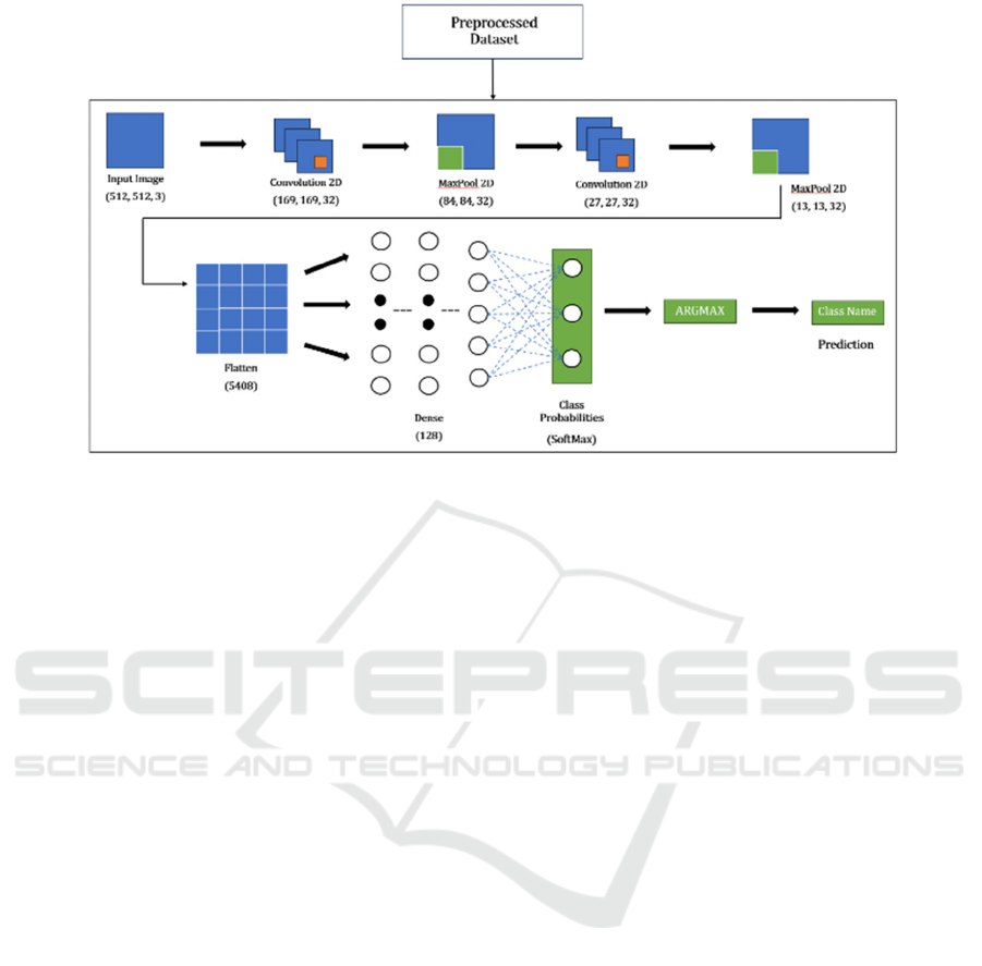
Figure 3: Neural network design diagram.
The model proposed by Mohd Mohsin Ali et al.
has several advantages. It uses a lightweight
architecture, and the storage size is compressed to
8.43MB through shallow convolution (2 layers),
large-step pooling, and low-dimensional fully
connected layers. The model can achieve efficient
feature extraction, and it uses 6×6 convolution kernel
to balance the receptive field and the amount of
computation. Finally, the model achieves a validation
accuracy of 99%, a training time of 1 minute, and a
model size of 8.43MB. Compared with mainstream
models such as XceptionNet and MobileNet, it
significantly outperforms in all three core metrics(Ali,
Jain and Chauhan et al, 2023).
The core contribution of the proposed model lies
in breaking through the trade-off between
performance and complexity of traditional deep -
learning models. While achieving a lightweight
design, the model maintains a high accuracy of 99%.
This achievement not only provides a new solution
for lung cancer screening but also points out the
development direction of "lightweight and efficient
model".
4.2 Hybrid Detection Model
of SMA-
CNN and Squeeze-Inception V3
Geethu Lakshmi G et al proposed a hybrid model of
"SMA-CNN feature extraction and Squeeze
Inception V3 classification" for the accuracy and
efficiency of CT image detection of lung cancer
(Lakshmi and Nagaraj, 2025). The goal of the
proposed model is to increase the detection rate of
early-stage lung cancer while maintaining
computational lightness. SMA-CNN uses the slime
mold algorithm to dynamically adjust the weight of
CNN's convolution kernel, which enhances the
feature capture of low-contrast tumors and replaces
the traditional CNN training with fixed parameters.
Squeeze-Inception V3 combines the lightweight
design of SqueezeNet with the multi-scale feature
extraction of Inception V3. The Fire Module of
SqueezeNet compresses the number of channels
through a 1×1 convolution kernel, and its parameter
number is 1/50 of the parameter number of AlexNet.
The average pooling layer of SqueezeNet replaces the
fully connected layer, and the decomposition
convolution of Inception V3 is combined to further
reduce the computational complexity. Inception V3
captures nodule contours and details of different
scales by using convolution kernels of different sizes
such as 1×1 and 3×3 in parallel. Traditional single-
scale convolutions tend to miss detecting small
tumors, while the multi-branch structure of Inception
V3 enables feature complementation through
different receptive fields, achieving a 12%
improvement in the recognition rate of small-sized
lesions. Finally, the proposed model achieved
maximum specificity, accuracy, and sensitivity on the
Chest CT-Scan dataset (1,001 cases, covering four
classes: adenocarcinoma, large cell carcinoma,
squamous cell carcinoma, and normal), with the
respective rates of 94%, 95%, and 98%. This model
reduces the missed diagnosis and misdiagnosis rates
for lung cancer, while the lightweight design
EMITI 2025 - International Conference on Engineering Management, Information Technology and Intelligence
142

enhances its adaptability to resource-constrained
environments.
5 APPLICATION OF CNN IN
SKIN CANCER DETECTION
Ashwani Kumar realize high precision of skin cancer
detection by improving the Falcon finch depth of
CNN. The core of the proposed model lies in the
combination of ResNet feature transfer and the hybrid
optimization algorithm, breaking through the
performance bottleneck of traditional CNNS in small
samples and complex scenes (Kumar, 2024). The
proposed model utilizes Resnet-101 to extract deep
features, combines statistical features for dimension
reduction processing, forms a 2048-dimensional
feature vector, and retains the subtle structural
differences in the high-dimensional space.
Subsequently, the features are fed into the improved
CNN. The Falcon Finch algorithm dynamically
adjusts the weights of the fully connected layer
through the echolocation mechanism. The FFO
algorithm is used to adjust the hyperparameters of the
deep CNN classifier, and the optimal combination is
determined through 100 iterations. The algorithm
optimizes the hyperparameters to improve the
efficiency and performance of the classifier, and then
improves the accuracy and speed of skin cancer
detection. Moreover, the FFO algorithm enhances the
robustness of the classifier and accelerates the
convergence speed, so that the model can complete
the training in a shorter time and achieve better
detection performance.
Finally, the experimental results of Ashwani
Kumar et al. show that the model optimized by FFO
performs well in terms of accuracy, sensitivity and
specificity. Ashwani Kumar et al. presented two-
index validation results: in k-fold cross validation
(k=8), the accuracy, sensitivity, and specificity of the
proposed model are 93.59%, 92.14%, and 95.22%,
which proves the robustness of the model in small
sample scenarios. In the training percentage test (80%
data training), the accuracy, sensitivity, and
specificity of the proposed model are 96.52%,
96.69%, and 96.54%, which verifies the efficiency
under large-scale data. In the comparison experiment,
compared with the traditional CNN (accuracy
80.78%), HHO-CNN (86.36%) and SSA-CNN
(86.88%), the accuracy of FFO-CNN was increased
by 12.81%, 7.23%, and 6.71%, respectively. Its
advantage in specificity (distinguishing benign
tumors) is even more significant. It proves the
effectiveness of FFO in improving the performance
of the model. The introduction of Falcon Finch
optimization provides a new solution to the problem
of parameter tuning of deep neural networks.
The proposed model also faces difficulties. Due to
the complex lesion structure, the similar appearance
of benign and malignant lesions can lead to
difficulties in visual analysis. In the future, hybrid
classifiers can be used for skin cancer detection and
classification to provide a more comprehensive
pathological classification solution.
6 CONCLUSIONS
Convolutional neural networks have promoted the
progress of cancer detection, tumor type
discrimination, and so on, and show that they still has
great potential for development in the medical field.
This study further analyzes various CNN-based
models for cancer detection image classification and
shows the results achieved by each model in each
case. This paper presents several solutions for
researchers aiming to use CNN models to address
cancer detection challenges, helping them understand
the corresponding models suitable for various types
of cancer detection. Some of these CNN-based
models bring higher cancer detection accuracy, some
realize the full automation of the detection process,
and some have lightweight architectures. While these
new CNN-based models have achieved such
successes, there are also many problems in their
development path: the demand for computing
resources of some models is too high, the training of
some models still requires a large numbers of data
sets to improve the accuracy. And some models can
only classify a limited number of categories, resulting
in incomplete pathological classification among other
issues.
To address these challenges, future research can focus
on how to improve network architecture to achieve
high accuracy while maintaining model
lightweighting, so that the model can adapt to
resource-limited grassroots scenarios. Future
research should also attempt to explore the transfer
learning strategy from skin cancer and breast cancer
models to other cancers, and establish a generalized
cancer detection framework, so as to make up for the
shortcomings of CNN in training medical images that
require massive data and extensive training time. to
improve the comprehensive judgment capacity of
difficult cases, future research might also try to design
a multimodal fusion architecture that integrates CT,
MRI, pathological pictures, and clinical data to create
Cancer Detection Using Improved CNN-Based Models
143

a full-dimensional cancer diagnostic model. Through
improvements in these aspects, CNN will continue to
provide better solutions for problems in the medical
field.
REFERENCES
Ali, M. M., & Jain, V., & Chauhan, A., & Ranjan, V., &
Raj, M. (2023). Automated Lung Cancer Detection
Using Lightweight Neural Network. 2023 International
Conference on Modeling, Simulation & Intelligent
Computing (MoSICom), 219-222.
Chen, C., & Mat Isa, N. A., & Liu, X. (2025). A Review of
Convolutional Neural Network Based Methods for
Medical Image Classification. Computers in Biology
and Medicine, 185, 109507.
Kumar, A., & Kumar, M., & Bhardwaj, V. P., & Kumar, S.,
& Selvarajan, S. (2024). A Novel Skin Cancer
Detection Model Using Modified Finch Deep CNN
Classifier Model. Scientific Reports, 14(1), 11235.
Lakshmi G, G., & Nagaraj, P. (2025). Squeeze-Inception
V3 with Slime Mould Algorithm-Based CNN Features
for Lung Cancer Detection. Biomedical Signal
Processing and Control, 100, 106924.
Lu, M., & Xiao, X., & Pang, Y., & Liu, G., & Lu, H. (2022).
Detection and Localization of Breast Cancer Using
UWB Microwave Technology and CNN-LSTM
Framework. IEEE Transactions on Microwave Theory
and Techniques, 70(11), 5085-5094.
Majumdar, S., & Pramanik, P., & Sarkar, R. (2023).
Gamma Function Based Ensemble of CNN Models for
Breast Cancer Detection in Histopathology Images.
Expert Systems with Applications, 213, 119022.
Mienye, I. D., & Swart, T. G., & Obaido, G., & Jordan, M.,
& Ilono, P. (2025). Deep Convolutional Neural
Networks in Medical Image Analysis: A Review.
Information, 16(3), 195.
Patel, S., & Khan, N. R. (2022). COVID-19 Detection by
Medical Images with Pretrained Transfer Learning-
Based Model Using CNN: A Systematic Review.
Proceedings of 2022 IEEE International Conference on
Current Development in Engineering and Technology,
CCET 2022.
Salehi, A. W., & Khan, S., & Gupta, G., & Alabduallah, B.
I., & Almjally, A., & Alsolai, H., & Siddiqui, T., &
Mellit, A. (2023). A Study of CNN and Transfer
Learning in Medical Imaging: Advantages, Challenges,
Future Scope. Sustainability, 15(7), 5930.
Sreelekshmi, V., & Pavithran, K., & Nair, J. J. (2024).
SwinCNN: An Integrated Swin Transformer and CNN
for Improved Breast Cancer Grade Classification. IEEE
Access, 12, 68697-68710.
EMITI 2025 - International Conference on Engineering Management, Information Technology and Intelligence
144
