
Detection of Brain Tumors Using Advanced Image Processing and
the Ensemble Model and YOLO Family
P. Poorna Priya and N. Vidhya Sree
Department of Electronics and Communication Engineering, DIET, Anakapalle, Visakhapatnam, Andhra Pradesh, India
Keywords: You Only Look once, CNN, Image Processing, Brain Tumor, Machine Learning, Deep Learning, Tumor
Detection, Classification.
Abstract: Effective diagnosis and therapy of brain tumors depend on their identification and classification. Using the
Brain Tumor dataset, this study makes use of sophisticated transfer learning models and deep convolutional
neural networks (DCNNs). When models like DCNN, ResNet152, EfficientNetB2, Exception, and
Nonmobile were tested, an ensemble of Exception and Nonmobile produced the best accuracy (98.1%). Grade
0 (no malignancy) to Grade III (big tumor) were the four grades into which tumors were divided. With a mean
average precision (map) of 78.9%, YOLOv9 fared better than other models for anomaly detection. A Flask-
based interactive interface was created for safe and easy access in order to improve usage.
1 INTRODUCTION
Unusual cell growths in the brain called brain tumors
raise the pressure inside the skull, impairing essential
processes including movement, speech, and thought.
They are categorized as either malignant (aggressive,
fast-growing, and invasive) or benign (slow-growing,
less invasive), and early identification is essential for
successful treatment. Conventional diagnosis uses
MRI and CT scans, which need to be interpreted by
experts and can be laborious and error-prone.
Improvements in deep learning (DL), especially
convolutional neural networks (CNNs), and artificial
intelligence (AI) have greatly enhanced tumor
categorization and detection (Kumar et al., 2022;
Ullah et al., 2022; Babu Vimala et al., 2023). Better
patient outcomes result from these models increased
diagnostic precision, accelerated analysis, and
support for early action. In order to increase
precision, effectiveness, and clinical decision-
making, this study investigates the use of deep
learning and machine learning for automated brain
tumor identification (Mathivanan et al., 2024; Das &
Goswami, 2024).
2 RELATED WORKS
MRI scans have been used in a number of researches
to investigate AI and deep learning methods for brain
tumor identification. Asif et al. used pre-trained CNN
architectures such as VGG16, Reset, and Inception to
introduce transfer learning-based models and show
increased classification accuracy. By improving CNN
models and focusing on feature extraction for
improved tumor classification, Srinivas et al. further
improved this methodology. CNN-based transfer
learning was used by Bairagi et al., who demonstrated
how effective it is at processing intricate medical
images. In order to improve detection accuracy with
little data, Anjum et al. optimized pre-trained
networks using ResNet50 and InceptionV3. By
comparing several CNN models, Khaliki and
Başarslan were able to verify that transfer learning
performed better in terms of classification accuracy
and computational efficiency than traditional three-
layer CNNs. By combining DenseNet169 with
machine learning classifiers such as Random Forest
and SVM, Khan et al. developed Crossover NET,
which improved tumor classification. CNNs and
transfer learning were integrated by Incir and
Bozkurt, who showed enhanced performance on
sizable and varied datasets. In order to highlight the
significance of flexible AI models in medical
imaging, Sadad et al. expanded deep learning
628
Priya, P. P. and Sree, N. V.
Detection of Brain Tumors Using Advanced Image Processing and the Ensemble Model and YOLO Family.
DOI: 10.5220/0013939700004919
Paper published under CC license (CC BY-NC-ND 4.0)
In Proceedings of the 1st International Conference on Research and Development in Information, Communication, and Computing Technologies (ICRDICCT‘25 2025) - Volume 5, pages
628-633
ISBN: 978-989-758-777-1
Proceedings Copyright © 2026 by SCITEPRESS – Science and Technology Publications, Lda.
applications to multi-class tumor classification
(benign, malignant, and metastatic) using VGGNet
and ResNet.
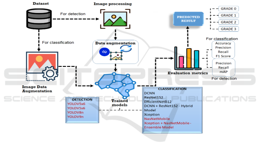
3 MATERIALS AND METHODS
The proposed brain tumour detection and
characterization framework fosters areas of strength
for a for-brain tumour identification and grouping by
utilization of a Brain Tumour dataset. To further
develop grouping accuracy, the framework totals
“deep convolutional neural networks (DCNNs)” with
cutting edge move learning models. It uses a half
breed model of DCNN and ResNet152 serving as the
baseline for growth grouping, utilizing modern
models like "DCNN, ResNet152, EfficientNetB2,
Exception, and Nonmobile." The four forms of cancer
are classified as "Grades 0 (no tumor), Grade I (little
tumor), Grade II (medium-sized tumor), and Grade III
(big tumor)”. To detect abnormalities in brain scans,
the invention makes use of cutting-edge YOLO
models, such as "YOLOv5x6, YOLOv5s6, YOLOv8,
YOLOv9."These models are trained and optimized to
enable precise cancer identification and
classification. Incorporated utilizing a Flask based
intelligent point of interaction, this offers an easy to
understand stage for clinical applications and safe
client recognizable proof.
Figure 1 shows The
Proposed Architecture.
Figure 1: The proposed architecture.
By integrating data augmentation, image
processing techniques, and deep convolutional neural
networks (DCNNs) for object detection and picture
sequencing, this architecture (fig.1) develops a deep
learning-based framework for image analysis.
Utilizing YOLO models “YOLOv5x6, YOLOv5s6,
YOLOv8n, and YOLOv9n” the identification module
exactly distinguishes objects inside pictures. The
framework makes use of cutting-edge models for
characterization, such as "ResNet152,
EfficientNetB2, Exception, and Nonmobile," in
addition to a proprietary cross-breed model that
combines "DCNN and ResNet152 and an outfit
model that combines Exception and Nonmobile."
Using metrics such as "mean Average Precision
(map), F1-score, recall, accuracy, and precision,
“execution is entirely surveyed areas of strength for
ensuring across both recognition and characterization
errands.
3.1 Dataset Collection
MRI images which fall into both harmless and
threatening tumour classes - make up the dataset
utilized for brain tumour detection and arrangement.
Publically available clinical picture files
remembering the Brain Tumour Dataset for Kaggle
give the dataset and there are many named MRI scans
that are pre-handled to guarantee uniformity in size
and quality. A complete rationale for model training
is provided by the division of the data into several
Detection of Brain Tumors Using Advanced Image Processing and the Ensemble Model and YOLO Family
629

growth grades: "Grade 0 (no tumour), Grade I (small),
Grade II (medium), and Grade III (big)".
3.2 Pre-Processing
3.2.1 Classification
Augmenting Image Data: Enhancement of image data
for characterization refers to different methods
designed to work on the dataset and then increase
model guesswork. Re-scaling the picture to normalize
the size, shear changes to add minor mathematical
bends, zooming in to get better subtleties, and level
flips to duplicate a few survey points. Changing the
picture likewise ensures consistency in extents, so
empowering great preparation. These strategies
together increment the dataset and empower the
model to more readily oversee variances in genuine
visual data.
3.2.2 Detection
Image Processing: Image processing for detection
comprises in a few significant stages intended to
prepare the information for model surmising. To
normalize the picture for input, it initially gets
transformed into a mass item. The class is hence
determined; next comes announcing the jumping box
for detection. To simplify dealing with, the picture is
transformed into a NumPy array. The organization
layers are perused for stacking the pre-trained model
and result layers are separated. Added are the picture
and explanation records, which make an
interpretation of BGR to RGB, produce a veil, and
resize the image to fit the information particulars for
the model.
3.2.3 Data Augmentation
In detection, data augmentation alludes to strategies
that further develop the summing up limit of the
model. To start with, the picture is randomized to add
preparing changeability. Arbitrary pivots then, at that
point, are utilized to imitate different directions,
consequently directing the model to learn invariant
properties. To additionally fluctuate datasets, picture
changes including mutilation, interpretation, or
scaling are likewise finished. These expansion
methods ensure the model's capacity to productively
recognize objects from a few points, positions, and
circumstances, consequently fortifying its
presentation and strength in reasonable settings.
3.3 Algorithms
3.3.1 For Classification
DCNN: Deep elements from brain tumour pictures
are extricated utilizing DCNN, which gains muddled
examples and isolates harmless from cancer cases
along these lines empowering dependable tumour
grouping.
ResNet152: Reset 152 is utilized since it can
oversee deep neural networks, consequently working
on the limit of the model to learn and classify growth
photographs with higher accuracy by residual
learning methods.
EfficientNetB2: Utilizing its adaptable
engineering to deal with brain tumour pictures with
less boundaries and quicker training times,
EfficientNetB2 is utilized to amplify arrangement
accuracy while safeguarding effectiveness.
DCNN + ResNet152 - Hybrid Model: Meaning to
involve the two plans' assets for further developed
highlight extraction and more prominent accuracy in
distinguishing brain tumour pictures, the crossover
model mixes DCNN and ResNet 152.
Exception: Exception’s powerful convolutional
design assists the organization with extricating
undeniable level data from growth pictures, hence
giving improved characterization results to brain
tumour identification.
NasNetMobile: Especially accommodating for
brain tumour picture examination with restricted
assets, Nonmobile gives lightweight execution while
keeping up with extraordinary grouping accuracy,
thus empowering successful component extraction in
versatile settings.
Exception + NasNetMobile - Ensemble Model:
Consolidating “Exception with NasNetMobile”
permits the group model to take utilization of the two
organizations' advantages, subsequently further
developing arrangement accuracy by joining different
component separating powers from the two models.
3.3.2 For Detection
YoloV5x6: Through continuous item discovery
abilities, YOLOv5x6 gives rapid handling and precise
distinguishing proof of cancer regions, consequently
empowering identification of irregularities in brain
tumor images.
YoloV5s6: More modest tumor regions can be
successfully distinguished utilizing YOLOv5s6,
which ensures quicker execution for constant
applications and jelly extraordinary accuracy in
anomaly recognition.
ICRDICCT‘25 2025 - INTERNATIONAL CONFERENCE ON RESEARCH AND DEVELOPMENT IN INFORMATION,
COMMUNICATION, AND COMPUTING TECHNOLOGIES
630
YoloV8: On account of its extended engineering
and component extraction strategies, YOLOv8 is
utilized for refined discovery occupations; it offers
higher accuracy and speed in growth finding,
especially in confounded pictures.
YoloV9: Coordinated for its "state-of- the-art
detection" abilities, YOLOv9 gives uncommon
accuracy in spotting brain tumor abnormalities with
few bogus up-sides, so ensuring steady outcomes for
use in centers.
The table 1 illustrate the performance
evaluation table for classification and table 2 shows
Performance Evaluation Table for Detection.
4 PERFORMANCE METRICS
Accuracy.
Accuracy
(1)
Precision.
Precision
(2)
Recall.
Recall
(3)
F1-Score.
F1 Score 2 ∗
∗ 1001 (4)
MAP.
"𝑚𝐴𝑃
∑
𝐴𝑃
" (5)
Where,
MAP- mean average precision
AP- the “average precision
K-over all clients or searches
For Classification.
Table 1: Performance evaluation table for classification.
Model Accuracy Precision Recall F1-Score
EfficientNetB2 89.7 89.7 89.7 89.7
ResNet152 77.5 82.3 74.2 76.9
DCNN 81.1 55.0 93.1 67.9
Exception 85.4 86.9 83.0 84.3
NASNetMobile 92.9 93.1 93.0 93.0
For Detection:
Table 2: Performance evaluation table for detection.
Model Precision Recall mAP
YOLOV5s6 86.3 79.3 86.9
YOLOV5x6 73.3 60.7 81.7
YOLOV8 73.2 80.5 86.7
YOLOV9 84.3 78.9 78.6
5 RESULTS
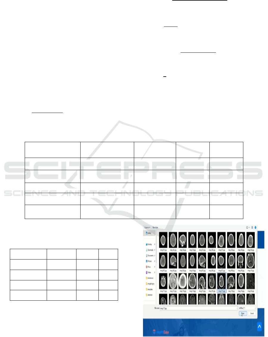
The figure 2 shows Uploading an Input Image for Detection
and figure 3 shows Final Outcome. The figure 4 and 5
illustrate Uploading an Input Image for Classification and
Final outcome.
Figure 2: Uploading an input image for detection.
Detection of Brain Tumors Using Advanced Image Processing and the Ensemble Model and YOLO Family
631
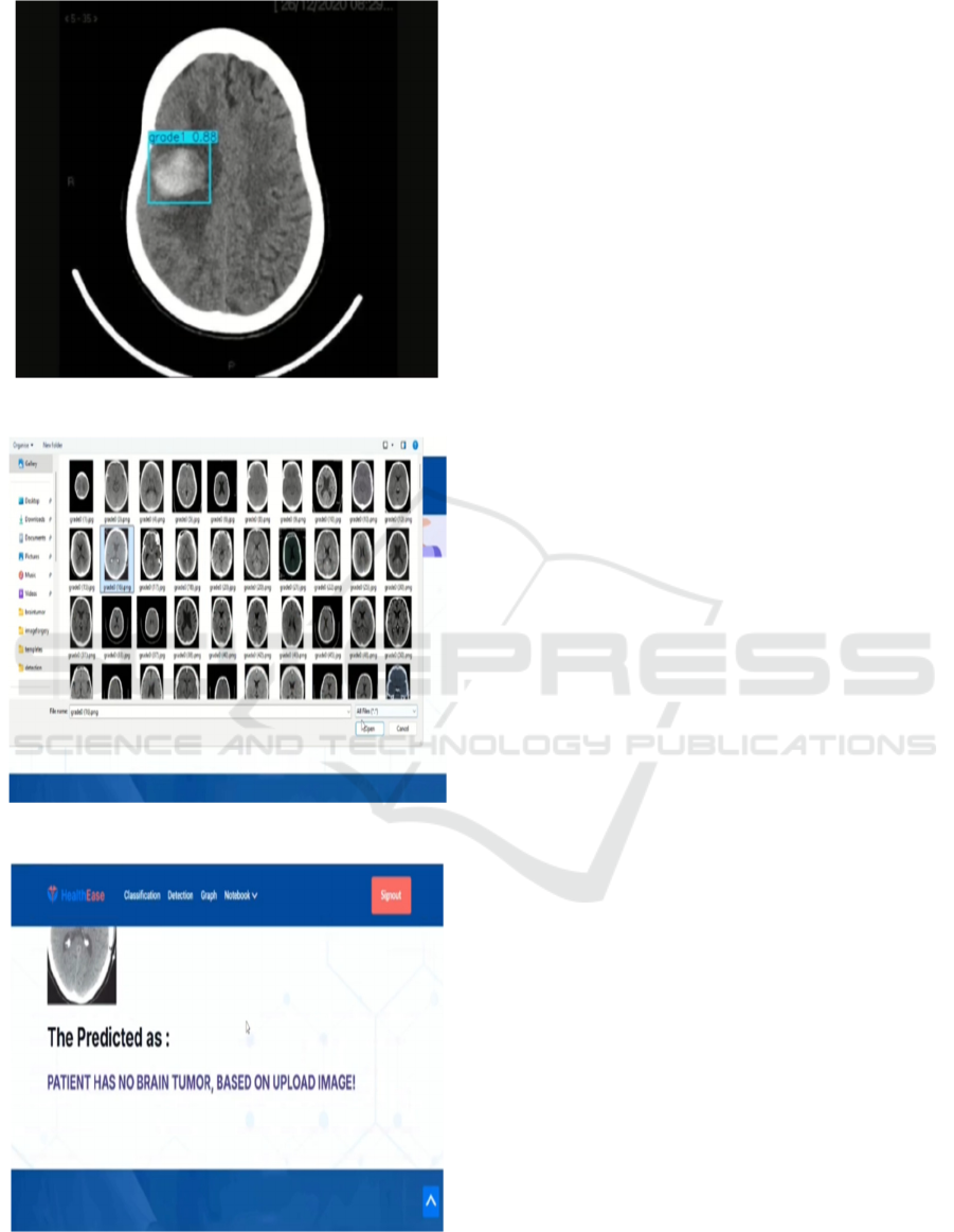
Figure 3: Final outcome.
Figure 4: Uploading an input image for classification.
Figure 5: Final outcome.
6 CONCLUSIONS
The suggested approach for brain tumor differential
verification and programming demonstrates a notable
increase in accuracy and reliability by utilizing state-
of-the-art machine learning and deep learning
techniques. Through the integration of "deep
convolutional neural networks (DCNNs)" with
cutting-edge transfer learning models, the approach
successfully divides mental diseases into four
different grades: "Grade 0 (no tumor), Grade I (little
tumor), Grade II (medium-sized tumor), and Grade III
(big tumor)." Although the combination model of
"Exception and NasNetMobile" achieves exceptional
performance with "accuracy of 98.1%, precision of
98.3%, recall of 97.9%, and F1 score of 98.1%," the
DCNN and ResNet152 mixing model provides a
respectable standard for cancer order. With a recall of
78.9%, precision of 84.3%, and mean average
precision (mAP) of 78.9%, These discoveries show
how definitively the framework recognizes and
arranges cancers, which makes it a significant
instrument for clinical navigation. The intuitive point
of interaction in light of Flask ensures safe
confirmation and effortlessness of purpose, in this
manner overcoming any issues among innovation and
helpful medical services applications. Extending the
dataset to integrate a more shifted range of tumour
sorts and imaging settings will assist with working on
model speculation and hence the future degree of this
brain tumour detection and grouping technique.
Clinically, coordination with ongoing MRI or CT
scan information for live tumour identification and
classification could further develop handiness.
Counting “explainable artificial intelligence (XAI)”
approaches would likewise assist with expanding
process transparency for deciding. Further refining of
the YOLO demonstrates for quicker and more exact
distinguishing proof could additionally work on
continuous relevance in clinical circumstances.
Data set link: https://www.kaggle.com/datasets/
masoudnickparvar/brain-tumor-mri-dataset/data.
REFERENCES
A novel method for detecting and identifying brain tumors
using transfer learning. Applied Sciences,
Issue:12(Volume :11), Pg-5645. Ullah, Nissar., Khan,
Junaid. Aamjad., Khan, Mohammed. Siraj., Khan,
Walleha., Hassan, Ismail., Obayya, Mourice., Salama,
Alka. Sharma., 2022.
A comprehensive review of machine learning, hybrid deep
learning, and transfer learning techniques for Magnetic
ICRDICCT‘25 2025 - INTERNATIONAL CONFERENCE ON RESEARCH AND DEVELOPMENT IN INFORMATION,
COMMUNICATION, AND COMPUTING TECHNOLOGIES
632

Resonance Imaginging- based classification & segme-
ntation in brain tumor analysis. Multi-media Tools &
Appls, Pg-1-38. Das, Sumit., & Goswami, Rahul.
Sandesh. ,2024.
An advanced Brain tumor Detection and Classification
system in medical image processing utilizing a hybrid
deep CNN-Cov-19-Res-Net Transfer learning model.
Biomedical Signal Processing and Control, Kumar,
Kiran. Arun., Prasad, Aja. Y., & Metan, Jonnes.,2022.
An efficient hybrid transfer learning model for medical
diagnosis of brain tumors within a classification
framework. Diagnostics, Issue 12(Volume 10), pg
2541. Samee, Niakath. Abrar., Mahmoud, Nouman.
Fatehi., Atteia, Golab., Abdallah, Haleed. Amir.,
Alabdulhafith, Mohsi., Al-Gaashani, Moin. Sheikh., &
Muthanna, Multan. Sialkot. Abbatabad.,2022.
CNN transfer learning approach for automated brain tumor
detection. Medical and Biological Engineering and
Computing, Issue 61(volume 7), Pg-1821-1836.
Bairagi, Viraha. Kumar., Gumaste, Pratima., Rajput,
Solace. Hulka., & Chethan, Ketan. S.,2023.
Employing deep learning convolutional neural network
with transfer learning for brain tumor detection. Internl.
Journ. of Imaging Systems & Tech., Anjum, Sahay.,
Hussain, Lukkal., Ali, Meer., Monagi H. Alkinani., &
Duong, T. Quresihsi.,2022.
Enhancing the efficacy of various deep transfer learning-
based models in brain tumor detection from MR
images. IEEE Access, Volume 10, Pg34716-34730.
Asif, Suraigha., Yi, Wien., Ain, Quaan. Ushu., Hou,
Jin., Yi, Tia., & Si, Juan.,2022.
Enhancing brain tumor classification using a combination
of CNN & transfer learning. Knowledge-Based
Systems, 111981. İncir, R., & Bozkurt, F.,2024.
Evaluating deep transfer learning approaches for brain
tumor classification performance using MRI images.
Journ.of Healthcare Engg., Issue 2022(Volume 1), Pg-
3264367. Srinivas, Chinna., Kumar Singh, Nutan .
Parashar., Zakariah, Mushir., Porashar Alothaibi,
Yall.Aali., Shaukat, Khaled., Partibane, Bhilal., &
Awal, Hulaik.,2022.
Hybrid ‐ NET: Combining DenseNet169 and advanced
machine learning classifiers for improved brain tumor
diagnosis. Internl. Journ. of Imaging Sys. & Tech.,
Khan, Sohail. Ushu . Rixckie., Zhao, Muah., Asif,
Sheikh., & Chen, Xian.,2024.
Image-based brain tumor detection and comparison of
transfer learning methods with 3-layer Convolutional
Neural Network. Khaliki, M. Z., & Muhammet Sinan
Başarslan.,2024.
Improving brain tumor classification through an extensive
analysis of transfer learning techniques & model
efficiency using Magnetic Resinance Imaging datasets.
Shamshad, Numaid., Sarwr, Delloitte., Almogren,
Archin., Saleem Khan, Khaled., Munawar, Ali.,
Rehman, Aamir. Umar., & Bharany, Shahid. ,2024.
Utilizing hybrid deep learning models for brain tumor
detection and classification. Scientific Reports,
Volume13(Issue - 1), Pg-23029. Babu Vimala, Balak.,
Srinivasan, Shohesh., Mathivanan, Sisir. Kumar.,
Mahalakshmi, Jayagopal, Prakash., & Dalu, Gopal.
Timor. ,2023.
Utilizing advanced machine learning techniques and
knowledge transfer for precise brain tumor
identification. Mathivanan, S. Kumar., Sonaimuthu,
Srinivasa., Murugesan, Shanmukha., Rajadurai,
Hitesh., Shivahare, Bolan. Duleep., & Shah,
Mohammaed. Aziz.,2024.
Detection of Brain Tumors Using Advanced Image Processing and the Ensemble Model and YOLO Family
633
