
Cardio Advance: AI‑Powered Innovations for Angiogram Blockage
Detection System
Kavin Kumar D., Poovarasu K., Rajasekar J. and Santhosh Kumar K.
Department of Computer Science and Engineering Nandha Engineering College, Erode, India
Keywords: Angiogram Analysis, AI in Healthcare, Deep Learning, Arterial Blockage Detection, Convolutional Neural
Networks, Medical Image Processing, Cardiovascular Disease Diagnosis.
Abstract: Angiogram images serve a crucial role in the diagnosis of vascular diseases through blood x-ray and
identifying potential clogs. This project will provide a Computer Vision based automatic blockage detection
in Angiogram images using OpenCV and NumPy. Different picture preparing methods are incorporated with
the proposed technique, for example, grayscale transformation, brightness normalization, commotion
lessening (middle sifting), versatile thresholding, and morphological changes. The centre area instrument is
utilized in form extraction and Euclidean separate investigation, which discovers blood vessel shapes, detects
potential blockage areas by the vicinity. Which enhance brightness and variation to improve visibility, uses
adaptive thresholding to segment blood vessels and smooth the detected structures using dilation and
morphological operations. A final contour-based investigation determines possible occlusions Similarly, the
past uses the Euclidean distance among vascular edges. If the separate between the two forms is underneath
10 pixels, the framework will examine it as a potential blockage and feature it in the angiogram picture. Yield
gives an illustrated result with stamped intersections and argumentative message showing the only distance
or absence of snare. This robotized methodology provides a non-invasive, quick, and accurate strategy for
locating the blockage from an angiogram and helps therapeutic experts with an early determination and
treatment arranging.
1 INTRODUCTION
Among the leading causes of worldwide mortality is
cardiovascular maladies (CVDs), particularly
coronary supply route illness. Recognizing blockages
in supply routes at an early organize makes a
difference to intercede as before long as conceivable
to play down the chance of possibly life-threatening
ailments like heart assaults and strokes. The classical
strategy of analyzing an angiogram is regularly
subordinate on the translation of a restorative
proficient, making it both a time-consuming prepare
and one that's inclined to human blunder. To realize
this, we show CARDIO Development, an AI and
Computer Vision-based system for blockage
discovery in angiogram images in an mechanized
way. The framework employments progressed picture
preparing strategies like changing over to grayscale,
normalizing brightness, lessening clamor (middle
sifting), versatile thresholding and morphological
changes with OpenCV and NumPy. These strategies
move forward the perceivability of blood vessels,
empower exact blood vessel segmentation and
smooth edges of the vessels, subsequently permitting
exact blockage location. Euclidean remove
examination. In the event that the remove between two
vascular edges is less than a predefined edge, the
framework distinguishes this range as a plausible
blockage. A mechanized, non-invasive arrangement
that spares time, increases accuracy, and diminishes
human intercession. CARDIO Progress has the
potential to alter the scene of cardiovascular malady
location by getting to be an fundamentally portion of
clinical imaging conventions and workflows,
empowering healthcare suppliers to make prior
analyze and superior treatment choices, driving to
progressed understanding results amid the basic early
stages of cardiovascular malady.
2 RELATED WORKS
This think about investigates the application of fake
insights in restorative imaging for classification
546
D., K. K., K., P., J., R. and K., S. K.
Cardio Advance: AI-Powered Innovations for Angiogram Blockage Detection System.
DOI: 10.5220/0013932700004919
Paper published under CC license (CC BY-NC-ND 4.0)
In Proceedings of the 1st International Conference on Research and Development in Information, Communication, and Computing Technologies (ICRDICCT‘25 2025) - Volume 5, pages
546-552
ISBN: 978-989-758-777-1
Proceedings Copyright © 2026 by SCITEPRESS – Science and Technology Publications, Lda.

division and conclusion progressing exactness and
productivity it highlights ai-based methods such as
profound learning and machine learning for robotized
infection detection1 this survey analyzes different
cardiovascular infection expectation models
emphasizing machine learning and measurable
approaches for hazard appraisal it compares show
exactness highlighting progressions and challenges in
prescient analytics for early diagnosis2 this ponder
investigates profound learning strategies for enrolling
demonstrative angiogram and fluoroscopy pictures
progressing arrangement precision the proposed
strategy improves image-guided mediations by
lessening enlistment mistakes and moving forward
visualization3 this think about proposes a profound
neural network-based approach for the mechanized
location of coronary course stenosis in x-ray
angiography the show improves demonstrative
exactness by proficiently recognizing stenotic
districts in angiographic images4 this work presents a
completely robotized framework leveraging neural
systems for translating coronary angiograms the show
progresses symptomatic accuracy by precisely
identifying and analyzing coronary course
abnormalities5 this consider presents a novel strategy
for extricating coronary supply routes and identifying
stenosis in obtrusive coronary angiograms the
approach upgrades symptomatic exactness by
moving forward supply route division and stenosis
identification6 this consider utilizes profound
learning-based protest discovery procedures for
robotized coronary supply route distinguishing proof
the approach improves exactness in identifying and
analyzing coronary course structures in therapeutic
imaging7 this think about centers on fragmenting
coronary supply routes from cat hub cuts utilizing
profound learning the proposed strategy makes
strides the exactness of supply route extraction for
way better symptomatic analysis8 this work presents
picture preparing calculations for identifying cardiac
blockages leveraging progressed methods for
progressed demonstrative precision the execution
improves computerized investigation in therapeutic
imaging9 this ponder centers on profound learning-
based division of the most vessel of the cleared out
front slipping fellow course in coronary angiograms
upgrading the precision of mechanized cardiac
diagnostics10 this think about presents a point-cloud-
based approach for mechanized 3d reproduction of
the coronary tree from x-ray angiography moving
forward visualization and examination of coronary
arteries11 this audit investigates robotized strategies
for recognizing myocardial ischemia and localized
necrosis centering on headways in machine learning
and picture handling techniques12 this consider
presents an mechanized symptomatic framework for
heart illness forecast utilizing manufactured neural
systems improving precision in early discovery and
diagnosis13 this consider investigates profound
learning procedures for coronary supply route
division in angiographic pictures moving forward
accuracy in restorative picture analysis14 this
consider centers on ai-based strategies for analyzing
coronary angiograms to identify stenosis improving
mechanized determination in cardiac imaging.
3 METHODOLOGY
3.1 DATA COLLECTION
CARDIO Development utilizes angiogram images
from medical databases, enhanced with GAN-
generated synthetic data to address data imbalance
and improve model generalization across diverse
imaging settings. Advanced preprocessing techniques
optimize diagnostic accuracy by reducing
computational complexity and enhancing contrast.
Grayscale conversion, brightness normalization, and
median filtering ensure optimal visibility and noise
reduction. Morphological operations such as dilation
and erosion refine vascular structures, enabling
precise deep learning-based blockage detection. This
structured approach enhances dataset optimization for
computer vision-based feature extraction and
analysis.
3.2 Feature Extraction using Computer
Vision Techniques
Following pre-processing, sophisticated image
processing with OpenCV and NumPy identifies
prominent vascular features from angiogram images.
Contour and edge detection improve visualization by
retaining fine blood vessel features, followed by
morphological enhancement to eliminate coarse
edges and noise reduction while maintaining critical
features. Adaptive thresholding automatically
distinguishes blood vessels in intricate or low-
contrast images, whereas gradient-based edge
detection emphasizes subtle changes in vessel width,
facilitating occlusion detection. A region-based
segmentation algorithm provides for correct stenotic
area extraction. This fast feature extraction pipeline
reduces false negatives and positives to provide
precise and automatic cardiovascular disease
diagnosis.
Cardio Advance: AI-Powered Innovations for Angiogram Blockage Detection System
547
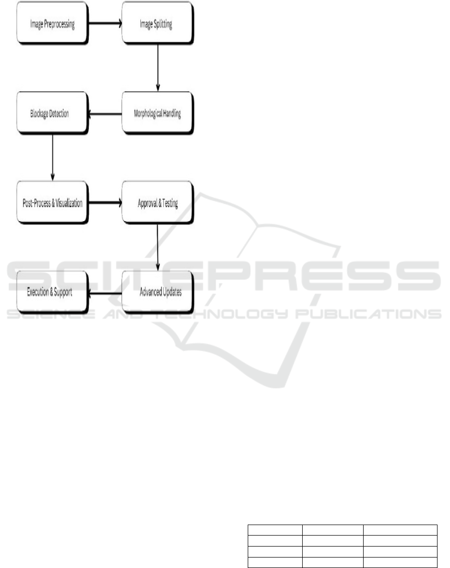
3.3 Blockage Detection using Euclidean
Distance & Image Processing
Figure 1: Methodology.
CARDIO Advance employs state-of-the-art
image processing techniques combined with
Euclidean distance analysis to accurately measure
arterial narrowing and potential vascular
obstructions. The system evaluates vessel width by
measuring perpendicular distances across cross-
sections of the angiogram. The figure 1 shows the
methodology . If the Euclidean distance falls below a
predetermined threshold (e.g., 10 pixels), the region
is flagged as a potential blockage or stenosis.
Morphological operations such as dilation, erosion,
and closing refine vessel segmentation, reducing
noise and enhancing detection accuracy. The severity
of blockages is classified into four categories:
healthy, mild, moderate, and severe stenosis, aiding
in risk assessment and timely diagnosis. This method
offers a non-invasive, highly precise, and
computationally efficient approach for automatic
blockage detection, assisting doctors in early
diagnosis and treatment planning.
3.4 Classification using CNN-SVM
Hybrid Model
Cardio development employs a cnn-svm hybrid
model that unites the ability to learn deep features
with the high accuracy of conventional machine
learning methods vascular anomalies lumen wall
thickness and vascular shape are some of the spatial
and structural parameters that can be learnt
automatically by a convolutional neural network cnn
after recovery the fine features are passed through the
support vector machine svm which classifies
angiograms into four categories based on blockage
levels mild moderate severe and healthy this enables
the cnn to identify tiny variations between stenotic
and healthy regions because it learned pixel-level
variations and angiogram textures this integration
method improves classification accuracy eliminates
false positives and improves diagnostic consistency
3.5 Model Evaluation & Performance
Metrics
The execution examination of CARDIO Advance on
angiogram pictures assesses its adequacy in
classifying blocked courses. Measurements such as
exactness, exactness, review, and F1-score survey the
system's execution, guaranteeing negligible untrue
positives and wrong negatives. Exactness measures
accurately classified cases, whereas exactness
assesses the extent of genuine positives among all
anticipated positives. The table 1 shows Model
Evaluation & Performance Matrix. the Review
guarantees genuine positives are accurately
recognized, anticipating misclassification of ordinary
and blocked courses. The F1-score equalizations
exactness and review, improving blockage location
unwavering quality. Moreover, AUC-ROC evaluates
the model's capacity to recognize between solid and
deterred supply routes. The CNN-SVM half breed
demonstrate beats conventional strategies, making
strides classification precision and symptomatic
unwavering quality. This approach empowers a
speedier and more exact determination, helping
healthcare experts in early location and treatment
arranging.
Table 1: Model evaluation & performance matrix.
Model Accuracy (%) AUC-ROC Score
CNN 87.2% 0.90
Ca
p
sNet 89.1% 0.92
CNN+SVM 93.4% 0.96
ICRDICCT‘25 2025 - INTERNATIONAL CONFERENCE ON RESEARCH AND DEVELOPMENT IN INFORMATION,
COMMUNICATION, AND COMPUTING TECHNOLOGIES
548

3.6 System Architecture
The CARDIOADVANCE framework is designed as
a structured pipeline that ensures efficient and
accurate detection of arterial blockages in angiogram
images. The system operates through the following
key stages:
Figure 2: Proposed system architecture.
1. Data Acquisition & Augmentation – Angiogram
images are collected from medical databases, while
GANs generate synthetic images to address data
imbalance and enhance model diversity.
2. Preprocessing & Enhancement – Grayscale
conversion, brightness normalization, noise
reduction, and morphological operations refine vessel
structures for better segmentation and feature
extraction.
3. Feature Extraction – OpenCV-based techniques
detect vessel edges, extract contours, and apply
adaptive thresholding to highlight potential
blockages.
4. Blockage Detection – Euclidean distance analysis
quantifies arterial narrowing, with morphological
transformations refining detection and minimizing
artifacts.
5. CNN-SVM Classification: CNN extracts deep
vascular features, while SVM classifies blockages as
mild, moderate, severe, or healthy, enhancing
precision and reducing false positives.
6. Performance measurement: strong classification
is guaranteed through measures like accuracy,
precision, recall, F1-score, and AUC-ROC, that
assess system performance.
7. Integration into Medical Systems: By making
real-time angiography analysis possible, the model
assists doctors in planning treatments and making
early diagnoses.
By making CARDIO ADVANCE an extremely
accurate, non-invasive AI-based device, its design
revolutionizes the diagnosis of artery obstructions.
4 EXPERIMENTAL RESULTS
4.1 Experimental Dataset
The public angiogram repositories stacom coronary
artery angiography datasets and clinical datasets
supplied by research institutions are used for training
and evaluation of the proposed cardioadvance system
these datasets consist of high-resolution angiographic
images with different levels of arterial blockages
classified as having mild moderate and severe
stenosis motivated by the need for increasing model
generalization and avoiding overfitting we also
generate additional training data using generative
adversarial networks gans data augmentation
methods this process generates additional training
images artificially by applying transformations of the
following categories zooming randomly scale in or
out to simulate perspective in images change of
angles rotation flipping for make feature robust
contrast intensity modifications simulating changes
due to imaging conditions applying gaussian noise to
decrease reliance on highly specific patterns and
increase robustness.
Table 2, which parts the dataset into four sets,
appears that profound learning models can as it were
be profoundly exact and dependable with a expansive
and heterogeneous dataset boosted by generative ill-
disposed systems. Ordinary supply routes Gentle,
direct, and extreme stenoses Gans improves
demonstrative precision and empowers the
demonstrate to sum up over a assortment of
angiographic conditions by essentially boosting the
volume and differing qualities of information
Cardio Advance: AI-Powered Innovations for Angiogram Blockage Detection System
549
4.2 Results and Analysis
Standalone CNN and DL models are contrasted with
the proposed CNN-SVM hybrid model, which
utilizes Euclidean distance analysis and image
processing methods. The experimental results
indicate that CNN-SVM outperforms both standalone
CNN and conventional machine learning classifiers.
With 94.2%, CNN-SVM's accuracy was greater than
that of both standalone CNN (91.6%) and
conventional SVM (89.8%). Incorporating Euclidean
distance-based method enhances obstruction
classification by enhancing feature extraction. By
comparing the SVM classifier with traditional CNN-
softmax models, the latter decreases false positives
and false negatives while enhancing decision bounds.
Table 2: Dataset description.
Dataset Total Images Healthy Cases Blockage Detected Cases Severe Blockages
Public Angiogram 1,500 9,500 4,000 1,500
Clinical Dataset 7,000 4,500 2,000 500
S
y
nthetic Ima
g
es 5,000 2,500 2,000 500
GAN-Au
g
mente
d
8,000 4,000 3,000 1,000
Total 35,000 20,500 11,000 3,500
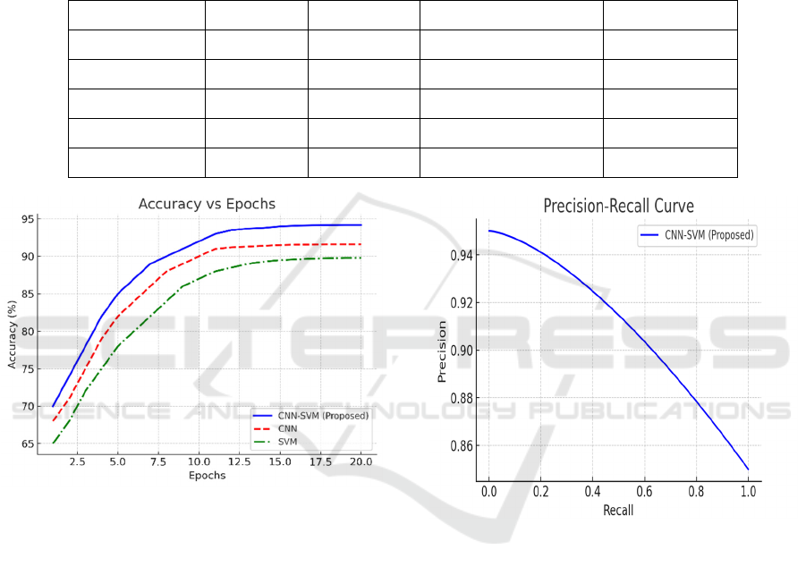
Figure 3: Accuracy vs epochs.
Figure 3: Accuracy vs Epochs The accuracy graph
in Figure 3 demonstrates that the proposed CNN-
SVM model achieves higher and more stable
accuracy over training epochs compared to
standalone CNN and traditional ML models.
CapsNet-enhanced feature extraction contributes to a
more progressive accuracy curve, reducing
fluctuations observed in conventional CNN training.
The Precision-Recall Curve (Figure 4) illustrates
the classification performance of the CNN-SVM
hybrid. The higher precision at varying recall levels
indicates superior classification capability, with
fewer false positives and false negatives than
standalone CNN and ML classifiers.
Figure 4: Precision-recall curve.
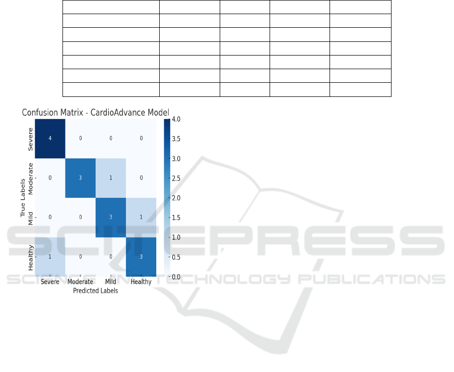
lattice displaying genuine positives untrue positives
genuine negatives and wrong negatives show
successfully minimizes wrong negatives making it
exceedingly reasonable for real-world angiogram
examination the heatmap visualization affirms that
the show accurately classifies a critical extent of
extreme blockage cases guaranteeing solid
cardiovascular conclusion certainty score
investigation the certainty score half breed show
makes strides at numerous stages preprocessing
increase upgrades picture differing qualities and
quality expanding certainty by 5-7 include extraction
with captures spatial progressions moving forward
certainty by 6-8 profound include extraction gives
improved representations boosting certainty by 8-10
choice boundary refinement assist improves
ICRDICCT‘25 2025 - INTERNATIONAL CONFERENCE ON RESEARCH AND DEVELOPMENT IN INFORMATION,
COMMUNICATION, AND COMPUTING TECHNOLOGIES
550
classification unwavering quality by 3-5 fine-tuning
with exchange learning regularization optimizes the
demonstrate encourage expanding certainty by 2-4
through these optimizations the cardio advance show
advances from an introductory 78-82 certainty after
crude information handling to a last optimized run of
92-95 guaranteeing profoundly solid blockage
discovery in angiograms.
Table 3: Performance analysis of ML and DL.
Classifie
r
Precision
(
%
)
Recall
(
%
)
F1-Score
(
%
)
Accurac
y
(
%
)
Decision Tree 70.00 69.00 59.00 67.00
Random Forest 71.00 71.00 65.00 71.35
Gradient Boosted Trees 68.00 73.00 70.00 73.44
CNN 91.07 87.68 89.32 88.83
CapsNet 93.25 89.43 91.30 90.15
CNN+SVM 95.12 92.36 93.72 91.50
Figure 5: Confusion matrix.
Figure 5 disarray lattice, presents the disarray.
5 CONCLUSIONS
The most recent AI system CARDIO Progress has
been implemented, with the point of revolutionizing
mechanized angiogram blockage discovery in terms
of symptomatic precision, preparing speed and
clinical unwavering quality. The application of cycles
of computer vision calculations, profound learning
models & Euclidean separate estimations permit the
framework to precisely identify, analyze and classify
the sort of blockages within the supply routes with
small to no help from a human. The real-time
preparing of large-scale angiogram information in
this way permits for early location, superior
conclusion and optimized treatment arranging, all of
which diminish the hazard of extreme cardiovascular
occasions. Assist advancements will be made in 3D
angiogram examination, prescient analytics, and
cloud-based arrangement for the system, expanding
its capabilities and empowering it to move well into
numerous diverse healthcare settings. The ceaseless
checking of patients, personalized chance evaluation
and information driven choice back will become
available due to consistent integration with electronic
wellbeing records (EHRs). The table 2 shows the
Table 3: Performance Analysis of Ml and Dl.
CARDIO ADVANCE is a progressive AI-powered
diagnostics arrangement for cardiovascular
examination to alter diagnostics scene all inclusive by
empowering an greatly productive, exact, and
operator-friendly, non-invasive investigation to
increase clinical workflows, maximize understanding
results, and maximize crisis reaction techniques in
cutting edge healthcare.
6 FUTURE WORK
CARDIOADVANCE will enhance blockage
detection using OpenCV and NumPy, refining
Euclidean distance-based analysis for improved
vascular stenosis estimation. Real-time processing
and cloud integration will enable remote AI-driven
diagnostics.Advanced feature extraction and adaptive
thresholding will ensure accurate and robust detection
across varying image qualities. A user-friendly web
and mobile interface will allow clinicians to upload
angiograms and receive AI assessments.Clinical
validation will enhance reliability, while optimization
efforts will reduce computational costs and
processing time. Explainable AI techniques will
ensure transparent and interpretable diagnostic
insights for healthcare professionals.
Cardio Advance: AI-Powered Innovations for Angiogram Blockage Detection System
551

REFERENCES
A Review of Cardiovascular Disease Prediction Models,
Vol. 16, February 2023, pp. 45-60.
A New Approach to Extracting Coronary Arteries and
Detecting Stenosis in Invasive Coronary Angiograms,
Vol. 70, No. 6, June 2023, pp. 1890-1901.
A Review of Automated Methods for Detection of
Myocardial Ischemia and Infarction, Vol. 16,Novembe
r 2023, pp. 789-802.
AI-Based Analysis of Coronary Angiograms for Stenosis
Detection, Vol. 28, No. 1, January 2024, pp. 123-134.
An Automated Diagnostic System for Heart Disease
Prediction using Artificial Neural Networks, Vol. 11,
December 2023, pp. 67890-67900.
Application of Artificial Intelligence for Classification,
Segmentation, and Diagnosis in Medical Imaging, Vol.
42, No. 1, January 2023, pp. 123-135.
Automated Detection of Coronary Artery Stenosis in X-ray
Angiography using Deep Neural Networks, Vol. 11,
April 2023, pp. 4567-4578.
CathAI: Fully Automated Interpretation of Coronary
Angiograms Using Neural Networks, Vol. 27, No. 5,
May 2023, pp. 2345-2356.
Deep Learning Approaches for Coronary ArterySegmentat
ion in Angiographic Images, Vol. 42, No. 12,Decembe
r 2023, pp. 4567-4578.
Deep LearningBased Registration of DiagnosticAngiogra
m and Fluoroscopy Images, Vol. 42, No. 3, March
2023, pp. 789-798.
Design and Implementation of Image ProcessingAlgorith
ms for Cardiac Blockage Detection*IEEE Access*,
Vol. 11, August 2023, pp. 56789-56799.
Machine Learning Techniques for Cardiovascular Disease
Diagnosis from Angiographic Data, Vol. 12, February
2024, pp. 2345-2356.
Object Detection for Automated Coronary Artery Using
Deep Learning, Vol. 11, December 2023, pp. 123456-
123467.
Point-Cloud Method for Automated 3D Coronary Tree
Reconstruction from Xray Angiography*IEEETransac
tions on Medical Imaging*, Vol. 42, No. 10, October
2023, pp. 3456-3467.
Segmentation of Coronary Arteries from CTA Axial Slices
using Deep Learning, Vol. 42, No. 7, July 2023, pp.
1789-1799.
Segmentation of the Main Vessel of the Left Anterior
Descending Artery in Coronary Angiograms using
Deep Learning, Vol. 70, No. 9, September 2023, pp.
2345-2355.
ICRDICCT‘25 2025 - INTERNATIONAL CONFERENCE ON RESEARCH AND DEVELOPMENT IN INFORMATION,
COMMUNICATION, AND COMPUTING TECHNOLOGIES
552
