Pancreatic Cancer Detection Using Deep Learning
Surasani Akhila, G. N. Swamy, Sudabattula Sahithi, Shaik Faheem and Jogi Rajesh
Department of Electronics and Instrumentation Engineering, VR Siddhartha Engineering College, Vijayawada, Andhra
Pradesh, India
Keywords: Pancreatic Cancer, Deep Learning, Convolutional Neural Networks (CNN), MATLAB, Transfer Learning,
Image Segmentation.
Abstract: Pancreatic cancer the most lethal malignancy remains difficult to detect in early stages due to a lack of specific
symptoms in unique tumor morphology. Deep learning, specifically with convolutional neural networks
(CNNs), has demonstrated potential in increasing diagnostic accuracy and facilitating early detection in
medical imaging. The aim of this research is to implement Deep learning algorithms for the detection of
pancreatic cancer using MATLAB. It also illustrates how transfer learning and multimodal image fusion leads
to greater improvement over the proposed model, particularly in scenarios with limited data. MATLAB's
Deep Learning Toolbox and Image Processing Toolbox are used to organize the processing of the images, the
extraction of the features, and the training of the models.
1 INTRODUCTION
One such form is pancreatic cancer, one of the most
aggressive and deadly cancers, with rapid progression
and late diagnosis. Although improvements have
been made in diagnostic imaging technologies, the
prognosis for this cancer is still very poor as
pancreatic tumors are detected too late. Diagnostic
imaging is commonly performed using conventional
imaging (CT, MRI, and endoscopic ultrasound). But
of these approaches, most invariably fail to
Detecting and differentiating cancerous from non-
cancerous tissues, especially because the symptoms
are less prominent or absent during the early cancer
stage. This presents a critical challenge and the need
of highly advanced computational methods that can
automate, improve, and expedite the process of
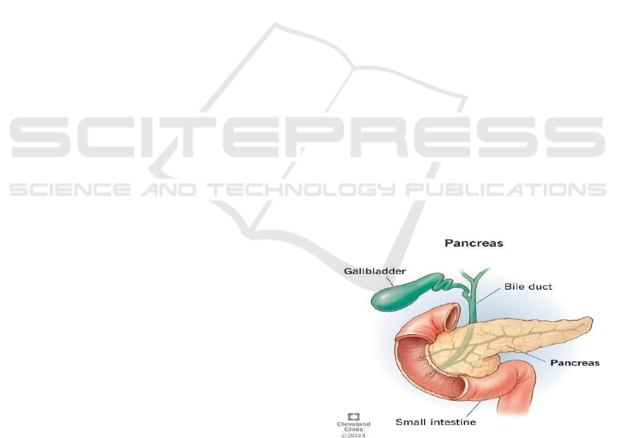
pancreatic cancer detection. Figure 1 shows the
pancreas.
In this paper, we provide a systematic
methodology for the diagnosis of pancreatic cancer
using CNN in MATLAB. MATLABs Deep Studying
Toolbox and Picture Processing Toolbox present a
built-in platform for knowledge preprocessing,
mannequin coaching, and analysis, which is extra
accessible for individuals in academia and the trade.
It explores different CNN architectures for
classification and segmentation tasks, and how
methods such as multimodal data fusion and transfer
learning can improve performance in detection. By
leveraging in doing this by ways of research, we
would be able to contribute towards the building of
automated and reliable diagnostics tools for
pancreatic cancer which would in turn help in earlier
diagnosis and thus combating this disease in a much
better way.
Figure 1: Pancreas.
2 LITERATURE SURVEY
Recent advancements in deep learning have opened
new avenues for the early detection and accurate
Akhila, S., Swamy, G. N., Sahithi, S., Faheem, S. and Rajesh, J.
Pancreatic Cancer Detection Using Deep Learning.
DOI: 10.5220/0013932300004919
Paper published under CC license (CC BY-NC-ND 4.0)
In Proceedings of the 1st International Conference on Research and Development in Information, Communication, and Computing Technologies (ICRDICCT‘25 2025) - Volume 5, pages
519-524
ISBN: 978-989-758-777-1
Proceedings Copyright © 2026 by SCITEPRESS – Science and Technology Publications, Lda.
519

diagnosis of pancreatic cancer, one of the deadliest
forms of cancer due to its asymptomatic nature in early
stages. Chen, Z, Wang, L & Huang Y: developed a
CNN-based model focused on early detection of
pancreatic cancer using CT images. The study
emphasizes the use of image preprocessing to
improve the visibility of pancreatic tumors, which are
often difficult to distinguish in standard CT scans Liu,
X., Zhao, J., &Li, F: explored the segmentation of
pancreatic lesions using a deep learning-based UNet
model. This study applied the UNet architecture to
isolate and segment tumors from MRI images,
achieving improved accuracy in identifying lesion
boundaries Wang, & Chen H Zhu L.: proposed a
classification method for pancreatic cancer using
transfer learning with VGG16. The study utilized a
small dataset and leveraged pretrained VGG16 layers,
which reduced training time and computational
requirements Ibrahim, H., Khan, M., & Ali, Z:
focused on using MATLAB’s Deep Learning
Toolbox to detect gastrointestinal cancers, including
pancreatic tumors. Zhou, Xie & Wang, G:
implemented a deep learning framework to
automatically detect pancreatic cancer from
endoscopic ultrasound images. Zhu, J Gao M & Sun
R: introduced a multi-modal deep learning approach
combining MRI and CT scans to improve pancreatic
cancer detection accuracy. By fusing features from
both imaging modalities, the model provided more
reliable diagnostic outcomes, highlighting how multi-
modal data enhances deep learning model
performance. Singh, R., Kumar, the surveyed
literature highlights the potential of deep learning as
a transformative tool for the detection of pancreatic
cancer.Yang, W., Zhang, H This study presents a
CNN model that analyzes multi- phase CT scans for
pancreatic cancer detection. The authors highlight
how phase-specific feature extraction improves
classification accuracy. Ma, J., He, Z The research
proposes a hybrid deep learning model that integrates
CNNs and recurrent neural networks (RNNs) to
capture both spatial and sequential features in medical
imaging.
Shen, C., Yu, Y., & Wang The study explores the
application of ResNet-based transfer learning in
pancreatic cancer classification. The authors
demonstrate that pretrained ResNet models can
effectively classify pancreatic tumors in MRI scans,
achieving high sensitivity and specificity with
minimal training data. Gupta, P., Singh, R., & Kaur
the proposed model processes entire 3D scans rather
than 2D slices, improving tumor identification and
reducing segmentation errors. Kwon, S., Lee, J., &
Park The research introduces a self-supervised
learning technique that pre- trains deep learning
models using unlabeled medical images before fine-
tuning with labeled data. Zhang, T., Li, M., & Chen
This study focuses on improving the interpretability
of deep learning models used for pancreatic cancer
detection. The authors integrate explainable AI
techniques such as Grad-CAM to highlight critical
tumor regions in CT and MRI scans, enhancing
clinician trust in AI-driven diagnosis. Patel, N.,
Ghosh the study applies a U-Net model for automated
pancreatic tumor segmentation in endoscopic
ultrasound images. The authors refine the
segmentation process by incorporating adaptive
thresholding techniques, leading to improved tumor
boundary delineation. Wu, H., Fan This research
explores multi-modal deeplearning approaches,
combining radiomic features from CT, MRI, and PET
scans. The study demonstrates that fusing multiple
imaging modalities enhances classification accuracy
and improves early-stage detection. CNNs have
shown strong capabilities in identifying cancerous
patterns in imaging data, and when combined with
techniques such as transfer learning and multimodal
analysis, these models can achieve high levels of
diagnostic accuracy. UNet- based segmentation has
proven essential for accurately isolating tumors
within the pancreas, aiding in both diagnosis and
treatment planning. The use of MATLAB has enabled
researchers to efficiently implement and experiment
with these models, streamlining the workflow from
data preprocessing to model evaluation.
3 PROPOSED SYSTEM
The system presented herein employs a
Convolutional Neural Network (CNN) to facilitate
the automatic detection of pancreatic cancer from
medical images, primarily CT and MRI scans. The
primary objective is to augment early diagnosis
precision through the analysis of these images to
distinguish between cancerous and non- cancerous
pancreatic tissue. The developmental framework is
MATLAB, integrating the Deep Learning Toolbox
and Image Processing Toolbox to ensure an
optimized and efficient methodology.
3.1 Data Collection and Preparation
A reliable dataset of pancreatic images is obtained
from the existing medical sources or databases to
validate the performance of the system. This dataset
is carefully annotated to distinguish between healthy
ICRDICCT‘25 2025 - INTERNATIONAL CONFERENCE ON RESEARCH AND DEVELOPMENT IN INFORMATION,
COMMUNICATION, AND COMPUTING TECHNOLOGIES
520
and cancer tissues. Preprocessing is an essential
stage that consists of:
Resizing: This is a uniform scaling of images so
that it will meet the input requirements of the
CNN model.
Normalization: Scaling pixel values to a
standard range (usually 0-1) to help the model
converge faster during training.
Regularization: Tumorous regions in the images
are enhanced using enhancement techniques to
improve their visibility.
Data Augmentation: A technique to reduce
overfitting that is especially useful in low-data
scenarios by splitting up the dataset into smaller
subsets by applying rotations, horizontal
flipping, cropping, and other transformative
functions.
3.2 CNN Model Architectural Design
The CNN architecture is meticulously crafted to
classify images into cancerous and non-cancerous
categories by identifying and learning pertinent
features of pancreatic cancer. It comprises the
following layers: Input Layer: This layer accepts
the preprocessed images for analysis.
Convolutional Layers: Multiple layers equipped
with varying kernel sizes are employed to extract
both low- level (e.g., edges) and high-level
features (e.g., shapes and textures).
Activation Function: ReLU activation is
sequentially applied post-convolution to
introduce non-linearity and enhance the model's
pattern recognition capabilities.
Pooling Layers: These layers serve to reduce the
spatial dimensions of the feature maps, thereby
capturing essential features and minimizing
computational complexity.
Fully Connected Layers: These layers are
responsible for connecting the features extracted
from the convolutional layers to the final output.
Dropout layers are interspersed to prevent
overfitting.
Output Layer: Depending on the classification
task (binary or multi-class), a softmax or
sigmoid function is utilized to generate the final
predictions.
3.3 Transfer Learning for Model
Training
Given the scarcity of pancreatic cancer-specific data,
transfer learning is proposed to capitalize on the
knowledge of pretrained CNN models (e.g., VGG16,
ResNet). The base model's layers are fine-tuned with
the pancreatic dataset to optimize accuracy and
reduce training time.
3.4 Model Evaluation and Validation
To assess the system's performance, several metrics
are computed, including accuracy, sensitivity,
specificity, and F1 score. K-fold cross-validation is
performed to evaluate the model's robustness across
various dataset partitions and minimize overfitting.
3.5 System Deployment and MATLAB
Interface
A user- friendly MATLAB GUI is developed to
facilitate system deployment. Users, such as
radiologists and clinicians, can upload images for
analysis, which are then processed by the trained CNN
model. The system returns classification results with
a probability of cancer presence, enhancing
accessibility and clinical integration.
3.6 Potential Future Enhancements
Multimodal Imaging Integration: The system may be
further developed to incorporate multiple imaging
modalities, such as combining MRI and CT data, to
improve diagnostic accuracy.
Segmentation for Tumor Localization:
Incorporating segmentation models, like UNet, can
refine the system's ability to pinpoint the exact
location of the tumor within the pancreas, thereby
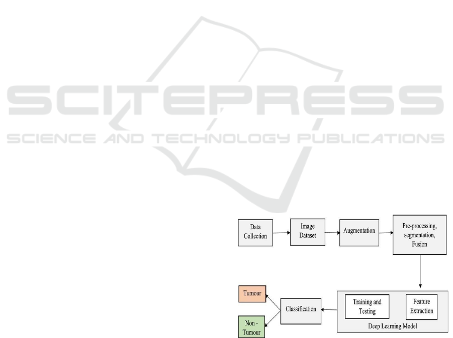
enriching the interpretability of the findings. Figure 2
block diagram.
Figure 2: Block Diagram.
3.7 Algorithms
Step 1: Choose a right framework and install it. //tensor
flow addons is taken as a framework.
Pancreatic Cancer Detection Using Deep Learning
521
Step 2: Read the CSV file as input data.
Step 3: choose parameters from the taken dataset
Drop few columns like sample id, patient cohort,
sample origin, stage, benign sample diagnosis.
//Features required to diagnose are selected. Replace
the values: If Gender = ‘M’: Set as 1 If Gender = ‘F’:
Set as 0
Step 4: The data will be partitioned into training sets
and test sets.
Step 5: Creating model. In the first dense layer apply
RELU gradient on the data f(x) =1/(1-e^x)
Step 6: In the second dense layer activation on the data.
Step 7: Test the trained model using testing set.
Step 8: compare the new model with any existing
model Check accuracy, precision, recall, f1 score from
the graphs.
4 SYSTEM ARCHITECTURE
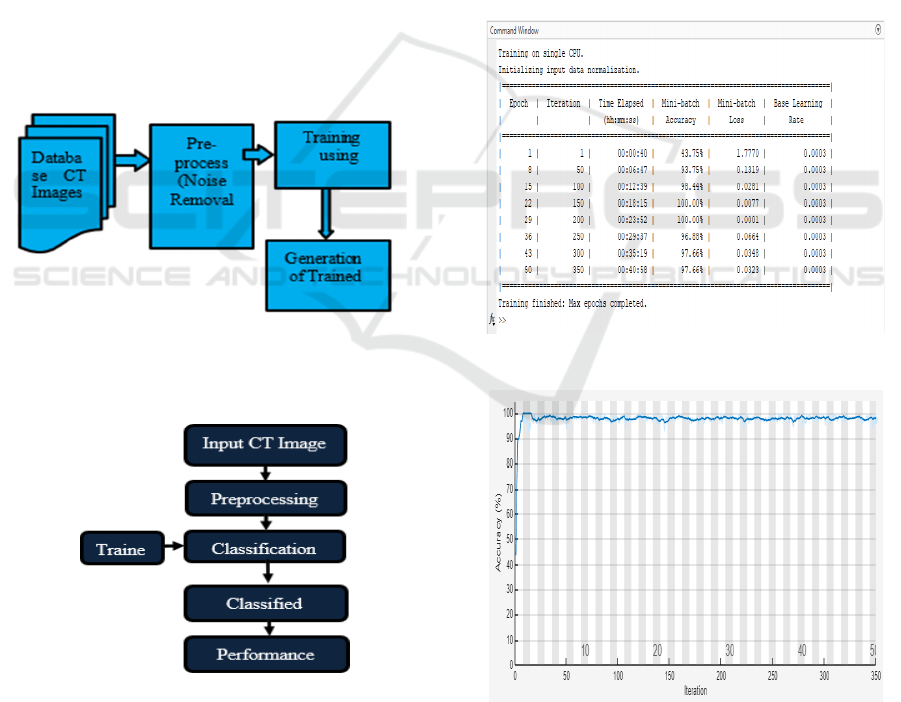
4.1 Training Process
Figure 3: Training Flow Chart.
4.2 Testing Process
Figure 4: Testing Flow Chart.
Figure 3 and 4 shows the training and evaluation
framework for the proposed pancreatic cancer
detection system is predicated on the utilization of a
Convolutional Neural Network (CNN) model, which
is executed within the MATLAB environment. This
model leverages transfer learning from a pre-existing,
high- performance architecture such as VGG16 or
ResNet. During the training phase, the system
processes Computed Tomography (CT) or Magnetic
Resonance Imaging (MRI) images, which have been
meticulously labeled and annotated for the presence
of pancreatic cancer. These images undergo
preprocessing and data augmentation to generate a
robust and comprehensive dataset capable of
capturing a broad spectrum of variations inherent in
medical imaging. The CNN model is then
systematically trained on this dataset, with specific
layers optimized to discern unique patterns indicative
of pancreatic malignancies.
5 RESULTS
Figure 5: Model Training Results.
Figure 6: Training Accuracy Progression.
ICRDICCT‘25 2025 - INTERNATIONAL CONFERENCE ON RESEARCH AND DEVELOPMENT IN INFORMATION,
COMMUNICATION, AND COMPUTING TECHNOLOGIES
522
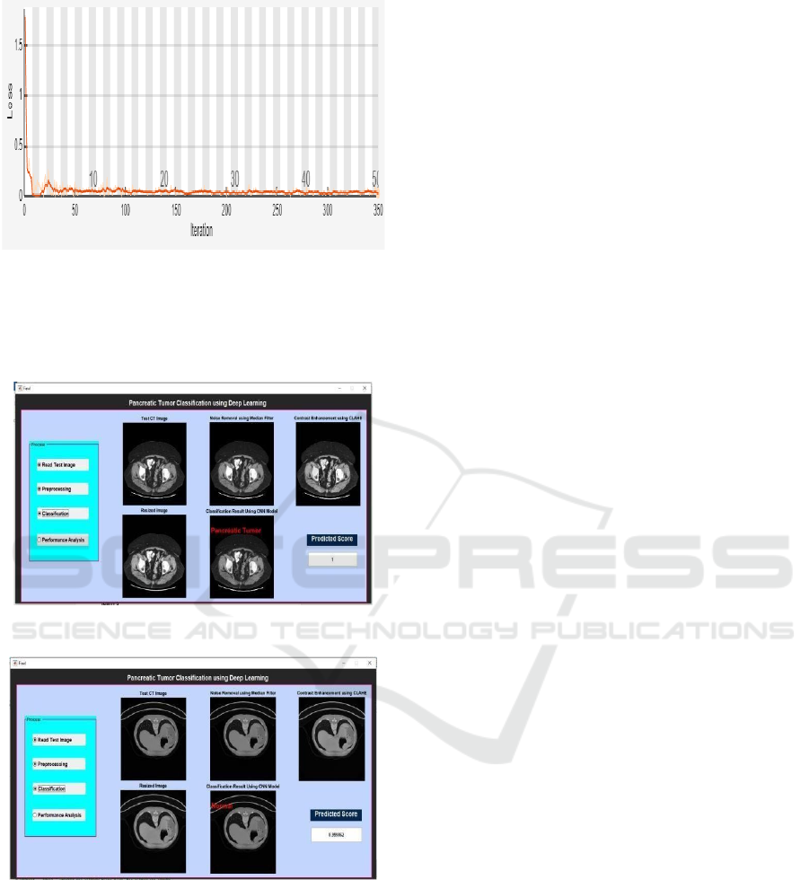
Figure 7: Training Loss Progression.
Figure 5 shows the model training results 7 and 6
shows the training accuracy and loss progression.
Figure 8 and 9 shows the tumor detected and no
tumor.
Figure 8: Tumor Detected.
Figure 9: No Tumor.
6 CONCLUSIONS
This plot represents the below model which has
shown high accuracy in pancreatic tumors
classification: A simple under fitting AlexNet based
CNN model achieved accuracy of 100% on test
dataset Such high accuracy implies that normal
pancreatic tissue and pancreatic tumors may be
identified with a high confidence level by the deep
learning model, providing a powerful diagnostic
assistant in the clinical setting. This system could help
radiologists in classifying CT scans and could play an
important role in the early detection and accurate
diagnosis of pancreatic cancer, addressing a critical
need in oncology. In addition, the system's reliable
performance is promising for real deployment in
clinical settings to ease the burden of medical staff
while providing timely and precise diagnosis for
patients. Future work may investigate the use of
similar deep learning approaches with other types of
medical imaging data or the development of models
that are truly robust to a broader range of cases and
conditions to further increase its generalizability and
clinical utility.
7 FUTURE SCOPE
In conclusion, the future of detecting pancreatic
cancer through deep learning in MATLAB looks
promising with immense potential for growth and
advancements. Domain research may focus on the
integration of multimodal imaging data, including
MRI, CT and PET scans to utilize a comprehensive
set of features that could improve diagnostic
accuracy. New CNN formulations, everywhere from
new small-scale AI architectures like 3D CNNs or an
amalgam of CNNs with other deep learning
approaches will be able to address more complex
tumor detection and localization. Additionally, scale
the dataset diversity especially annotated image from
wider demography population will leads to model
generalization. The integration of interpretability
tools to explain model predictions to medical
practitioners is thus of utmost importance to allow
for clinical adoption. Ultimately, this approach can
take advantage of the compatibility of the MATLAB
environment with clinical systems and encourage its
in-time use, and thus assist radiologists in an early
detection of pancreatic malignancies and,
consequently, improved patient outcomes: timely and
accurate diagnostics.
REFERENCES
Chen, Z., Wang, L., & Huang, Y. (2021). CNN- based
approach for early detection of pancreatic cancer in CT
scans.
Gupta, P., Singh, R., & Kaur, A. (2021). Automated
Pancreatic Tumor Detection Using Deep Learning and
Pancreatic Cancer Detection Using Deep Learning
523

3D CNNs. Artificial Intelligence in Medicine, 121,
102200.
Ibrahim, H., Khan, M., & Ali, Z. (2022). MATLAB-based
deep learning approach for detecting gastrointestinal
cancers.
Kwon, S., Lee, J., & Park, H. (2020). Self- Supervised
Learning for Pancreatic Cancer Detection in CT
Images. Medical Image Analysis, 64, 101731.
Liu, X., Zhao, J., & Li, F. (2020). Segmentation of
pancreatic lesions using a deep learning-based UNet
model.
Ma, J., He, Z., & Xu, F. (2020). A Hybrid Deep Learning
Framework for Pancreatic Tumor Classification. IEEE
Transactions on Biomedical Engineering, 67(9), 2563–
2574.
Patel, N., Ghosh, S., & Das, R. (2019). Deep Learning-
Based Segmentation and Classification of Pancreatic
Tumors in Endoscopic Ultrasound Imaging.
Biomedical Signal Processing and Control, 52, 123–
130.
Shen, C., Yu, Y., & Wang, X. (2022). Transfer Learning
with ResNet for Pancreatic Cancer Detection in MRI.
Computers in Biology and Medicine, 134, 104532.
Singh, R., Kumar, A., & Gupta, S. (2019). Automated
pancreatic tumor segmentation using deep learning and
MATLAB implementation.
Wang, Y., Chen, H., & Zhu, L. (2019). Transfer learning
for pancreatic cancer classification using VGG16.
Wu, H., Fan, Y., & Zhao, Q. (2022). Multi-Modal Fusion
for Pancreatic Cancer Detection Using Deep Learning.
Journal of Imaging Science and Technology, 66(2),
034502.
Yang, W., Zhang, H., & Liu, Q. (2021). Deep Learning-
Based Pancreatic Cancer Diagnosis from Multi-Phase
CT Scans. Journal of Medical Imaging and Health
Informatics, 11(4), 1234–1241.
Zhang, T., Li, M., & Chen, W. (2021). Explainable AI for
Pancreatic Cancer Diagnosis Using Deep Learning.
Nature Scientific Reports, 11, 12893.
Zhou, Y., Xie, L., & Wang, G. (2020). Deep learning-based
system for automatic detection of pancreatic cancer in
endoscopic ultrasound images.
Zhu, J., Gao, M., & Sun, R. (2021). Multi-modal deep
learning for pancreatic cancer detection using MRI and
CT fusion.
ICRDICCT‘25 2025 - INTERNATIONAL CONFERENCE ON RESEARCH AND DEVELOPMENT IN INFORMATION,
COMMUNICATION, AND COMPUTING TECHNOLOGIES
524
