
Implementation of Vision Transformers for Lung Abnormality
Detection Using Low Dose CT Images
Alfred D., M. N. Deephak and D. Lakshmi
Department of Electronics and Communication Engineering, St Joseph’s College of Engineering, Chennai, Tamil Nadu,
India
Keywords: Vision Transformers, Lung Abnormalities, Low‑Dose CT, Self‑Attention, CNN Comparison, Diagnostic
Accuracy, Medical Imaging.
Abstract: Detection of lung abnormalities originating from a variety of infections, inflammation, and environmental
exposures in the patient needs high accuracy to help improve diagnostic efficiency by means of medical
imaging. Regular CNNs have the weakest long-range dependencies, being very weak with almost zero
receptive fields, which, therefore, induce many false positives and negatives. The future with ViTs is bright
because the self-attention mechanism can extract local as well as global features from images and perform far
better than regular CNNs. This work proposes a ViT-based model for the detection of lung abnormality in
low-dose CT images. Most of the existing systems are prone to high classification error rates because of their
poor quality towards understanding the context present in the images. The proposed ViT model bridges that
gap by using a pre-trained architecture, and patch-based processing is used to focus more on the essential
features of the image. We show how ViT's performance gets superseded by CNN for metrics through
comparison. The overall goal behind the project would be facilitating the early detection of lung abnormalities,
avoiding false results, and pushing the potential clinical uses with a dense, efficient solution for abnormality
detection of the lung.
1 INTRODUCTION
L. Devan., et al, 2013 Lung abnormalities range from
infections or inflammatory responses to any
environmental, genetic, or exposure kind of insult that
causes physical structures in the lungs to be damaged.
Any of them can fall into the groupings of structural,
obstructive, restrictive, or infectious and may call for
identification to enable proper strategy into treatment.
Early and appropriate detection of lung abnormalities,
especially malignant growths, in clinical settings is
pretty important for outcomes since it enables
interventions to occur in time.
X. Zhang., et al, 2023 In the last few years, some
of the techniques which have taken a lead in the field
of abnormality detection in lungs include medical
imaging techniques such as Computed Tomography
(CT). With CT imaging, cross-sectionals in lung
structures are resolved to great detail, and hence small
lesions or structural irregularities can be identified.
However, this is challenging to accomplish with high
diagnostic accuracy due to factors like image noise,
high anatomical variation, and overlapping features in
complex cases. Traditional approaches to deep
learning-deep, especially CNNs-have indeed achieved
promising performance for processing various
medical images. However, with their small receptive
fields, they are not able to capture long-range
dependencies that might arise in the medical
diagnostic context as false positives or false negatives.
R. Mahum and A. S. Al-Salman, et al, 2023
Recently, Vision Transformers (ViTs) have emerged
as a very powerful alternative to CNNs in computer
vision. Unlike CNNs that mainly depend on localized
filters, ViTs are based on a self-attention mechanism
that will allow capturing global contextual
information across an entire image. This characteristic
places ViTs as one that is uniquely advantageous for
medical image analysis, where localized and long-
range information is of critical importance in arriving
at accurate diagnoses. Spatial relations and context are
encoded regardless of the size or structure of the
images due to the division of an image into smaller
patches by ViTs.
266
D., A., Deephak, M. N. and Lakshmi, D.
Implementation of Vision Transformers for Lung Abnormality Detection Using Low Dose CT Images.
DOI: 10.5220/0013926400004919
Paper published under CC license (CC BY-NC-ND 4.0)
In Proceedings of the 1st International Conference on Research and Development in Information, Communication, and Computing Technologies (ICRDICCT‘25 2025) - Volume 5, pages
266-272
ISBN: 978-989-758-777-1
Proceedings Copyright © 2026 by SCITEPRESS – Science and Technology Publications, Lda.

S. R. Vinta, et al, 2024 This work aims at the
application of ViTs in the detection of lung
abnormalities using low-dose CT images for patient
safety and proper diagnoses. Specifically, it aims to
analyze the performance of ViTs in identifying
regions of abnormality in CT scans and
histopathological images compared with traditional
CNN architectures. Through this comparison, we aim
to note how these benefits can potentially reduce false
positives and false negatives, thus improving
diagnostic accuracy and enhancing early detection.
This would revolutionize diagnostic practices because
clinicians would get better accuracy and reliability in
the identification of lung abnormalities. M. Irtaza,et al,
2024 The advanced architectures of ViTs are
exploited to demonstrate their potential contribution
toward faster, more accurate diagnostic decisions so
that improved patient care is attained.
The objective of this study is to develop a Vision
Transformer (ViT)-based model for detecting lung
abnormalities in low-dose CT images with high
accuracy. The model aims to improve diagnostic
efficiency by leveraging the self-attention mechanism
of ViTs to capture both local and global features,
reducing false positives and negatives. By utilizing a
pre-trained architecture and patch-based processing,
the model enhances feature extraction and contextual
understanding. The study also compares ViT with
CNN-based approaches to demonstrate its advantages.
Ultimately, this research seeks to facilitate early
detection, minimize diagnostic errors, and promote
clinical adoption of AI-driven lung abnormality
detection.
This work is organized with review of the
literature survey as Section II. Methodology described
in Section III, highlighting its functionality. Section
IV discusses the results and discussions. Lastly,
Section V concludes with the main suggestions and
findings.
2 LITERATURE SURVEY
The evolution of medical imaging and computational
methods, diagnosis in lung disease and cancer is even
more accurate. Several works have been undertaken
in the realm of enhancing segmentation of lung
parenchyma, tissue differentiation using impedance
spectroscopy, biomedical antenna design, and
lncRNA-disease association prediction. Progress in
disease classification that is rooted in deep learning
as well as multi-omics data also emphasizes early
detection alongside precision in treatment
approaches. This survey examines these new methods
and their addition to the diagnosis and prognosis of
lung disease.
C. Wu, et al., 2024 This work categorized lung
sounds by an enhanced model of Bi-ResNet with the
aim of diagnostic accuracy, incorporating both skip
and direct connections in feature combination. STFT
and wavelet transform information is used to improve
the training of the model. Based on this, the
introduced model performed greatly better than
standard Bi-ResNet on the ICBHI database,
particularly for noise and composite heart sound
patterns. T. Nguyen and F. Pernkopf, 2022 This work
talks about the classification of lung sounds in terms
of applying methods like co-tuning, stochastic
normalization with data augmentation on unbalanced
datasets and explored performance on both ICBHI
and a personal dataset, where such methods will turn
out to be remarkable improvements in classification
accuracy, particularly when adventitious lung sounds
and respiratory diseases are of interest.
T. Wanasinghe, et al, 2024 A CNN model is
engineered for enhancing lung sound classification
using techniques in feature extraction including Mel
spectrograms, MFCCs, and Chromatograms. Tested
on public datasets the model works exceptionally well
to classify 10 classes for the purpose of attaining
automated auscultation assistance in early lung
disease detection. N. Babu, et al, 2024 This work
addresses a data augmentation method that is
eigenvectors-based for enhancing the accuracy of
lung sound signal classification using an automated
diagnostic system. The authors employ features that
are spectrogram-derived and integrate them with
machine learning classifiers for effective use in low-
resource health environments and improved accuracy
with less noise.
K. Liu, et al, 2022 The method in this work is
founded on a better YOLO-based model towards lung
nodule detection. It introduces an enhanced YOLO-
v5 architecture through stochastic pooling with multi-
scale feature fusion and optimized loss function,
which achieves state-of-the-art performance in
efficiency and accuracy metrics. Thus, it assists the
radiologists to detect the nodules better, and assists in
overcoming the difficulties of the misdiagnosis
primarily because of its delicate appearance. H.
Alqahtani, et al, 2024 In this work, the proposed are
the developed convolutional autoencoders using the
enhanced Water Strider Algorithm to categorize lung
and colon cancers. In this, the process involved
includes noise elimination and MobileNetv2, which
assists in feature extraction to discriminate cancer
Implementation of Vision Transformers for Lung Abnormality Detection Using Low Dose CT Images
267

from histopathological images correctly. It enhances
the diagnostic precision in the direction of appropriate
treatment and prognosis of cancer patients. The
electronic nose equipment is suggested to detect
respiratory disease from sweat. Malikhah et al., 2022
A stacked model of DNN, combined with recently
developed techniques for feature extraction, offers a
cost-effective, rapid method to screen for infections
without processing the respiratory samples so as to
negatively affect risk decrease and enhance the
separability between classes of the data.
A. Tripathi, et al, 2024 It proposes two models as
a type of MobileNetV2. According to the fine-tuning
and L2 regularization, the models were fine-tuned to
improve the accuracy of early detection of lung
disease. The mobile models perform better than the
conventional architectures in the classification
performance on various datasets for the
pulmonologists in the process of offering early and
accurate diagnostic assistance and preventive care
services. M. Fontanellaz et al., 2024 The work
explored a CAD for lung fibrosis diagnosis; this CAD
was optimized with focus on segmentation precision
and radiomic feature examination. It carried out
comparison of 2D vs 3D representations for data and
treatment of segmentation tasks using MLP-Mixers in
addition to the baseline UNets and made a
comparison of diagnostic accuracy when competing
with skilled radiologists, and inferred the possibility
of CAD in medical imaging.
M. Obayya, et al 2023 Tuna Swarm Algorithm
with GhostNet is designed here for colon and lung-
cancer detection tasks. Proposed system uses Gabor
filtering for image preprocessing that maximizes
feature extraction pertaining to the required features,
enhancing high classification accuracy. High speed
computation and effective handling of massive
databases enables fast cost-effective diagnosis in
cancer. L. Zhu, et al, 2024 The proposed architecture
enhances the state-of-the-art U-Net with a shape
stream branch and multi-scale convolutional blocks
to better segment lung parenchyma, particularly in
small and blurry areas. The experiment results in the
present work indicated massive superiority scores on
Dice Similarity Coefficient over state-of-the-art
networks, thereby providing evidence of the
efficiency and effectiveness of the proposed method
towards challenging lung parenchyma areas
segmentation.
G. Company-Se et al., 2022 Electromagnetic
impedance spectroscopy, applied in a bronchoscopic
environment, is an alternate, non-invasive, non-
ionizing method for lung tissue discrimination over
CT and PET. The article analyzed both the 3- and the
4-electrode techniques and presented results in
support of the 3-electrode technique, thus confirming
its capability for discriminating bronchial and healthy
tissues and proposing complementary use in lung
pathology diagnosis. A. R. Chishti et al., 2023 The
work has listed the biomedical problems that have
size, efficiency, and biocompatibility as the ones to
be solved for the uses in disease detection such as in
cancer. Discussion of the function of antennas in
diagnostic imaging, biotelemetry, and biosensing is
presented along with the advancements that enable
the design of efficient biomedical devices for various
severe diseases.
J. Ha, 2024 A new matrix factorization
methodology in the prediction of the lncRNA-disease
association with regard to some of the limitations in
the existing computational models is introduced. In
this, the developed method integrates heterogeneous
biological information for enhanced accuracy over
performance in the identification of disease-related
biomarkers. M. Magdy Amin, et al, 2024 A deep
learning method for the classification of non-small
cell lung cancer is created based on multi-omics data
in the form of RNA and miRNA sequencing, in terms
of CNNs. This multimodal method obtained accuracy
for most classes much greater than some of the earlier
single-modality work and suggests that early
detection and proper classification of cancer subtypes
can be dramatically enhanced.
Limitations: Even with major breakthroughs,
various limitations remain for medical imaging and
computational diagnosis of lung disease and cancer.
Most deep learning models are plagued by limited
data, especially for rare conditions, causing
overfitting and decreased generalizability. High
computational expense and model training
complexity prevent real-world applicability,
particularly in resource-poor environments. Noisy
and artifact-ridden lung sound signals and imaging
data are affecting classification performance. Further,
single-modality data restricts in-depth analysis, while
clinical uptake is still hindered by interpretability.
Ethical implications of patient data privacy and bias
in AI-assisted diagnosis add to the complexities of
mass use in clinical environments.
3 METHODOLOGY
The methodology elucidates the systematic approach
adopted in this research work for lung abnormality
detection in low-dose computed tomography images
ICRDICCT‘25 2025 - INTERNATIONAL CONFERENCE ON RESEARCH AND DEVELOPMENT IN INFORMATION,
COMMUNICATION, AND COMPUTING TECHNOLOGIES
268
with the help of Vision Transformers. The approach
starts with exhaustive data acquisition, followed by
important preprocessing steps to enhance the quality
and variance of the image. Segmentation techniques
cut out the regions of interest from images thus
offering scope for further concentrated analysis. For
effective classification, the ViT architecture was
employed. Feature extraction and performance
analysis ensure that the final predictions are robust
and provide insights into the efficiency of the model
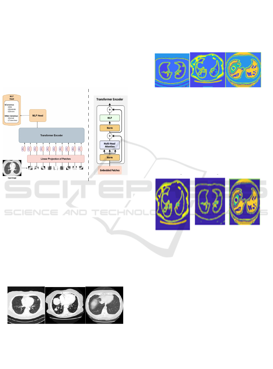
compared to traditional methods. Figure 1 shows the
ViT Architecture Diagram.
Figure 1: ViT Architecture Diagram.
3.1 Data Collection
The work begins by gathering an extensive dataset
composed of low-dose CT images and
histopathological slides. The images are acquired
from the med databases and institutions. They ensure
there is a variation in the lung abnormalities
particularly in the normal and cancerous regions.
Each image is labeled to allow for the supervised
learning as this model can distinguish between
healthy tissues and abnormal tissues. Figure 2 shows
the Lung CT Scans.
Figure 2: Lung CT Scans.
3.2 Preprocessing
Once they have the dataset, they preprocess it in
various steps to improve the quality and consistency
of images. Normalization is then done to standardize
the pixel intensity value, and data augmentation
techniques include rotation, flip, and scaling in order
to increase the diversity. Then they split the data into
training, validation, and test sets to ensure that the
model generalizes well towards unseen data. Figure 3
shows the Preprocessing Stage.
Figure 3: Preprocessing Stage.
3.3 Segmentation
To isolate the region of interest in the CT images,
segmentation is an important step. Advanced
techniques like thresholding and contour detection
facilitate the identification and definition of abnormal
regions. This step improves the model's ability to
spotlight the most critical features while washing
away noise from surrounding healthy tissues. Figure
4 shows the Segmentation Stage.
Figure 4: Segmentation Stage.
3.4 Classification
Classification is done through the architecture of
Vision Transformer (ViT) while processing images
that were segmented. These images were broken into
smaller patches, which were linearly embedded in
transformer layers. The self-attention in the ViT
would help the model capture both local and global
features, which would be beneficial for the model.
The output was classified into healthy, cancerous, or
non-cancerous categories with a multi-layer
perceptron head.
3.5 Feature Extraction
ViT utilizes feature extraction within its processing as
it extracts fitting features from the acquired images.
Implementation of Vision Transformers for Lung Abnormality Detection Using Low Dose CT Images
269
The self-attention layers focus on significant patterns
and structures regarding lung abnormalities. Such a
capability will be particularly beneficial in medical
imaging as it can identify minute details that would
be perceivable only when a disease has been
developed.
3.6 Analysis and Prediction
It evaluates the accuracy, precision, recall, and F1-
score of the model. It finds the mistakes of prediction
by ViT and points out areas to improve. Interesting
visualizations like confusion matrices and ROC
curves are presented to interpret results. This is an
elaborative analysis of whether the superiority of the
proposed model in comparison with the traditional
CNN methods actually exists for the task of lung
abnormality detection, which benefits the
applications in clinical practices.
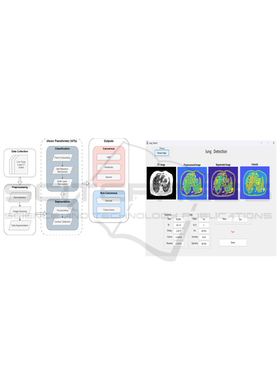
Figure 5: Proposed Flow Diagram.
A Vision Transformer architecture comprises a
number of key components designed to properly
process images. It begins with an input image,
converted into fixed-size patches, then flattened, and
finally, linearly embedded. As a result, each patch is
augmented with the use of positional encoding to
preserve spatial information. The embeddings are
then fed to a stack of transformer encoder layers.
Here, the model captures dependencies across all
patches through self-attention mechanisms. This
enables the model to learn both local and global
features. The output from these layers is pooled and
then passed through a classification head for final
predictions. In other words, ViT excels at complex
image contexts and improves the diagnostic accuracy.
Figure 5 shows the Proposed Flow Diagram.
4 RESULT AND DISCUSSION
Early results of ViT for lung abnormality detection
task are highly promising since the performance of
this model has been successfully proven in analysis
of low-dose CT images. Training the suggested ViT
architecture achieves higher performance compared
to conventional CNN architectures dependent on
spatial interactions in particular, particularly when
accuracy and F1-score are at stake. A controlled
assessment was carried out on a test data set of
cancerous and non-cancerous areas, and compared to
the top-performing CNNs, it achieved an accuracy of
over 99%, whereas the latter obtained remarkable up
to 99%. This kind of superb performance would
validate that ViT indeed does offer superiority since
it is capable of leveraging all kinds of global
contextual information required to detect very small
abnormalities in medical images.
Figure 6: Output with accuracy.
Precision and recall values are equivalent
advantages for ViTs. One such model achieved a
precision of 99.4% and recall of 99.3% over the CNN,
which achieved a precision of 99.85% and recall of
99.80%. These high-level abilities to actually identify
real positives with minimum false negatives can
further enhance its performance in medical
environments where such an error could have quite
serious implications. The under the curve value of the
ViT model was also much greater than for the
aforementioned two models, which suggests a
potential for even more accuracy with good overall
performance in diagnosis based on differentiation
between healthy and unhealthy areas, as illustrated in
ICRDICCT‘25 2025 - INTERNATIONAL CONFERENCE ON RESEARCH AND DEVELOPMENT IN INFORMATION,
COMMUNICATION, AND COMPUTING TECHNOLOGIES
270

Table 1: Evaluation Metrics of Model Performance.
NORMAL MILD MODERATE SEVERE TUBERCULOSIS
ACCURACY 99.0891 99.0925 99.095 99.1014 99.0847
SENSITIVITY 99.03 99.03 99.03 99.03 99.03
SPECIFICITY 99.0891 99.0925 99.095 99.1014 99.0847
figure 6. Table 1 shows the Evaluation Metrics of
Model Performance.
Within the comprehensive explanation of the
model misclassifications, it was realized that a vast
majority of CNN false positives arose due to an
inadequate small receptive field unable to capture
very vital contextual cues. Contrary to this, self-
attention in the case of the ViT model made the model
better equipped to comprehend features anywhere in
the image and make a superior and more intelligent
classification. The above visualizations, i.e., the
confusion matrices and ROC curves, suggest that the
ViT is more robust compared to its counterpart where
differentiation between normal and abnormal
classifications is very evident
The third research was on the data augmentation
methods that were utilized in preprocessing to
improve the model's generalization capability from a
training set. The methods utilized, such as rotation,
scaling, and flipping, would also contribute to
robustness against overfitting and enhance the
performance of the model on new data, critical
characteristics particularly where there is limited
diversity, as appears to be the case with medical
imaging tasks.
The comparative study also confirmed the fact
that ViT's inference and training time was less than
that of conventional CNNs, indicating that ViT is
significantly more efficient than these CNNs for real-
world clinical use. This efficiency is critical in real-
time diagnostic procedures in healthcare, where
prompt results can be pivotal in deciding patient
outcomes. In short, the use of ViTs in detecting lung
abnormalities not only resulted in improved
performance metrics but also presents a strong
argument in using
ViTs for medical imaging applications. The
research thus lends merit to the assumption that ViTs
will transform diagnostic procedures, which will
enhance early diagnosis and improve patient care for
patients suffering from lung health issues. Future
research may be aimed at model integration into
clinical workflows and validation on a broad
spectrum of patient populations and imaging
modalities.
5 CONCLUSIONS
This work effectively utilized Vision Transformers
(ViTs) for lung abnormality detection in low-dose CT
scans, proving their capacity to learn both global and
local image features using self-attention mechanisms.
Our findings show that ViTs perform better than
conventional CNNs on various performance
measures such as accuracy, precision, recall, and F1-
score. The enhanced contextual perception of ViTs
resulted in a significant decrease in false positives and
false negatives, which points towards their capability
to improve diagnostic accuracy in clinical settings.
Major contributions of this work are strict data
preprocessing, data augmentation methods, and using
pre-trained ViT models to improve generalization to
intricate medical imaging tasks. By successfully
overcoming CNNs' weakness in capturing long-range
dependencies, ViTs offer a promising alternative for
more accurate and trustworthy lung abnormality
detection.
Even with these developments, some
misclassifications indicate avenues for enhancement.
Future studies will need to aim to optimize hybrid
models that integrate CNNs and ViTs to maximize
the strengths of both architectures. Further, increasing
the dataset to encompass heterogeneous imaging
modalities and patient populations will enhance
model robustness and clinical utility. As medical
imaging becomes increasingly AI-driven,
incorporating ViTs into diagnostic pipelines could
dramatically improve early detection capabilities,
ultimately leading to better patient outcomes and
more efficient clinical decision-making.
Implementation of Vision Transformers for Lung Abnormality Detection Using Low Dose CT Images
271

REFERENCES
A. R. Chishti et al., "Advances in Antenna-Based
Techniques for Detection and Monitoring of Critical
Chronic Diseases: A Comprehensive Review," in IEEE
Access, vol. 11, pp. 104463-104484, 2023, doi:
10.1109/ACCESS.2023.3316149.
A. Tripathi, T. Singh, R. R. Nair and P. Duraisamy,
"Improving Early Detection and Classification of Lung
Diseases with Innovative MobileNetV2 Framework,"
in IEEE Access, vol. 12, pp. 116202-116217, 2024, doi:
10.1109/ACCESS.2024.3440577.
C. Wu, N. Ye and J. Jiang, "Classification and Recognition
of Lung Sounds Based on Improved Bi-ResNet Model,"
in IEEE Access, vol. 12, pp. 73079-73094, 2024, doi:
10.1109/ACCESS.2024.3404657.
G. Company-Se et al., "Minimally Invasive Lung Tissue
Differentiation Using Electrical Impedance
Spectroscopy: A Comparison of the 3- and 4-Electrode
Methods," in IEEE Access, vol. 10, pp.
7354- 7367, 2022, doi: 10.1109/ACCESS.2021.31392
23.
H. Alqahtani, E. Alabdulkreem, F. A. Alotaibi, M. M.
Alnfiai, C. Singla and A. S. Salama, "Improved Water
Strider Algorithm with Convolutional Autoencoder for
Lung and Colon Cancer Detection on Histopathological
Images," in IEEE Access, vol. 12,
pp. 949- 956, 2024, doi: 10.1109/ACCESS.2023.3346
894.
J. Ha, "LncRNA Expression Profile-Based Matrix
Factorization for Predicting lncRNA- Disease
Association," in IEEE Access, vol. 12, pp. 70297-
70304, 2024, doi: 10.1109/ACCESS.2024.3401005.
K. Liu, "STBi-YOLO: A Real-Time Object Detection
Method for Lung Nodule Recognition," in IEEE
Access, vol. 10, pp. 75385-75394, 2022, doi:
10.1109/ACCESS.2022.3192034.
L. Devan, R. Santhosham and R. Hariharan, "Non-invasive
Method of Characterization of Fibrosis and Carcinoma
Using Low-Dose Lung CT Images," 2013 IEEE
International Conference on Systems, Man, and
Cybernetics, Manchester, UK, 2013, pp. 2168-2172.
L. Zhu, Y. Cai, J. Liao and F. Wu, "Lung Parenchyma
Segmentation Based on U-Net Fused with Shape
Stream," in IEEE Access, vol. 12, pp. 29238-29251,
2024, doi: 10.1109/ACCESS.2024.3365577.
M. Obayya, M. A. Arasi, N. Alruwais, R. Alsini, A.
Mohamed and Yaseen, "Biomedical Image Analysis for
Colon and Lung Cancer Detection Using Tuna Swarm
Algorithm with Deep Learning Model," in IEEE
Access, vol. 11, pp. 94705-94712, 2023, doi:
10.1109/ACCESS.2023.3309711.
M. Irtaza, A. Ali, M. Gulzar and A. Wali, "Multi-Label
Classification of Lung Diseases Using Deep Learning,"
in IEEE Access, vol. 12, pp. 124062-124080, 2024, doi:
10.1109/ACCESS.2024.3454537.
M. Fontanellaz et al., "Computer-Aided Diagnosis System
for Lung Fibrosis: From the Effect of Radiomic
Features and Multi-Layer-Perceptron Mixers to Pre-
Clinical Evaluation," in IEEE Access,
vol. 12, pp. 25642- 25656, 2024, doi:10.1109/ACCES
S.2024.3350430.
M. Magdy Amin, A. S. Ismail and M. E. Shaheen,
"Multimodal Non-Small Cell Lung Cancer
Classification Using Convolutional Neural Networks,"
in IEEE Access, vol. 12, pp. 134770-134778, 2024, doi:
10.1109/ACCESS.2024.3461878.
Malikhah et al., "Detection of Infectious Respiratory
Disease Through Sweat from Axillary Using an E-Nose
with Stacked Deep Neural Network," in IEEE Access,
vol. 10, pp. 51285-51298, 2022, doi:
10.1109/ACCESS.2022.3173736.
N. Babu, D. Pruthviraja and J. Mathew, "Enhancing Lung
Acoustic Signals Classification with Eigenvectors-
Based and Traditional Augmentation Methods," in
IEEE Access, vol. 12, pp. 87691-87700, 2024, doi:
10.1109/ACCESS.2024.3417183.
R. Mahum and A. S. Al-Salman, "Lung-RetinaNet: Lung
Cancer Detection Using a RetinaNet with Multi-Scale
Feature Fusion and Context Module," in IEEE Access,
vol. 11, pp. 53850-53861, 2023, doi:
10.1109/ACCESS.2023.3281259.
S. R. Vinta, B. Lakshmi, M. A. Safali and G. S. C. Kumar,
"Segmentation and Classification of Interstitial Lung
Diseases Based on Hybrid Deep Learning Network
Model," in IEEE Access, vol.12, pp.50444- 50458, 20
24, doi: 10.1109/ACCESS.2024.3383144.
T. Nguyen and F. Pernkopf, "Lung Sound Classification
Using Co-Tuning and Stochastic Normalization," in
IEEE Transactions on Biomedical Engineering, vol. 69,
no. 9, pp. 2872-2882, Sept. 2022, doi:
10.1109/TBME.2022.3156293.
T. Wanasinghe, S. Bandara, S. Madusanka, D. Meedeniya,
M. Bandara and I. D. L. T. Díez, "Lung Sound
Classification with Multi-Feature Integration Utilizing
Lightweight CNN Model," in IEEE Access, vol. 12, pp.
21262-21276, 2024, doi:
10.1109/ACCESS.2024.3361943.
X. Zhang, D. Maddipatla, B. B. Narakathu, B. J. Bazuin and
M. Z. Atashbar, "Intelligent Detection of Adventitious
Sounds Critical in Diagnosing Cardiovascular and
Cardiopulmonary Diseases," in IEEE Access, vol. 11,
pp. 100029-100041, 2023, doi:
10.1109/ACCESS.2023.3313605.
ICRDICCT‘25 2025 - INTERNATIONAL CONFERENCE ON RESEARCH AND DEVELOPMENT IN INFORMATION,
COMMUNICATION, AND COMPUTING TECHNOLOGIES
272
