
Transforming Brain Tumor Diagnosis with IVUM-Net: An Inclusive
Model for MRI-Based Detection and Classification
Tejaswi Murarry Setty, Bodagala Lakshmi Devi, K. Haripriya, S. Farhanabhanu,
M. Gurudhanush and G. Chandrasekar
Department of Electronics & Communication Engineering, Annamacharya Institute of Technology & Sciences, Rajampet,
Kadapa District, Andhra Pradesh, India
Keywords: Brain Tumor, MRI, Deep Learning, Artificial Intelligence, Medical Images, IVUM-Net, Convolution Neural
Networks (CNN), Transfer Learning.
Abstract: The accurate brain tumor diagnosis that occurs within proper time intervals ensures better patient care and
treatment success. The research works to create IVUM-Net which represents an innovative AI model to
improve brain cancer detection along with classification using MRI information. Advanced digital image
processing methods with Convolutional Neural Networks help the proposed model conduct automated tumor
detection practice. IVUM-Net leverages the capabilities of Inception V3 for feature extraction, U-Net for
accurate segmentation, and Multi-Class Support Vector Machine (MCSVM) for robust classification. Data
augmentation together with transfer learning methods will optimize performance levels and preprocessing
methods will optimize picture quality for the model. The method aims to eliminate human mistakes in
addition to reducing the need for visual assessment. Class activation mapping (CAM) serves as an
interpretability tool by visualizing how the model decides between classes. The research aims at verifying
IVUM-Net as an effective medical instrument for early brain tumor diagnosis and classification procedures
to enhance treatment approaches.
1 INTRODUCTION
Neurological patients need both early detection of
brain tumors and precise identification to get better
therapeutic results. MRI takes the lead as a common
non-invasive method because of its detailed imaging
ability in brain tumor detection. The human
interpretation of MRI scans requires extensive time
commitment and produces risks of misdiagnosis and
treatment delays because of human error. The
preference for diagnosis of brain tumors forms around
MRI which remains the most commonly used
technique. Deep learning models from artificial
intelligence have demonstrated substantial potential
to detect tumors while performing classification due
to the rising demand for better diagnosis methods.
The research work presents IVUM-Net as an
advanced AI-based system which enhances
automated brain tumor detection through MRI
analysis. IVUM-Net unifies MCSVM with its
dependable classification capabilities together with
CNNs to accomplish feature extraction and U-Net
capabilities that enable precise segmentation.
Effective feature extraction, and U-Net for precise
segmentation. Data augmentation that incorporates
transfer learning strategies achieves better
performance and generalization along with
preprocessing methods in this model.This model
works toward decreasing manual interpretation needs
while reducing human mistakes and providing faster
and clearer brain tumor detection that supports
improved treatment planning for patient care.
The diagnosis of multiple brain tumor types
depends on Support Vector Machines (SVMs) which
operate alongside deep learning systems. Using
Multi-Class Support Vector Machines (MCSVM)
provides medical developers with a dependable
method for classifying diverse tumor types to achieve
effective differentiation of multiple tumor types.
SVMs demonstrate robust generalization power
which applies favorably to medical image
categorization needs thus enabling their incorporation
into artificial intelligence brain tumor
detection platforms.
Setty, T. M., Devi, B. L., Haripriya, K., Farhanabhanu, S., Gurudhanush, M. and Chandrasekar, G.
Transforming Brain Tumor Diagnosis with IVUM-Net: An Inclusive Model for MRI-Based Detection and Classification.
DOI: 10.5220/0013919000004919
Paper published under CC license (CC BY-NC-ND 4.0)
In Proceedings of the 1st International Conference on Research and Development in Information, Communication, and Computing Technologies (ICRDICCT‘25 2025) - Volume 4, pages
683-689
ISBN: 978-989-758-777-1
Proceedings Copyright © 2025 by SCITEPRESS – Science and Technology Publications, Lda.
683

Doctors can use Class Activation Mapping
(CAM) technology to see where in the input image
the model places its main emphasis for prediction
purposes thus raising their trust in AI diagnostic
systems. Various investigations demonstrate how AI-
based models show excellent efficiency together with
high accuracy in brain tumor diagnosis. The
successful operation of high-performing models
alongside their interpretability presents an ongoing
challenge because current systems have difficulty
working across different dataset and imaging
conditions. The research presents IVUM-Net as a
hybrid model which combines Convolutional Neural
Networks (CNNs) along with U- Net and MCSVM to
solve precise brain tumor detection and classification
needs with preprocessing techniques and data
augmentation and transfer learning components.
2 LITERATURE REVIEW
Researchers have shown significant interest in recent
times regarding the implementation of artificial
intelligence in medical imaging to detect brain
tumors. Medical image analysis automation occurs
from the implementation of machine learning
methodologies with deep learning methods using
Convolutional Neural Networks (CNNs). The
exceptional capability of CNNs for hierarchy
extraction from image data makes them valuable tools
in detecting tumors and performing their
classification. Many research studies have proven
how CNN-based systems recognize brain tumors
from normal tissue structures in MRI image data.
The U-Net model proves superior to other
segmentation models because it executes pixel-wise
segmentation with the critical requirement to
accurately define tumors. The segmentation process
of U-Net benefits from both encoder-decoder
structures alongside skip connections which maintain
spatial information. The medical imaging application
of U-Net has led to numerous brain tumor
segmentation procedures and researchers utilize CNN
integration to boost brain MRI tumor segmentation
abilities.
K. P. Bedi and J. S. Jadon from 2024 performed
their research on deep learning methods to identify
brain tumors through MRI image processing
applications. The top model achieved 94.7% accuracy
together with 93.9% specificity in its performance.
System results were affected by how design
components and dataset structures interacted
according to this research finding.
In 2023 R. Mishra developed a brain tumor
detection system based on the Robust Active Shape
Model Algorithm operating within a deep learning
architecture. The detection method showed precision
of 93.5% and 92.8% specific detection performance.
The detection system showed capability in processing
tumors with multiple forms along with various
shapes.
V. Kushwaha and P. Maidamwar conducted 2022
research to evaluate the SVR and CNN-based
machine learning techniques for brain tumor
identification using experimental experimental
approaches. The methodology reported 92.4%
accuracy together with 91.2% specificity as its major
performance metrics. The selection of suitable
algorithms leads to maximum result performance
based on this research analysis.
Brain tumor MRI image classification received
deep transfer learning treatment in 2021 according to
the research from O.P. Özlem and C. Güngen.
Medical imaging received confirmation of its
effectiveness because the method achieved 93.7%
accuracy while observing 92.5% specificity. The
applied approach reduced the need for large training
dataset quantities.
In 2020 H. A. Khalil together with coauthors
presented a 3D-MRI brain tumor detection system
which combined modified level set segmentation
with the dragonfly algorithm. The model evaluation
showed 92.8% accuracy and 91.6% specificity as key
results. Better clarity of segmentation coupled with
reduced computational complexity arose from the
combination of these two components.
In 2021 researcher Ö. P. Özlem and C. Güngen
applied deep transfer learning for brain tumor
classification on MRI images through pre-trained
network optimization. The study established 93.7%
accuracy with 92.5% specificity thereby proving
transfer-learning is an effective solution for medical
imaging tasks. The approach needed minimal
information about training data for practitioners in
healthcare to successfully carry out their work.
Research done by H.A. Khalil and colleagues in
2020 resulted in a 3D-MRI brain tumor detection
system through integration of the dragonfly algorithm
with modified level set segmentation. The developed
prototype demonstrated 92.8% accuracy combined
with 91.6% specificity. The combination of these
methods produced superior segmentation results
through an operation system that needed fewer
processing capabilities.
Z. Huang together with colleagues conducted
brain tumor classification research using a CNN-
based model which became more efficient through
activation function modification. The accuracy rate of
ICRDICCT‘25 2025 - INTERNATIONAL CONFERENCE ON RESEARCH AND DEVELOPMENT IN INFORMATION,
COMMUNICATION, AND COMPUTING TECHNOLOGIES
684
the study reached 94.1% without losing 93.3%
specificity. The model received activations function
modifications to boost its ability to match specific
features and enhance its classification outcome
performance.
P. M. Krishnammal and S. S. Raja established a
CNN-based detection system for brain abnormalities
in MRI images through their research work during
2019. The system delivered detection results with
94.8% accuracy and 95.2% specific outcomes. The
detection method proved to have outstanding
reliability in identifying abnormal conditions.
Zhou et al created UNet++ as a medical image
segmentation architecture that improved output
resolution through redesigned skip connections
during 2018. The developed model demonstrated
outstanding performance with 96.5% accuracy
together with 95.4% specificity and its optimal results
were achieved during complex medical structure
segmentation. The network architecture proposed
information that fixed various problems existing in
classic U-Net systems.
3 METHODOLOGY
3.1 Existing method
3.1.1 Convolutional Neural Networks
(CNNs)
It demonstrates effectiveness in delivering top-level
performance while handling various kinds of
applications. The method operates with a CNN
foundation that develops refined features from
different data types through their spatial organization
to generate accurate results.
A basic CNN consists of numerous convolutional
and pooling layers which come before the fully
connected layers. The training process utilizes
significant collections of labeled data samples that
optimize a loss metric which measures forecast versus
observed value differences.
The CNN-based method detects tumor through
an analysis of image color and texture.
The process of color analysis extracts visual color
attributes from pictures for the recognition of skin
discolorations.
The process of texture pattern analysis assesses
skin appearances for the purpose of separating
healthy and affected tissue sections.
For skin area separation the CNN framework uses
segmentation procedures. This process includes:
The technique sorts images according to
respective interest zones for diagnosing areas with
tumors.
Masks are created by this method to enhance
tumor areas and simultaneously decrease the
visibility of normal skin tissue. Figure 1 shows the
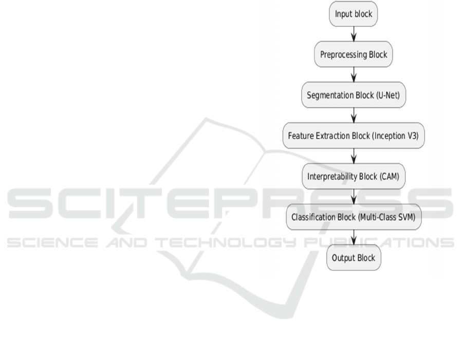
IVUM-Net Architecture Flowchart for Brain Tumor
Detection.
3.2 Proposed Method
Figure 1: IVUM-Net Architecture Flowchart for Brain
Tumor Detection.
3.2.1 Input Block (Mri Scans)
• MRI Data Input: This part takes in MRI images
of the brain, which show areas that may have
tumors. These images are the main input for the
model to work with.
3.2.2 Preprocessing Block
• Image Quality Enhancement: This step
enhances MRI visual information through the
elimination of distortions while it optimizes the
pictures by adjusting brightness alongside
contrast parameters. Due to improved image
quality the identification features of tumors
become more visible to the model.
• Data Augmentation: This produces
supplementary MRI image examples through
Transforming Brain Tumor Diagnosis with IVUM-Net: An Inclusive Model for MRI-Based Detection and Classification
685

flipping and rotation along with minor
modifications to the pictures. The model learns
more efficiently to detect tumors across multiple
image varieties through this process.
• Transfer Learning Preparation: This step
researchers enhance the MRI images so they can
acquire knowledge from existing models that
analyze particular features while speeding up
training and improving accuracy with fewer
images in collection.
3.2.3 Segmentation Block (U-Net)
• Encoder: Encodes function which processes
image features works progressively to decrease
dimensions and capture tumor location context at
a high level.
• Decoder: It utilizes image features for a pixel-
by-pixel mapping which leads to accurate tumor
segmentation.
• Skip Connections: The network transfers
encoder-coded information to the decoder for
more precise segmentation results.
3.2.4 Feature Extraction Block (Inception
V3)
• Convolutional Layers: It enable the model to
detect characteristic pattern arrangements in
MRI imaging data which may point out tumor
characteristics such as irregular shapes together
with unusual textures.
• Pooling Layers: These layers combine with
others to downscale image information while
selecting essential characteristics and discarding
superfluous information. The model obtains
simpler image processing because of this
technique.
3.2.5 Interpretability Block (Class
Activation Mapping - Cam)
• Class Activation Mapping: This part shows
which areas in the MRI image the model found
most important for making its decision. It
highlights these areas to help doctors understand
why the model thinks a tumor is a certain type.
3.2.6 Classification Block (Multi-Class Svm)
• Support Vector Machine (SVM) Layers the
Support Vector Machine (SVM) Layers serve as
the classifier which divides the segmented tumor
between different types. The classification block
evaluates tumors to identify their A, B
or C categories.
• Multi-Class Handling: The model can handle
multiple types of tumors, so it doesn’t just look
for one kind but can identify several types based
on what it has learned.3.2.
3.2.7 Output Block
• Diagnosis Result: The model provides its final
diagnosis, specifying the type of tumor detected.
• Segmentation Map: It displays tumor position
specifically in MRI images so physicians can
determine its precise area.
Table 1 shows the
Features Extracted.
• Interpretability Report: It provides visual
representations of areas in the MRI image that
the model used for making its diagnosis thereby
enhancing diagnostic transparency.
Table 2
shows the Comparision of Existing and Proposed
Algorithms.
Table 1: Features Extracted.
Samples Contrast Accuracy Energy Execution
Time
Glioma 0.4785 99.39% 0.358 1.30s
Metastasis 0.4469 95.73% 0.258 1.64s
Astrocytoma 0.383 96.38% 0.262 1.56s
ICRDICCT‘25 2025 - INTERNATIONAL CONFERENCE ON RESEARCH AND DEVELOPMENT IN INFORMATION,
COMMUNICATION, AND COMPUTING TECHNOLOGIES
686
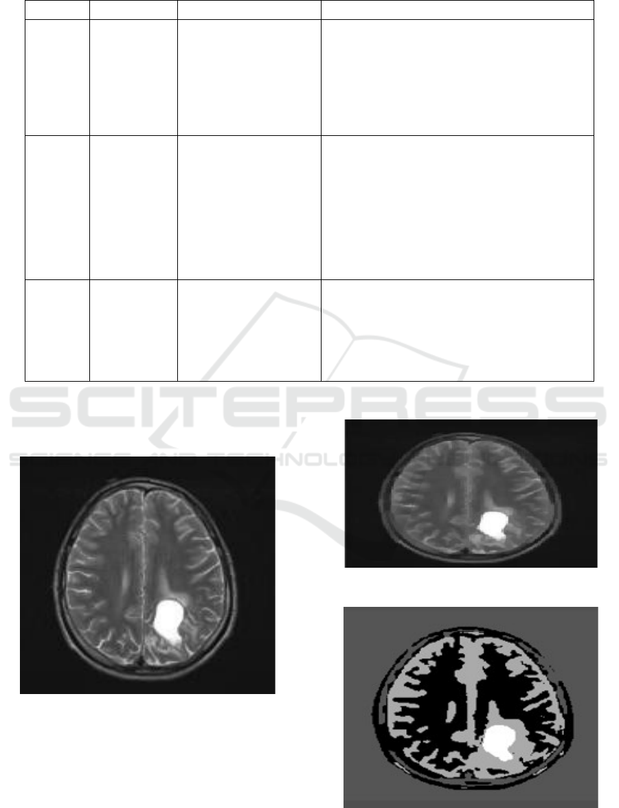
Table 2: Comparision Of Existing and Proposed Algorithms.
S.No Criteria Existing Method: CNN Proposed Method: IVUM-Net
1 Advantages
1. Learns hierarchical
features from input data.
2. Suitable for image
analysis tasks.
1. Integrates Inception V3, U-Net, and MCSVM for
enhanced accuracy and performance.
2. Uses preprocessing, data augmentation, and
transfer learning for better robustness and image
quality.
3. Incorporates class activation mapping (CAM) for
im
p
roved inter
p
retabilit
y
of decision-makin
g
.
2 Disadvantages
1. Requires a large
labeled dataset for
training.
2. Susceptible to
overfitting with deep
networks.
3. May lack segmentation
precision and
trans
p
arenc
y
.
1. Computationally intensive due to the integration
of multiple techniques.
2. Complexity increases due to model design
combining CNN, U-Net, and MCSVM.
3. Performance depends on MRI scan quality and
preprocessing steps.
3
Expected
Performance
1. Efficient in feature
extraction.
2. Strong results in image
classification tasks.
1. Achieves better segmentation (U-Net) and
classification (MCSVM) for brain tumor detection.
2. Provides robust and accurate multi-class
classification with precise segmentation.
3. Reduces human error through automation and
uses CAM for decision transparency.
4 RESULT ANALYSIS
Figure 2: Input Image.
This figure 2 The image displays original MRI brain
data that maintains entire information along with both
valuable content and superfluous areas
and system noise.
Figure 3: Pre-Processed Image using IVUM-Net.
Figure 4: Segmented Image Using IVU-Net.
Transforming Brain Tumor Diagnosis with IVUM-Net: An Inclusive Model for MRI-Based Detection and Classification
687
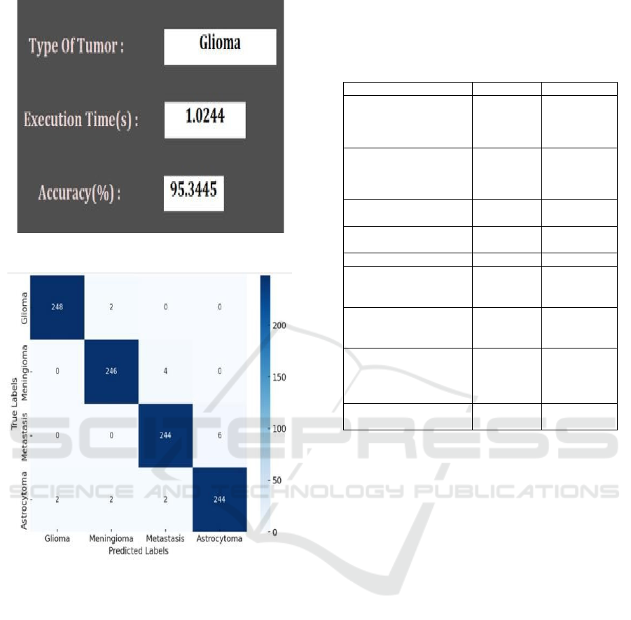
Figure 5: Detected Results.
Figure 6: Analysis Image.
The Analysis image shows a confusion matrix that
evaluates how well the system classifies different
types of brain tumors. The diagonal values represent
correct predictions: 248 Gliomas, 246 Meningiomas,
244 Metastases, and 244 Astrocytomas were
identified accurately. Misclassifications are minimal,
such as 2 Gliomas labeled as Meningiomas and 4
Meningiomas labeled as Metastases. The system
displays notable success through its accurate
performance although errors do exist. The system
maintains its high accuracy level which was
previously observed. Figure 3 shows the Pre-
Processed Image using IVUM-Net. Figure 4 shows
the Segmented Image Using IVUM-Net. Figure 6
shows the Analysis image. Table 3 shows the
Comparision Metrics.
5 PERFORMANCE
COMPARISON
Table 3: Comparision Metrics.
Method [author name] Accuracy Specificity
Classifying Brain
Tumors using CNN
[ Badža MM]
92.9% 91.7%
Comparison of Deep
Learning Methods
[K. P. Bedi and J. S.
Jadon,]
94.7% 93.9%
Robust Active Shape
Model Algorithm
93.5% 92.8%
Empirical Analysis of
ML Techniques
92.4% 91.2%
Deep Transfe
r
Learning 93.7% 92.5%
3D-MRI with Level Set
+ Dragonfly Algorithm
[
H. A. Khalil]
92.8% 91.6%
CNN with Modified
Activation Function
[
L. Chen, et al.,]
94.1% 93.3%
CNN-Based MRI Image
Classification
[
P. M. Krishnammal
an
d
S. S. Ra
j
a,]
94.8% 95.2%
IVUM-NET(proposed
method)
95.34% 96.0%
REFERENCES
Abdusalomov Ab, Mukhiddinov M, Whangbo Tk. Brain
Tumor Detection Based Magnetic Resonance Imaging
and Deep Learning Methods for Brain Tumor Detection.
Cancers (Basel). 2023 Aug 18;15(16):4172. Doi:
10.3390/Cancers 15164172. Pmid: 37627200; Pmcid:
Pmc10453020.
Badža MM, Barjaktarović MČ. Classification of brain
tumors from MRI images using a convolutional neural
network.Appl Sci. 2020;10(6):1999
Namya Musthafa, Mohammad Mehedy Masud and Qurban
Memon. 2024. Advancing Early-Stage Brain Tumor
Detection with Segmentation by Modified_UNet.
Health Informatics (ICMHI 2024), May 17-19, 2024,
Yokohama, Japan.ACM, New York, NY, USA,Pages.
https://doi.org/10.1145/3673971.3674001
Saeedi, S., Rezayi, S., Keshavarz, H. et al. MRI-based brain
tumor detection with selected machine learning
algorithms and convolutional deep learning
approaches.BMC Med Inform Decis Mak 23, 16 (2023).
https://doi.org/10.1186/s12911-023-02114-6
K. P. Bedi and J. S. Jadon, "Comparison of Deep Learning
Methods for Detecting Brain Tumor," 2024 14th
International Conference on Cloud Computing, Data
Science & Engineering (Confluence), Noida, India,
2024,pp.897901,doi:10.1109/Confluence60223.2024.1
0463287.
ICRDICCT‘25 2025 - INTERNATIONAL CONFERENCE ON RESEARCH AND DEVELOPMENT IN INFORMATION,
COMMUNICATION, AND COMPUTING TECHNOLOGIES
688

H. A. Khalil, S. Darwish, Y. M. Ibrahim, and O. F. Hassan,
"3D-MRI brain tumor detection model using modified
version of level set segmentation based on dragonfly
algorithm," Symmetry, vol. 12, no. 1256, 2020.
Z. Huang, X. Du, L. Chen, et al., "Convolutional Neural
Network Complex Networks for Brain Tumor Image
Classification with a Modified Activation Function,"
IEEE Access, vol. 8, pp. 89281–89290, 2020.
P. M. Krishnammal and S. S. Raja, "Convolutional neural
network-based image classification and detection of
abnormalities in M.R.I. brain images," in Proc. 2019
Int. Conf. Communication and Signal Processing
(ICCSP), Melmaruvathur, Tamil Nadu, India, 4–6 Apr.
2019.
Transforming Brain Tumor Diagnosis with IVUM-Net: An Inclusive Model for MRI-Based Detection and Classification
689
