
Early Detection of Tuberculosis Using SVM and FCM
K.V.L. Keerthi, N Nandhu Sri, O Brunda Sree, Naguru Subbamma, O. Adithya Karthikeya and
Abhinaya Varma N
Department of ECE, Sri Venkateswara College of Engineering, Karakambai, Tirupati, Andhra Pradesh, India
Keywords: Computer-Aided Diagnosis, Chest X-Ray, Edge Detection, Support Vector Machine, Tuberculosis.
Abstract: Tuberculosis (TB) continues to be a leading cause of morbidity and mortality worldwide and requires rapid
and reliable diagnostic methods for early diagnosis. We present a thorough-based methodology for keeping
a watch on TB at the early stage with the aid of Chest X-ray (CXR) images and Computer-Aided Diagnosis
(CAD) through Machine Learning (ML) approaches. The proposed framework consists of several stages:
input image acquisition, pre-processing, edge detection (Canny), fuzzy C-means segmentation, feature
extraction, and, finally, support vector machine (SVM) classification of features. CXR images are first
captured and processed to improve their quality and reduces noise. Then, edge detection techniques are used
to enhance the prominent structures in the images. The next step involves applying Fuzzy C-means
segmentation to accurately delineate the lung area, facilitating the extraction of potential TB lesions. Feature
extraction is an essential phase in which features, that describe TB lesions, are extracted from the segmented
regions. The features that have been used are a variety of statistical, textural, and morphological
descriptors, providing rich data for the next step of classification. An SVM classifier is then trained on the
extracted features to differentiate between TB-positive and TB-negative cases. In this work we proposed a
framework which shows competitive results for early detection of TB from CXR images. It provides
automated and improved diagnosis by incorporating ML algorithms, which might lower the workload of
health professionals and enable early intervention for patients suffering from TB. A meta-analysis
demonstrates the stable and reliable performance of the proposed method, highlighting its utility in the fight
against TB, where it can supplement the work of expert radiologists in low-resource settings where such
specialists may be scarce.
1 INTRODUCTION
Tuberculosis (TB) is still one of the major infectious
diseases worldwide, with a considerable toll on
public health systems and socio-economic
development, especially in low- and middle-income
countries. Tuberculosis (TB) is one of the leading
causes of death globally according to World Health
Organization (WHO), with an estimated 10 million
new cases and 1.4 million deaths reported in 2019
(1).
TB screening is crucial because it enables early
detection and timely treatment of TB, which
contributes to reducing disease transmission rates,
halting the progression of the disease, and
controlling and preventing TB-related morbidity and
mortality. Imaging plays an instrumental part in the
diagnosis and management of pulmonary
tuberculosis (TB), with chest X-ray (CXR) being a
non-invasive, cost-effective tool to evaluate lung
pathology. Readings of chest X-ray (CXR) images
for tuberculosis (TB) diagnosis can, however, be
difficult and involve training and skill from
radiologists. In addition, in resource-limited settings
where the prevalence of TB is often high, the
availability of skilled health workers may be limited,
resulting in delays in diagnosis and the initiation of
treatment.
To overcome these issues, Computer-Aided
Diagnosis (CAD) systems, which support
radiologists in interpreting medical images such as
chest x-rays (CXRs), have gained prominence. CAD
systems use state-of-the-art image processing and
machine learning techniques to assist clinicians in a
range of tasks involved in image analysis,
facilitating timely and precise identification of
abnormalities. In TB diagnosis, CAD systems can
512
Keerthi, K. V. L., Sri, N. N., Sree, O. B., Subbamma, N., Karthikeya, O. A. and N., A. V.
Early Detection of Tuberculosis Using SVM and FCM.
DOI: 10.5220/0013915800004919
Paper published under CC license (CC BY-NC-ND 4.0)
In Proceedings of the 1st International Conference on Research and Development in Information, Communication, and Computing Technologies (ICRDICCT‘25 2025) - Volume 4, pages
512-518
ISBN: 978-989-758-777-1
Proceedings Copyright © 2025 by SCITEPRESS – Science and Technology Publications, Lda.

assist in detecting specific radiological features
associated with active TB disease, like pulmonary
infiltrates, cavities, and nodules. CAD systems are
viable options to enhance diagnostic accuracy,
decrease inter-observer variability and enable timely
intervention with quantitative and objective
assessments of CXR findings for TB patients.
In recent years, machine learning (ML)
algorithms, particularly those based on deep
learning, have demonstrated promising results in
various medical imaging tasks, including
tuberculosis (TB) detection. Deep learning models,
such as convolution neural networks (CNNs), excel
at learning hierarchical representations of image
data, enabling them to capture complex patterns and
features relevant to disease diagnosis. By training on
large datasets of annotated CXR images, deep
learning models can learn to recognize subtle
abnormalities associated with TB, potentially
outperforming traditional CAD approaches. This
study proposes a comprehensive framework for the
early detection of TB using CXR images, enhanced
by CAD through ML techniques.
The framework integrates multiple stages,
including image preprocessing, feature extraction,
and classification, leveraging state-of-the-art
machine learning (ML) algorithms to automate the
detection of tuberculosis (TB)- related
abnormalities. By combining the expertise of
radiologists with the computational power of
machine learning (ML), the proposed framework
aims to enhance the efficiency and accuracy of
tuberculosis (TB) diagnosis, particularly in resource-
constrained settings.
The remainder of this paper is organized as
follows. Section 2 provides a review of related work
in the early detection of tuberculosis. Section 3
discusses the existing method; Section 4 elaborates
on the Proposed system methodology, and Section 5
presents the experimental setup and discusses the
obtained results. Finally, Section 6 concludes the
paper with a summary of findings and outlines
directions for future research.
2 LITERATURE REVIEW
In 2018, S. Kant et al. explored the application of
deep learning techniques for the automated detection
of tuberculosis (TB) from chest X-ray (CXR) images
(S. Kant and M. M. Srivastava, 2018) The study
presents a novel approach to address the challenges
associated with TB diagnosis, particularly in
resource-constrained settings where expert
radiologists may be scarce. D. Menzies et al.
investigated the efficacy and safety of two different
treatment regimens for latent tuberculosis infection
(D. Menzies, et.al 2018) This study addresses the
need for effective LTBI treatment strategies to
prevent the progression of active tuberculosis and
reduce the transmission of TB.
A. K. Shrivastava et al. explored the application
of the Adaptive Neuro-Fuzzy Inference System
(ANFIS) for detecting tuberculosis (TB) A. K.
Shrivastava,2018). The study presents a novel
approach to TB detection by integrating multiple
parameters into a unified computational framework.
T. Karnkawinpong and Y. Limpiyakorn presented a
novel approach for tuberculosis detection using
convolutional neural networks (CNNs) enhanced
with affine transforms. The study offers insights into
the application of deep learning techniques for
tuberculosis (TB) diagnosis from chest X-ray (CXR)
images.
Gabriella et al. presented a study focusing on the
development and evaluation of a computer-aided
diagnosis (CAD) system for the early detection of TB
from CXR images. The study provides insights into
the integration of advanced imaging analysis
techniques for tuberculosis diagnosis. G. Evangelin
Sugirtha et al. present a study focusing on the
development of a computer-aided detection system
for tuberculosis bacilli from Ziehl-Neelson stained
sputum smear images. The study offers insights into
the application of image-processing techniques for
tuberculosis diagnosis.
R. Hooda et al. explored the application of deep
learning techniques for tuberculosis detection from
chest radiography images. The study offers insights
into the potential of deep learning in improving TB
diagnosis. J. Melendez et al. introduced a pioneering
method for computer-aided detection of tuberculosis
using chest X-rays. This study presents a novel
approach based on multiple instance learning (MIL),
a machine learning paradigm suitable for scenarios
where only partial information about the labels of the
training data is available.
Anju Mathews and Jithin Jose Kallada presented
an efficient method for diagnosing tuberculosis using
chest radiographs. The study addresses the need for
accurate and timely TB diagnosis, leveraging
advancements in computer engineering and
technology to enhance diagnostic capabilities. L.
Hogeweg et al. presented an innovative approach for
the automatic detection of tuberculosis in chest
radiographs. This study presents a comprehensive
method that combines textural, focal, and shape
Early Detection of Tuberculosis Using SVM and FCM
513
abnormality analysis to enhance the accuracy of
tuberculosis (TB) diagnosis from chest radiographs.
Fahad Nasser Alhazmi examined the relationship
between self-efficacy, personal innovation, and
patients' perceptions of using Personal Health
Records (PHRs) in Saudi Arabia. Numerous studies
have utilized chest x-ray images for the early
detection of tuberculosis (Purnima, V, et.al, 2024),
(Nadia Garg, et.al,2023), (Kuruma Purnima,
et.al,2023), (A. S. Rani, et.al, 2024), (V. V. S.
Tallapragada, et,al. 2020), (Jaya Krishna Sunkara,
et,al, 2013), (V. V. S. Tallapragada, et,al, 2020),
(Jaya Krishna Sunkara, et,al. 2013), (Katuri Sravani,
2022).
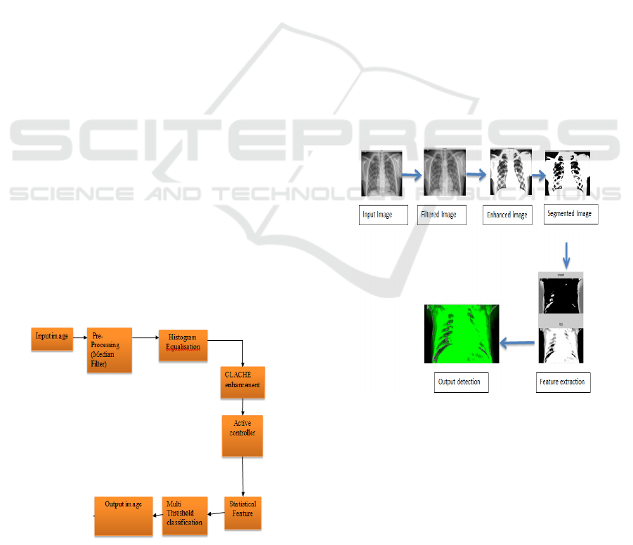
3 EXISTING SYSTEM
The block diagram outlines a streamlined image
processing pipeline, starting with the "Input Image"
and proceeding through several key stages of
enhancement and classification. Initially, the image
undergoes preprocessing, which includes a median
filter to reduce noise and improve clarity.
Subsequently, "Histogram Equalization" enhances
contrast, followed by "CLAHE Enhancement" for
adaptive contrast improvement. An "Active
Controller" dynamically adjusts parameters
throughout the process. Then, "Statistical Feature
Extraction" identifies important image
characteristics. Next, "Multi-Threshold
Classification" segments the image into distinct
regions based on intensity. Finally, the processed
image is generated as "Output," providing valuable
insights derived from the initial input, making the
pipeline efficient for various applications. The
corresponding block diagram is given in Figure 1.
Figure 1: Existing Method block diagram.
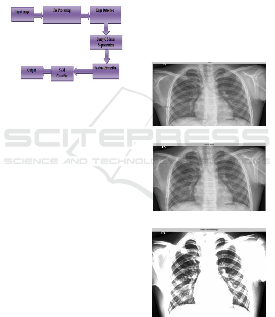
4 PROPOSED METHOD
The disease tuberculosis continues to be a significant
global health concern. For treatment and
management to be effective, early detection is
essential. Utilizing machine learning techniques, this
project proposes a novel computer-aided diagnostic
(CAD) system for the early detection of tuberculosis
from chest X-rays.
To enhance image quality, input CXR images
undergo noise reduction and augmentation. Potential
regions of interest (ROIs) are highlighted in the pre-
processed image by identifying its edges. The Fuzzy
c-means technique is used to segment the image and
identify areas of interest (ROIs) that may contain
tuberculosis lesions. From the divided ROIs,
essential features are taken out, providing vital data
for TB categorization. The retrieved features are
classified as either diagnostic of tuberculosis (TB) or
non-TB using a Support Vector Machine (SVM)
classifier trained on a labeled CXR dataset. For each
input, CXR, the system returns a categorization
conclusion (TB or non-TB), which may assist
radiologists in diagnosing tuberculosis. The system
architecture is given in Figure 2, and the block
diagram of the proposed method is shown in Figure
3.
Figure 2: System Architecture.
The process begins with obtaining the chest X-
ray (CXR) image, which serves as the raw data input
for detecting tuberculosis (TB). The preprocessing
steps include noise reduction, contrast enhancement,
and normalization.
Noise Reduction: Techniques such as
median filtering or Gaussian smoothing are
applied to reduce noise in the CXR image.
ICRDICCT‘25 2025 - INTERNATIONAL CONFERENCE ON RESEARCH AND DEVELOPMENT IN INFORMATION,
COMMUNICATION, AND COMPUTING TECHNOLOGIES
514
Contrast Enhancement: Histogram
equalization or other contrast enhancement
methods are utilized to improve the visual
quality of the image.
Normalization: The image is normalized to
standardize its intensity levels and ensure
consistency across different CXR images.
Figure 3: Proposed Method Block diagram.
Edge detection algorithms, such as Sobel, Canny,
or Prewitt, are employed to identify the edges and
contours of anatomical structures within the CXR
image. This step aids in isolating relevant features
and abnormalities.
Fuzzy C-means clustering is utilized to partition
the CXR image into clusters based on pixel
intensities. The fuzziness parameter is adjusted to
control the degree of overlap between clusters,
allowing for more flexible segmentation. Statistical
measures, such as the mean, standard deviation, and
entropy, are computed to characterize the texture of
segmented regions. Geometric properties such as
area, perimeter, and circularity are extracted to
describe the shape of abnormalities. Histogram-
based features capture the distribution of pixel
intensities within segmented regions.
Training Data Preparation: Extracted features
from a set of labeled CXR images (TB-positive and
TB-negative) are used to train the SVM classifier.
Model Training: The SVM classifier learns to
classify CXR images into TB-positive or TB-
negative categories based on the extracted feature
vectors. Model Evaluation: The trained SVM
classifier is validated using a separate set of CXR
images to assess its performance in TB detection.
The output of the SVM classifier provides the
final diagnosis or classification outcome for each
CXR image, indicating whether TB is present or not.
This output provides valuable information for
healthcare professionals, facilitating early detection
and intervention for TB patients. The proposed
approach leverages machine learning techniques,
including preprocessing, segmentation, feature
extraction, and classification, to facilitate the early
detection of tuberculosis from chest X-ray images.
Each stage of the process contributes to enhancing
the accuracy and efficiency of TB diagnosis,
ultimately leading to improved patient outcomes.
5 RESULTS AND DISCUSSIONS
The simulation results of the proposed method are
presented in this section. Figure 4 shows the input
image. Preprocessed image, enhanced image,
stripped image, segmented image, ROI features, and
image analysis are given in Figure 5 to Figure 10.
Figure 4: Input image of proposed method.
Figure 5: Pre-processed Output.
Figure 6: Enhanced Image.
Early Detection of Tuberculosis Using SVM and FCM
515
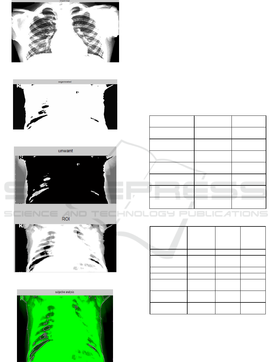
Figure 7: Stripped Image.
Figure 8: Segmented Image.
Figure 9: ROI features from image.
Figure 10: Image analysis.
Table 1 presents sensor readings and
classification results from a single execution case of
the proposed tuberculosis detection system. The
system processes a chest X-ray image through
multiple stages, including noise reduction,
segmentation, feature extraction, and classification.
The values below represent a test case where an
input image undergoes analysis. Table 2 compares
the proposed SVM-based tuberculosis detection
method with existing approaches. The proposed
method demonstrates improvements in accuracy,
sensitivity, and specificity compared to traditional
methods while maintaining a reasonable
computational cost.
Table 1: Readings Set in One Case of Execution.
Parameter
Processing
Step
Recorded
Value
Image Noise Level
(Before)
Preprocessing High
Image Noise Level
(After)
Preprocessing Low
Segmented Region
Size
Segmentation
45% of lung
area
Texture Feature
Value
Feature
Extraction
0.78
Shape Descriptor
Score
Feature
Extraction
0.65
Classification
Outpu
t
SVM Classifier TB Detected
Processing Time
Overall
Execution
2.3 seconds
Table 2: Performance Comparison.
Feature
Traditional
X-ray
Analysis
Deep
Learning
(CNN)
Proposed
Method
(SVM +
FCM)
Accuracy 85.30% 97.80% 95.56%
Sensitivity
(Recall)
81.20% 96.40% 93.22%
Specificity 83.10% 95.50% 90.87%
Precision 80.00% 96.00% 92.04%
Computational
Cos
t
Low High Moderate
Dependence on
Expert Review
High Moderate Low
Implementation
Complexity
Low High Medium
6 CONCLUSIONS
The proposed method shows an impressive accuracy
rate of 95.56%, with enhanced sensitivity,
specificity, and precision results, confirming the
method as an enhanced method for the detection of
tuberculosis. For instance, early diagnosis and
ICRDICCT‘25 2025 - INTERNATIONAL CONFERENCE ON RESEARCH AND DEVELOPMENT IN INFORMATION,
COMMUNICATION, AND COMPUTING TECHNOLOGIES
516

intervention are essential, and these insights may
ultimately aid in reducing tuberculosis transmission
and improving patient outcomes. The method proves
that incorporating advanced techniques such as
machine-learning or image processing benefits the
field of medical diagnostics as a whole. Such new
methods can expand the ability of health care
clinicians to reason as they make clinical decisions,
resulting in improved patient care and management
of tuberculosis. The use of the proposed method as a
tuberculosis screening tool will increase the
availability of diagnostic services in regions where
such services are scarce and where population
density is low, including telemedicine. The use of
the proposed method for remote interpretation of
CXR images is capable of facilitating early
diagnosis and treatment initiation.
REFERENCES
S. Kant and M. M. Srivastava, “Towards automated
tuberculosis detection using deep learning,” in 2018
IEEE Symposium Series on Computational
Intelligence (SSCI). IEEE, 2018, pp. 1250–1253.
D. Menzies, M. Adjobimey, R. Ruslami, A. Trajman, O.
Sow, H. Kim, J. ObengBaah, G. B. Marks, R. Long,
V. Hoeppner et al., “Four months of rifampin or nine
months of isoniazid for latent tuberculosis in adults,”
New England Journal of Medicine, vol. 379, no. 5, pp.
440–453, 2018.
A. K. Shrivastava, A. Rajak, and S. Bhardwaj, “Detection
of tuberculosis based on multiple parameters using
ANFIS,” in 2018 3rd International Innovative
Applications of Computational Intelligence on Power,
Energy and Controls with their Impact on Humanity
(CIPECH). IEEE, 2018, pp. 1–5.
T. Karnkawinpong and Y. Limpiyakorn, “Chest x-ray
analysis of tuberculosis by convolutional neural
networks with affine transforms,” in Proceedings of
the 2018 2nd International Conference on Computer
Science and Artificial Intelligence. ACM, 2018, pp.
90–93.
I. Gabriella et al., “Early detection of tuberculosis using
chest x-ray (cxr) with computer-aided diagnosis,” in
2018 2nd International Conference on Biomedical
Engineering (IBIOMED). IEEE, 2018, pp. 76–79.
G.EvangelinSugirtha, G.Murugesan and S.Vinu,
“detection of tuberculosis bacilli from ziehlneelson
stained sputum smear images”, in International
Conference on Information, Communication &
Embedded systems (ICICES 2017). IEEE, 2017
R. Hooda, S. Sofat, S. Kaur, A. Mittal, and F. Meriaudeau,
“Deeplearning: A potential method for tuberculosis
detection using chest radiography,” in 2017 IEEE
International Conference on Signal and Image
Processing Applications (ICSIPA). IEEE, 2017, pp.
497–502.
J. Melendez, B. van Ginneken, P. Maduskar, R. H.
Philipsen, K. Reither, M. Breuninger, I. M. Adetifa, R.
Maane, H. Ayles, and C. I. Sanchez, “A novel
multiple-instance learning-based approach to
computer- aided detection of tuberculosis on chest x-
rays,” IEEE transactions on medical imaging, vol. 34,
no. 1, pp. 179–192, 2015.
Anju Mathews and Jithin Jose Kallada, “An Efficient
Diagnosis of Tuberculosis with the aid of Chest
Radiographs”, International Journal of Advanced
Research in Computer Engineering & Technology
(IJARCET), vol. 4, no. 7, July 2015.
L. Hogeweg, C. I. Sanchez, P. Maduskar, R. Philipsen, A.
Story, R. Dawson, G. Theron, K. Dheda, L. Peters-
Bax, and B. Van Ginneken, “Automatic detection of
tuberculosis in chest radiographs using a combination
of textural, focal, and shape abnormality analysis,”
IEEE transactions on medical imaging, vol. 34, no. 12,
pp. 2429–2442, 2015.
Fahad Nasser Alhazmi. (2023). Self-Efficacy and Personal
Innovation: Conceptual Model Effects on Patients’
Perceptions of PHR Use in Saudi Arabia. International
Journal of Intelligent Systems and Applications in
Engineering, 11(4s), 369–376. Retrieved from
https://ijisae.org/index.php/IJISAE/article/view/2676.
K. Purnima, V. V. Satyanarayana Tallapragada, B. Devi,
M. S. Kumar, K. Pavithra and T. G. Rao,
"Enhancement of Low-Light Images using Structure-
Aware Illumination Mapping: A LIME
Approach," 2024 15th International Conference on
Computing Communication and Networking
Technologies (ICCCNT), Kamand, India, 2024, pp. 1-
6, doi: 10.1109/ICCCNT61001.2024.10725294.
Nadia Garg, Ravi Jain, Raksha Sharma, “Color Cast
Correction Mechanisms: Techniques and Innovations
for Image Enhancement,” International Journal of
Emerging Research in Engineering, Science, and
Management, vol. 2, no. 4, pp. 5-16, 2023. doi:
10.58482/ijeresm. v2i4.2
Kuruma Purnima and C. Siva Kumar, "Gradient-Based
Design Metrics for Assessment of Underwater Image
Enhancement," 2023 International Conference on Self
Sustainable Artificial Intelligence Systems (ICSSAS),
Erode, India, 2023, pp. 783-788, doi:
10.1109/ICSSAS57918.2023.10331789
A. S. Rani, K. Venkata Lakshmi Keerthi, M. V. N. Rao,
G. V. P. Kumar, V. V. S. Tallapragada and K.
Purnima, "Efficient Non-Local Similarity-based Image
Dehazing: A Pixel-Level Approach for Enhanced
Performance and Robustness," 2024 8th International
Conference on Electronics, Communication and
Aerospace Technology (ICECA), Coimbatore, India,
2024, pp. 826829, doi:10.1109/ICECA63461.2024.10
800755.
V. V. S. Tallapragada, N. A. Manga, G. V. P. Kumar, and
M. V. Naresh, “Mixed image denoising using
weighted coding and non-local similarity,” SN Applied
Early Detection of Tuberculosis Using SVM and FCM
517

Sciences, vol. 2, no. 6, May 2020, doi:
10.1007/s42452-020-2816-y.
Jaya Krishna Sunkara, Uday Kumar Panta, Nagarjuna
Pemmasani, Chandra Sekhar Paricherla, Pramadeesa
Pattasani, Venkataiah Pattem, “Region Based Active
Contour Model for Intensity Non-uniformity
Correction for Image Segmentation”, International
Journal of Engineering Research and Technology, vol.
6, no. 1, pp. 61-73, 2013.
V. V. S. Tallapragada, B. B. Reddy, V. Ramamurthy, and
J. K. Sunkara, “Effective Compression of Digital
Images Using SPIHT Coding with Selective
Decomposition Bands,” in Lecture notes in electrical
engineering, 2020, pp. 955–961. doi: 10.1007/978-
981-15-5558-9_80.
Jaya Krishna Sunkara, Sundeep Eswarawaka, Kiranmai
Darisi, Santhi Dara, Pushpa Kumar Dasari, Prudhviraj
Dara, “Intensity Non-uniformity Correction for Image
Segmentation”, IOSR Journal of VLSI and Signal
Processing, vol. 1, Issue 5, pp.49-57, Jan-Feb 2013.
Katuri Sravani, S.V. Padmavathi Devi, “Light Attenuation
Prior based Underwater Image Enhancement,”
International Journal of Emerging Research in
Engineering, Science, and Management, vol. 1, no. 2,
pp. 12-17, 2022. doi: 10.58482/ijeresm. v1i2.3
ICRDICCT‘25 2025 - INTERNATIONAL CONFERENCE ON RESEARCH AND DEVELOPMENT IN INFORMATION,
COMMUNICATION, AND COMPUTING TECHNOLOGIES
518
