
Performance Comparison of Deep Learning‑Based Classification of
Skin Cancer
D. Gowthami, C. Gowri Shankar, K. Kathiravan, K. Dhanush, K. M. Dharshni and C. Lavanya
Department of Electronics and Communication Engineering, KSR College of Engineering, Thiruchengode, Tamil Nadu,
India
Keywords: Skin Cancer Classification, Deep Learning, NASNet, CNN.
Abstract: Skin cancer is among the most prevalent and perilous diseases globally, necessitating accurate and efficient
categorisation methods for early detection. Deep learning methodologies have demonstrated significant
potential in the automated identification of skin cancer by enhancing diagnostic precision and minimising
human error. The main motive of this work is Deep Learning (DL)-based skin cancer classification, this work
proposed NASNet DL model, compared to conventional Convolutional Neural Networks (CNNs). NASNet
is a neural architecture designed through automated architecture search that optimizes feature extraction and
classification accuracy while maintaining computational efficiency. Experimental results verify that NASNet
surpasses most traditional CNNs in classification precision, recall, and F1-score. Consequently, the
supremacy of NASNet will pave its way for usage in real applications in the domain of medicine that will
eventually translate into early, improved patient diagnoses.
1 INTRODUCTION
Millions and millions of humans are diagnosed yearly
with skin cancers, which occur when the ordinary
cells in human skin grow or multiply in very
abnormal ways usually due to intense exposure to
radiation from the ultraviolet rays produced by the
sun or artificial machines such as a tanning bed. It is
crucial to identify skin cancer in time and classify it
correctly to provide the best treatment and improve
the survival chances of patients. The epidermis is the
main type of tissue in the cutaneous membrane, and it
acts as the outer covering of the body. It plays a
significant role in the immune system's defense
against infections and excessive water loss in the
body. It provides a flexible, mechanical, physical, and
protective barrier against all external assaults,
including harmful chemicals, pathogenic bacteria,
ultraviolet (UV) radiation, and mechanical forces.
Moreover, it modulates immunological,
thermoregulatory, and sensory coordination
responses. The body’s living cells grow, divide, and
eventually pass away. The cell cycle replaces dead
cells constantly in the human body. Contrarily,
uncontrolled cell division and the growth of abnormal
cells cause cancer. It originates in the skin and is
brought on by cells that develop atypically and are
more likely to be distributed to several bodily regions.
Skin cancer can be mainly divided into three biggest
categories, viz. Basal cell carcinoma (BCC),
Melanoma, Squamous Cell Carcinoma (SCC).
In dermatology, skin cancer classification is a
significant responsibility that involves accurate
identification of malignant and benign lesions on the
skin to allow their early detection and treatment.
Since skin cancers are increasingly common
worldwide, effective classification systems are
needed to improve the chances of survival among
patients as well as to reduce healthcare costs.
Traditional methods of diagnosing skin cancer
include clinical examination, dermoscopy, and
histological testing. However, these treatments
sometimes require a lot of time, involve the expertise
of trained dermatologists, and are prone to human
error. Automated skin cancer classification using
Deep Learning (DL) has become a feasible solution
to overcome these limitations.
DL models, specifically CNNs, have been pretty
efficient in the analysis of medical images and in
classifying different types of skin cancer. New
designs such as NASNet, a Neural Architecture
Search Network, have been recently designed to
improve classification performance through the
146
Gowthami, D., Shankar, C. G., Kathiravan, K., Dhanush, K., Dharshni, K. M. and Lavanya, C.
Performance Comparison of Deep Learning-Based Classification of Skin Cancer.
DOI: 10.5220/0013909400004919
Paper published under CC license (CC BY-NC-ND 4.0)
In Proceedings of the 1st International Conference on Research and Development in Information, Communication, and Computing Technologies (ICRDICCT‘25 2025) - Volume 4, pages
146-152
ISBN: 978-989-758-777-1
Proceedings Copyright © 2026 by SCITEPRESS – Science and Technology Publications, Lda.

optimization of feature extraction and efficiency of
the model.
Automated skin cancer classification accelerates
the diagnostic process and provides a reliable, non-
invasive, and cost-effective method for early
detection. Ongoing breakthroughs in AI and deep
learning are anticipated to significantly assist
dermatologists and enhance patient care.
2 LITERATURE REVIEW
In 2021, Chu et al. proposed two levels of fairness:
expectation fairness and rigorous fairness. These
measures are put in place to reduce supernet bias and
enhance evaluation competence. Both methods
performed better than existing unfair methods,
including back-propagation and direct parameter
updates for every model. Strict Fairness performs best
when it is a combination of back-propagation
operations (BPs) and a single parameter change as a
supernet step.
Res-Unet, a combination of pre-trained CNNs
ResNet and UNet, was introduced by Zafar et al.
(2020) to detect lesion boundaries and significantly
enhance classification performance. The proposed
solution employed computer vision-based supervised
learning methods. The model was validated and
trained utilizing the ISIC-2017 and PH2 datasets for
detecting epidermal tumours accurately, segmenting
them, and classifying them. Jaccard index was
employed to identify how close the datasets were to
one another. Aziz et al. (2020) employed complex
features that were derived from a pre-trained AlexNet
network and classified them with SVM. This
approach yielded correct outcomes for tumour
classification.
A novel technique for data analysis was proposed
by Arivuselvam et al. (2021). The data were then
separated into training and testing sets and then
classified via the SVM algorithm. Statistical
measures are utilized to quantify how similar the
features of two images are, which enhances the
accuracy of the classifications. Lesions are eliminated
and subsequently standardized into symmetrical Grey
Co-occurrence Level Matrices (GLCM) for the sake
of texture analysis. The Multi-Class SVM Classifier
was applied in the classification. Here, Support
Vector Machine classifiers are employed to compare
the characteristics of the Test image and those of the
data set.
Houssein et al. (2020) proposed a Levy flight
distribution (LFD) metaheuristic ´ method based on
Levy flight for handling practical optimization issues.
The Levy flight random walk serves as the model for
the LFD algorithm in traversing unfamiliar extensive
search domains. A variety of optimisation test bed
issues are considered to evaluate the performance of
the LFD method. The statistical simulation findings
indicated that the LFD technique surpasses several
prominent metaheuristic algorithms in the majority of
tests, producing superior outcomes.
Praveena et al. (2020) the hazardous infection
which causes the prime resaon for death rate is skin
malignant growth. The reason for skin disease is
because of the unusual development in melanocytic
cells. Because of hereditary elements and openness of
bright radiation, melanoma shows up on the skin as
brown or dark in variety. Early conclusion can fix this
skin malignant growth totally. The conventional
strategy to distinguish the skin disease is Biopsy
which is obtrusive and difficult. This strategy for
research facility testing consumes additional time. To
determine the above issues, finding of skin disease is
created in light of systematic supported. The
proposed framework utilizes four stages to
distinguish the skin disease. In the first place, it
utilizes Dermoscopy to catch the skin picture.
Subsequent stage is to pre-process the picture. After
the progression of pre-handling, it is sectioned which
is trailed by include extraction with novel highlights
from the divided sore. A last, these highlights were
given to a directed classifier named SVM to group
whether the given picture is as typical picture or
melanoma infected skin picture.
Arivuselvam et al. (2021) human disease is the
perilous infections existing which are mainly
achieved by genetic feebleness of various nuclear
changes. Among the various sorts of infection, skin
disease is potentially the most generally perceived
kinds of threat. Skin malignant growth recognition
innovation is broadly confined into four basic parts
starting from social affair dermoscopic picture
informational collection, dermoscopic picture data
set, picture pre-handling which incorporates hair
expulsion, commotion evacuation, resize, honing,
contrast extending of the input data.
Priya Choudhary et al. (2022) based on the
circumstance in the residing become more unpleasant
constantly which appears to make a few issues to
people where one of the most hazardous issue is
malignant growth that should be recognize in
beginning phase and afterward to fix it too. In this
paper the demonstrative apparatuses for the
recognition of skin disease sores as Dermoscopic
pictures that will eventually help in decreasing the
melanoma-actuated mortality. We have presented
division procedure which helps in the mechanized
Performance Comparison of Deep Learning-Based Classification of Skin Cancer
147
skin sore determination pipe-line. Here, we have
introduced quick and completely programmed
calculation for the location of the skin disease in
Dermoscopic pictures which are concocted by past
statements. In this paper, we have likewise introduced
the issues which were identified in the past work. The
obtained skin pictures are preprocessed by middle
channel and portioned by Edge-based division,
Morphological division and K-implies strategies. The
factual elements mean, and standard deviation, and
the surface highlights difference, and energy are
determined for all the fragmented skin injury pictures.
The presentation of the three division strategies are
thought about and found that the K-Means calculation
creates improved outcomes.
Kavitha et al. (2020) because of the rising
intricacies in human discernment challenges and
subjectivity, the dermatological problems are as yet
staying as one of the best clinical issues. Lately, a
melanocytic malignant growth is becoming as a most
dangerous disease in the mankind. Dermatologists are
expecting a PC supported framework that can
distinguish it in beginning phase. So, the doctors
genuinely must recognize disease in its beginning
phase. This paper has been introducing an overview
on promptly open picture handling procedures for
melanoma discovery as picture handling has a huge
impact on the pictures got from the computerized
center in distinguishing and characterizing the
sicknesses. This paper learns about the different
accessible harmless procedures that are should give a
mechanized picture. Various classification
techniques perform certainly for the conclusion of
skin injuries is related and the comparing discoveries
are described.
3 SYSTEM METHODOLOGY
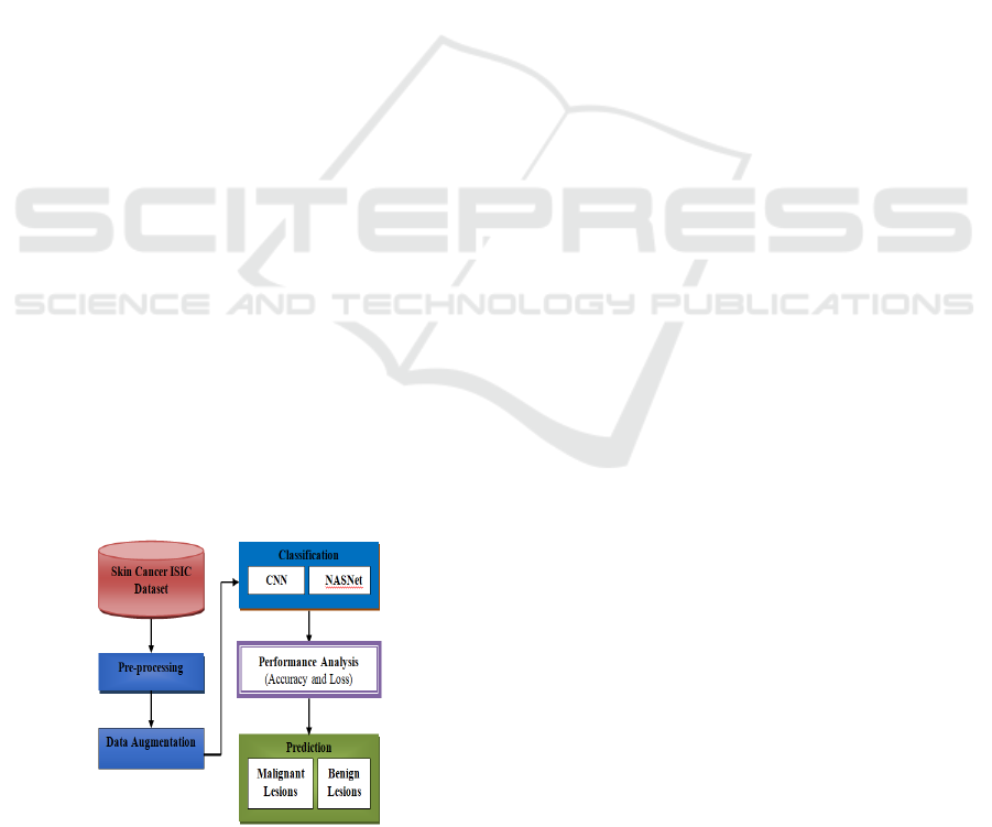
Figure 1: System methodology.
Figure 1 illustrates the proposed System
Methodology for skin cancer classification.
3.1 Pre-Processing
ISIC image pre-processing for skin cancer
classification involves essential steps to enhance
image quality and improve classification accuracy.
First, images are resized and normalized to a standard
resolution (e.g., 224×224) to ensure uniform input for
deep learning models like NASNet. Noise reduction
techniques, such as median filtering, are applied to
remove unwanted artifacts like hair, reflections, and
uneven lighting. Additionally, color normalization is
performed to maintain consistency in color
distribution across images, improving lesion feature
extraction. These preprocessing procedures enhance
dermoscopic pictures, facilitating improved feature
representation and more precise skin cancer
classification.
3.2 Data Augmentation
After preprocessing, data augmentation techniques
are applied on ISIC images to enhance generalization
in a model and hence increase the precision of skin
cancer classification. A few of these common
augmentation techniques include rotation, flipping,
scaling, and cropping, used in improving the diversity
of lesion orientation and size. It has the ability to
adjust to several lighting conditions with contrast and
brightness adjustments, although Gaussian noise and
blurring are used to emulate the fluctuations found in
images during real-world scenes. Additionally,
elastic transformations as well as affine distortions
indirectly change the geometry of lesions while not
changing the diagnostic features of the lesions. This
type of augmentation has the impacts of minimizing
class imbalance, minimizing overfitting, and
maximizing the robustness of deep learning models
like NASNet when it comes to diagnosing skin
cancer.
3.3 Classification
Deep learning models, especially Convolutional
Neural Networks (CNN), are commonly used to
classify skin cancers since they can learn
automatically hierarchical features from dermoscopic
images with or without human involvement. Existing
methods primarily use CNN models such as as
VGG16, ResNet, or Inception for skin lesion
identification and classification. The performance of
these models is impressive; however, accuracy and
ICRDICCT‘25 2025 - INTERNATIONAL CONFERENCE ON RESEARCH AND DEVELOPMENT IN INFORMATION,
COMMUNICATION, AND COMPUTING TECHNOLOGIES
148
efficiency have shortcomings, particularly with
complicated lesion types. To address this issue, the
improved version is NASNet, or the Neural
Architecture Search Network. NASNet uses
automated search to improve network design,
enabling better feature extraction and achieving high
classification accuracy while maintaining computing
efficiency. This study reveals that the NASNet model
far outperforms conventional CNN models in skin
cancer diagnosis, making it a better and more
effective method.
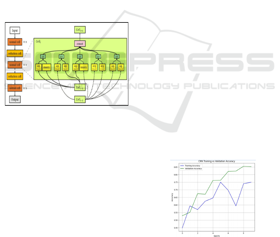
3.4 NASNet
The Neural Architecture Search Network (NASNet)
is a state-of-the-art deep learning architecture created
through automated neural architecture search. It
improves accuracy as well as computational
efficiency. NASNet has proven to be quite effective
in image classification, making it a good candidate for
skin cancer classification.
Figure 2: NASNet architecture diagram.
Figure 2 shows the architecture of NASNet, which is
automatically built to optimize the layers and
parameters of the network and determine the most
efficient architecture. The process entails the search
for the best convolutional layer arrangement,
activation functions, pooling, and skip connections to
create a better and more robust architecture with the
capability to extract the important information from
images and improve classification accuracy.
NASNet is able to learn hierarchical features of
incoming images independently without any human
intervention. Skin cancer classification identifies
essential features like texture, colour patterns, and
morphology that distinguish malignant lesions from
benign lesions.
NASNet uses a modular architecture with several
convolutional layers and cell blocks. The cells in
NASNet are engineered to collect both detailed and
overarching information, which are crucial for
differentiating various types of skin diseases. The
architectural components comprise
• Convolutional Layers for detecting edges,
textures, and patterns.
• Max Pooling Layers for down sampling the
image, preserving important features while
reducing computational complexity.
• Batch Normalization for stabilizing and
accelerating training.
• Dense Layers at the final stage to perform
classification based on the learned features.
This transfer learning method allows the model to use
previously acquired attributes (such as edges and
textures) and adapt them for the specific aim of
detecting skin cancer. By training on the ISIC dataset,
NASNet improves its ability to extract features and
classify skin lesions. While being trained, NASNet
decreases loss (for example, category cross-entropy)
and improves its performance by employing back-
propagation and gradient descent.
4 EXPERIMENTAL RESULTS
The effectiveness of the proposed model is assessed
using measures such as validation accuracy and loss.
4.1 Dataset Details
The ISIC dataset is a well-known standard for
classifying skin cancer and segmenting lesions. It
consists of 25,000 annotated dermoscopic
photographs, as well as high-resolution dermoscopic
JPEG and PNG photos. Dermatologists have
annotated it, and it includes a wide range of skin
problems, such as benign lesions and malignant types.
Figure 3: Accuracy of CNN training versus validation
Performance Comparison of Deep Learning-Based Classification of Skin Cancer
149
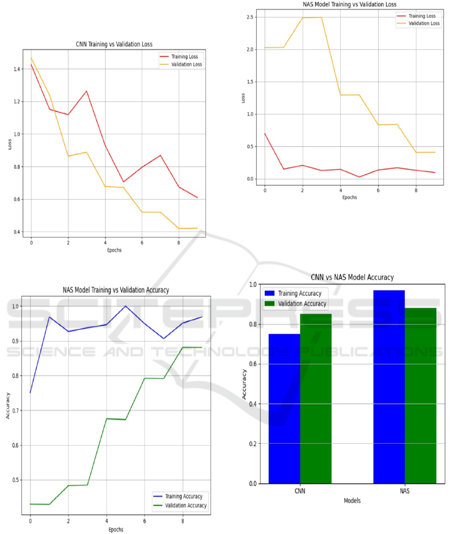
Figure 3 presents Accuracy of CNN Training versus
Validation and Figure 4 shows the Loss of CNN
Training versus Validation.
Figure 4: Loss of CNN training versus validation.
Figure 5: Accuracy of NASNet training versus validation.
Figure 6: Loss of NASNet training versus validation.
Figure 5 represents Accuracy of CNN Training versus
Validation and Figure 6 illustrates the Loss of CNN
Training versus Validation.
Figure 7: Performance analysis – accuracy.
Figure 7 depicts the Performance Analysis that uses
Accuracy. NASNet is better than typical CNN models
since it automatically finds the ideal architecture for
classifying skin cancer. Figure 8 gives Output skin
cancer classification image.
ICRDICCT‘25 2025 - INTERNATIONAL CONFERENCE ON RESEARCH AND DEVELOPMENT IN INFORMATION,
COMMUNICATION, AND COMPUTING TECHNOLOGIES
150
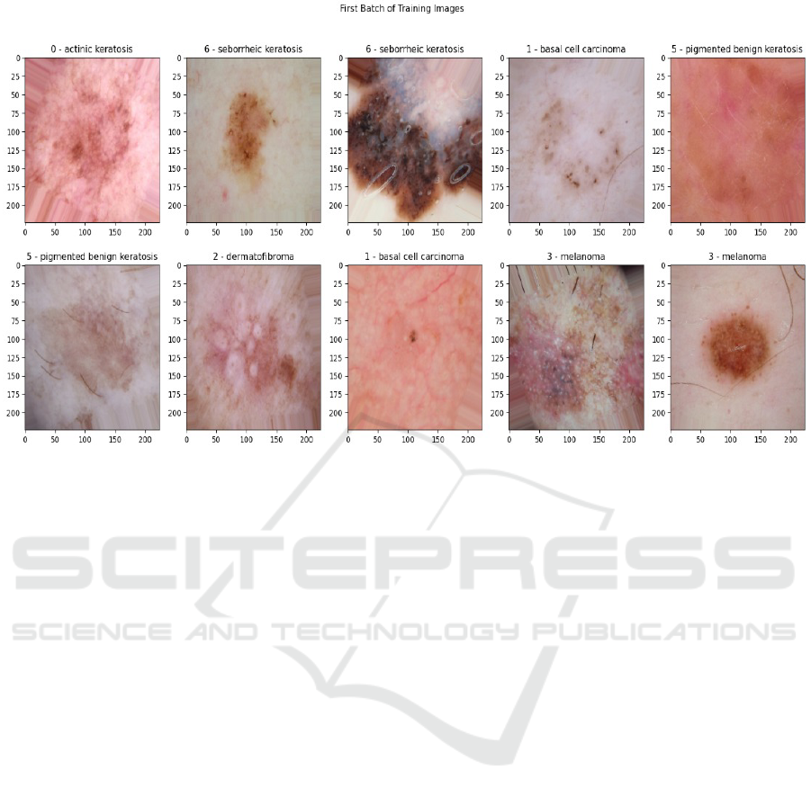
Figure 8: Sample output image.
5 CONCLUSIONS
This work highlights the efficacy of deep learning
models in skin cancer classification, with NASNet's
design delivering major enhancements over standard
CNNs. It is extremely effective for automated skin
cancer detection, especially when trained on ISIC
datasets, because to its advanced search for the best
design. The study, which is based on validation
accuracy and loss, shows that NASNet is significantly
better than traditional CNN models when it comes to
classification accuracy and efficiency. The automated
architectural optimisation of NASNet improves its
ability to gather complex features of skin lesions,
leading to more accurate predictions and lower loss
values during training. The findings show that
NASNet is a suitable model for detecting skin cancer.
It performs better and is more reliable than current
CNN-based approaches, which leads to better early
diagnosis and patient outcomes.
REFERENCES
Ajmal, M., Khan, M. A., Akram, T., Alqahtani, A.,
Alhaisoni, M., Armghan, A., Althubiti, S. A. and
Alenezi, F. (2023), ‘Bf2sknet: Best deep learning
features fusion-assisted framework for multiclass skin
lesion classification’, Neural Computing and
Applications 35(30), 22115– 22131.
Arivuselvam, B. et al. (2021), ‘Skin cancer detection and
classification using svm classifier’, Turkish Journal of
Computer and Mathematics Education (TURCOMAT)
12(13), 1863–1871.
Bassel, A., Abdulkareem, A. B., Alyasseri, Z. A. A., Sani,
N. S. and Mohammed, H. J. (2022), ‘Automatic
malignant and benign skin cancer classification using a
hybrid deep learning approach’, Diagnostics 12(10),
2472.
Cassidy, B., Kendrick, C., Brodzicki, A., Jaworek-
Korjakowska, J. and Yap, M. H. (2022), ‘Analysis of
the isic image datasets: Usage, benchmarks and
recommendations’, Medical image analysis 75,
102305.
Chu, X., Zhang, B. and Xu, R. (2021), Fairnas: Rethinking
evaluation fairness of weight sharing neural
architecture search, in ‘Proceedings of the IEEE/CVF
International Conference on Computer Vision’, pp.
12239–12248.
Houssein, E. H., Saad, M. R., Hashim, F. A., Shaban, H.
and Hassaballah, M. (2020), ‘Levy flight distribution:
A new metaheuristic algorithm for solving engineering
opti- ´ mization problems’, Engineering Applications of
Artificial Intelligence 94, 103731.
Iqbal, I., Younus, M., Walayat, K., Kakar, M. U. and Ma, J.
(2021), ‘Automated multi-class classification of skin
lesions through deep convolutional neural network with
dermoscopic images’, Computerized medical imaging
and graphics 88, 101843.
Performance Comparison of Deep Learning-Based Classification of Skin Cancer
151

Kaur, R. and Ranade, S. K. (2023), ‘Improving accuracy of
convolutional neural networkbased skin lesion
segmentation using group normalization and combined
loss function’, International Journal of Information
Technology pp. 1–9.
Kavitha, P & Jayalakshmi, V 2020, Survey of Skin Cancer
Detection using Various Image Processing Techniques,
Proceedings of the Third International Conference on
Intelligent Sustainable Systems, IEEE Xplore Part
Number: CFP20M19-ART; ISBN: 978-1-7281-7089-3,
pp. 1062-1069.
Khan, M. A., Akram, T., Zhang, Y.-D. and Sharif, M.
(2021), ‘Attributes based skin lesion detection and
recognition: A mask rcnn and transfer learning-based
deep learning framework’, Pattern Recognition Letters
143, 58–66.
Khouloud, S., Ahlem, M., Fadel, T. and Amel, S. (2022),
‘W-net and inception residual network for skin lesion
segmentation and classification’, Applied Intelligence
pp. 1–19.
Li, X., Zheng, J., Li, M., Ma, W. and Hu, Y. (2022), ‘One-
shot neural architecture search for fault diagnosis using
vibration signals’, Expert Systems with Applications
190, 116027.
Mehwish Dildar, Shumaila Akram, Muhammad Irfan,
Hikmat Ullah Khan, Muhammad Ramzan, Abdur
Rehman Mahmood, Soliman Ayed Alsaiari, Abdul
Hakeem M Saeed & Mohammed Olaythah Alraddadi
2021, ‘Skin cancer detection: A review using deep
learning techniques’, International Journal of
Environmental Research and Public Health, vol. 18, no.
01, pp. 1-22.
Mohakud, R. and Dash, R. (2022), ‘Designing a grey wolf
optimization based hyperparameter optimized
convolutional neural network classifier for skin cancer
detection’, Journal of King Saud University-Computer
and Information Sciences 34(8), 6280–6291.
Nawaz, M., Mehmood, Z., Nazir, T., Naqvi, R. A., Rehman,
A., Iqbal, M. and Saba, T. (2022), ‘Skin cancer
detection from dermoscopic images using deep learning
and fuzzy k-means clustering’, Microscopy research
and technique 85(1), 339–351.
Ragab, M., Choudhry, H., Al-Rabia, M. W., Binyamin, S.
S., Aldarmahi, A. A. and Mansour, R. F. (2022), ‘Early
and accurate detection of melanoma skin cancer using
hybrid level set approach’, Frontiers in Physiology 13,
2536.
Ragumadhavan, R, Aravind Britto, KR & Vimala, R 2022,
‘Melanoma skin cancer detection using wavelet
transform and local ternary pattern’, Journal of Medical
Imaging and Health Informatics, vol. 12, no. 1, pp. 15-
19, ISSN: 2156-7018,
Sangers, T, Reeder, S, Vander Vet, S, Jhingoer, S,
Mooyaart, A, Siegel, DM & Wakkee, M 2022,
‘Validation of a market-approved artificial intelligence
mobile health app for skin cancer screening: A prosp-
ective multicenter diagnostic accuracy study’,
Dermatology: Skin CancerResearch Article, vol. 11,
no. 8, pp. 1-8.
Wu, Y. P., Parsons, B., Jo, Y., Chipman, J., Haaland, B.,
Nagelhout, E. S., Carrington, J., Wankier, A. P., Brady,
H. and Grossman, D. (2022), ‘Outdoor activities and
sunburn among urban and rural families in a western
region of the us: Implications for skin cancer preventi-
on’, Preventive Medicine Reports 29, 101914.
Zafar, K., Gilani, S. O., Waris, A., Ahmed, A., Jamil, M.,
Khan, M. N. and Sohail Kashif, A. (2020), ‘Skin lesion
segmentation from dermoscopic images using convol-
utional neural network’, Sensors 20(6), 1601.
ICRDICCT‘25 2025 - INTERNATIONAL CONFERENCE ON RESEARCH AND DEVELOPMENT IN INFORMATION,
COMMUNICATION, AND COMPUTING TECHNOLOGIES
152
