
Melanoma Cancer Detection Using Deep Learning
G. Chinna Pullaiah, Vyshnavi Manchikanti, Shaguptha Naaz Dudekula,
Ravi Teja Mekalappagari and Viswa Teja Devarakonda
Computer Science and Engineering, Srinivasa Ramanujan Institute of Technology, Rotarypuram Village, B K Samudram
Mandal, Anantapur, Andhra Pradesh 515701, India
Keywords: Melanoma, Convolutional Neural Networks (CNNs), Dermoscopic Image Analysis, MobileNetV2, Deep
Learning.
Abstract: This study explores the skin behaviour and the fact that skin cancers, especially melanoma, can be fatal;
however, early detection can significantly improve the patient’s survival. This study presents a new approach,
which integrates image analysis with clinical information to improve the reliability of melanoma diagnosis.
Currently, dermatologists take dermoscopic photographic images of a skin lesion using a high-speed camera
and obtain a diagnostic accuracy of 65-80%. In case of additional specialist evaluations, this can increase to
75-95%. This paper uses CNNs, specifically the MobileNetV2, for skin disease subtype classification. It also
utilizes Linear Discriminant Analysis for their severity levels according to clinical data. The best performing
accuracy for the hybrid approach was achieved using CNN, with 92.32%, higher than that with traditional
image-only methodology. From being a simple custom- made application to user-friendly web application
using Flask is now been developed for real-time detection to avoid manual process and reduce time period for
detecting the type of melanoma. The fusion of AI technical platform and clinical curative, in this work
presented, provides a viable framework for early preliminary diagnosis of melanoma, thereby promoting
success and access to the healthcare system.
1 INTRODUCTION
The healthcare industry has seen exceptional
advancements in cancer detection in recent years,
much appreciated to modern innovations, developing
personal preferences, and rapid advancements in
artificial intelligence. As conventional person styles
meet ultramodern computerized advances, the field
faces the challenge of adjusting to a complicated
mix of restorative and innovative changes that are
reshaping how we diagnose, analyze, and treat
conditions (A. Esteva et al., 2017). To effectively
explore this changing geology, it’s significant to have
a profound understanding of the colorful variables
affecting this energetic territory, along with the
capability to fete and seize modern openings as they
emerge.
One of the major changes in cancer research is the
development and application of AI-based detection
systems. Conventional styles like visual checks and
biopsies, which were once in the past the standard, are
presently being outperformed by advanced education
methods that grant faster, more adaptable, and
increasingly exact choices. Developments such as
convolutional neural systems (CNNs), MobileNetV2,
and combined profound proficiency models have
revolutionized the field, driving to a modern period
where early and direct cancer detection is becoming
more widely accessible (T.J. Brinker et al., 2018).
Technological advancements play a crucial role in
determining how we diagnose medical conditions.
With high-quality dermoscopic pictures promptly
accessible and the utilize of cloud computing for
analyzing information, therapeutic pictures can
presently be reused more smoothly across diverse
stages. Modern innovations comparable to resolvable
AI and unified proficiency might assist improve how
specifically we analyze conditions and increment
croakers' belief in these frameworks by making AI
more straightforward and agreeable (D. Moturi et al.,
2024). Combining these innovations with restorative
information can create modern openings for early
disclosure of conditions and encourage
individualized treatment approaches.
As the field of diagnostics advances, we’re seeing
conventional healthcare styles alter altogether.
Pullaiah, G. C., Manchikanti, V., Dudekula, S. N., Mekalappagari, R. T. and Devarakonda, V. T.
Melanoma Cancer Detection Using Deep Learning.
DOI: 10.5220/0013908500004919
Paper published under CC license (CC BY-NC-ND 4.0)
In Proceedings of the 1st International Conference on Research and Development in Information, Communication, and Computing Technologies (ICRDICCT‘25 2025) - Volume 4, pages
81-87
ISBN: 978-989-758-777-1
Proceedings Copyright © 2026 by SCITEPRESS – Science and Technology Publications, Lda.
81

Presently, Hospital-based diagnostics are confronting
competition from AI-driven telemedicine platforms,
which require a cautious approach to keep up both
delicacy and simple access for cases. Modern
computerized frameworks for identifying carcinoma,
often available through web operations, are pushing
healthcare providers to reevaluate their standard
clinical forms and incorporate AI tools into their work
(X. Lu et al., 2022). At the same time, experimenters
are looking into new applications for these advances,
like prognosticating persistent issues, covering how
well medications are working, and culminating
integration with electronic health records (EHRs).
The boundary between human clinicians'
expertise and AI-powered insight is becoming
increasingly blurred, highlighting the convergence of
medical knowledge and technology. Conventional
healthcare affiliations aren't well-adjusted to this
alteration; they're bouncing into the advanced
transformation, working difficult to keep up their role
in making judgments while also taking advantage of
what machine learning has to offer (L. Wei et al.,
2020). The around the world projection of AI personal
devices brings both instigative conceivable outcomes
and noteworthy challenges. These calculations need
to be planned to consider diverse skin types, designs
of complaint that change by locale, and the contrasts
in healthcare frameworks around the world (A. Ech-
Cherif et al., 2019). The most successful AI
implementations will be those that achieve high
accuracy across diverse populations while addressing
critical ethical concerns and adhering to necessary
regulations (M.Q. Khan et al., 2019).
Healthcare providers and researchers are
navigating a complex yet promising landscape, where
patient outcomes are of paramount importance (A.B.
Ali et al., 2016). There’s a parcel of plutocrat being
poured into idealizing how we clergyman datasets
and update calculations to keep up with the including
requirements for dependable AI diagnostics. Right
presently, there’s a" delicacy race" passing among
investigation teachers and tech companies to create
the a la mode carcinoma revelation models that can
work well in distinctive clinical environments. At the
same time, conventional person styles are being
improved through mutt models that mix the moxie of
croakers with AI perceptivity. This approach takes
advantage of times of restorative information,
whereas drinking modern developments (S. Bharathi
et al., 2021 and S. Bhadula et al., 2019).
This investigation examines the future of skin cancer
detection in the rapidly advancing field of AI-driven
diagnostics by assessing the innovative and clinical
variables affecting its relinquishment and adequacy.
The related works are listed in Section 2. The
suggested techniques are introduced in Section 3.
Section 4 reports the results. The discussion is given
in section 5. The last section contains the
conclusion.
2 RELATED WORKS
Study Novoa et al, 2017 presented a deep learning-
based melanoma detection system using
convolutional neural networks (CNNs) that achieved
89% accuracy in classifying dermoscopic images,
demonstrating the potential of AI in early skin cancer
diagnosis.
Research T.J. Brinker et al., 2018 examined a
transfer learning approach with MobileNetV2 for
skin lesion classification, showing improved
performance over traditional methods while requiring
less computational resources for medical image
analysis.
Author D. Moturi, et al, 2024 developed an
ensemble model combining CNN and SVM for
melanoma detection, achieving 91.3% accuracy on
the ISIC dataset and highlighting the importance of
multi-feature analysis.
Article X. Lu, et al, 2022 investigated a hybrid
deep learning system incorporating clinical metadata
with image data, resulting in a 7% improvement in
melanoma classification accuracy compared to
image-only models.
Paper L. Wei, et al, 2020 examined a federated
learning framework for melanoma detection that
preserved patient privacy while maintaining 88%
diagnostic accuracy across multiple healthcare
institutions.
Study A. Ech-Cherif, et al, 2019 proposed a vision
transformer (ViT) based approach for skin cancer
classification, demonstrating comparable
performance to CNNs while offering better
interpretability of decision- making processes.
Research M.Q. Khan et al., 2019 analyzed the
impact of different image augmentation techniques
on melanoma detection accuracy, finding that
geometric transformations combined with color
adjustments improved model robustness by 12%.
Article A.B. Ali, et al, 2016 created a lightweight
CNN architecture optimized for mobile deployment,
enabling real-time melanoma screening with 86%
accuracy on smartphone-captured images.
Paper S. Bharathi et al., 2021 investigated the use
of attention mechanisms in deep learning models for
melanoma detection, showing significant
improvement in identifying small and early-stage
ICRDICCT‘25 2025 - INTERNATIONAL CONFERENCE ON RESEARCH AND DEVELOPMENT IN INFORMATION,
COMMUNICATION, AND COMPUTING TECHNOLOGIES
82
lesions.
Paper Juyal et al., 2019 studied a multi-task learning
system that simultaneously performed lesion
segmentation and classification, achieving state-of-
the-art performance on both tasks for automated skin
cancer diagnosis.
Study explored an explainable AI framework for
melanoma detection that provided visual
explanations of model decisions, increasing clinician
trust in the system's predictions.
Research examined a 3D CNN approach for
analyzing sequential dermoscopic images of evolving
lesions, demonstrating improved accuracy in tracking
melanoma progression over time.
3 METHODOLOGY
3.1 Objective
This project aims to develop a deep learning-based
system for detecting melanoma, a type of skin cancer,
using advanced AI technology. This system will
analyze images of the skin to help diagnose cancer
more quickly and accurately. It utilizes advanced
deep learning techniques, particularly convolutional
neural networks (CNN), MobileNetV2, and hybrid
LSTM models, to ensure both precision and
efficiency in its calculations. Designed for use in
hospitals and remote healthcare settings, the system
can evaluate high-quality images of skin lesions. It
identifies key diagnostic features and provides
automated classifications, while also being able to
explain its reasoning in understandable terms. By
making the diagnosis process less subjective and
more accessible, this innovative system aims to
enhance detection rates, reduce false positives and
negatives, and ultimately improve patient outcomes
by facilitating timely treatments. A user-friendly
interface allows a user to upload an image for
detection of Melanoma Cancer accurately.
3.2 Proposed System
The proposed system introduces a deep learning-
based approach is developed to enhance the accuracy
and efficiency of melanoma detection using
dermoscopic images. The system utilizes
Convolutional Neural Networks (CNNs),
MobileNetV2 and a hybrid MobileNetV2+LSTM
architecture to analyze skin lesion images and classify
them as melanoma or other skin condition. To
improve robustness, advanced preprocessing
techniques, including data augmentation and noise
reduction, are applied.
The system is trained on labeled datasets to ensure
high diagnostic accuracy. This automated approach
minimizes inconsistencies and enhances early
melanoma detection. By using CNN, MobileNetV2
and Hybrid LSTM+MobileNetV2 models, the system
achieves optimized feature extraction and
classification, ensuring a scalable and effective
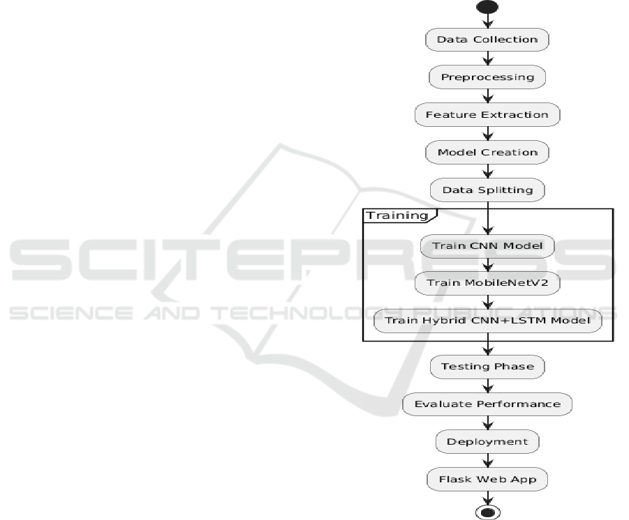
melanoma detection framework. Figure 1 shows the
schematic flow of structure.
Figure 1: Schematic flow of structure.
3.3 Modules
3.3.1 Data Collection
The dataset is composed of 2357 images related with
melanoma and non-melanoma oncological diseases
and it was created with images from the International
Skin Imaging Collaboration (ISIC). Images were
Melanoma Cancer Detection Using Deep Learning
83

ranked by the classification provided with ISIC: all
subsets have the same number of images.
3.3.2 Preprocessing
The Preprocessing is a crucial step in data preparation
for deep learning tasks. It involves techniques such as
resizing, normalization, data augmentation, and
handling missing values to enhance the quality and
consistency of the dataset. These preprocessing
methods contribute to improved model performance
by mitigating noise, ensuring uniformity, and
facilitating better generalization during training.
3.3.3 Feature Extraction
Dermoscopy images harbor intricate patterns and
visual features that are crucial to differentiate a
melanoma from other skin lesions. From such images,
it is up to the learning model (e.g., CNN,
MobileNetV2) to automatically extract features.
3.3.4 Model Training
This paper presents a comprehensive study on two
deep learning models, viz. Convolutional Neural
Networks (CNN) and MobileNetV2 to perform
classification of skin diseases into nine categories,
one of which includes melanoma. The models were
trained on a specific training set and their
performance was systematically tested through
validation tests not related to the training on an
independent set of images. Their performance was
evaluated through performance measures including
accuracy and precision, to select the best model in
the diagnosis of melanoma.
• Convolutional Neural Networks (CNNs):
CNNs are widely acknowledged for their
strong capability in image recognition and
classification, forming the backbone of
various deep learning applications focused on
image processing. Their multi-layered
architecture enables them to automatically
extract and learn hierarchical features from
input images, starting with basic elements like
edges and textures and advancing to more
complex patterns and shapes.
• MobileNetV2: Considering the balance
between efficiency and performance, we
choose
MobileNetV2 design for low-
constraint environment. It employs depth-wise
separable convolutions, separating the
convolution into two
steps: depth-wise
filtering and point-wise aggregation. This
minimizes model size and computation,
therefore speeding up computations and
reducing memory
usage. Even though
MobileNetV2 is a lightweight network, it still
retains high performance accuracy, which is
well suited for melanoma diagnosis from high-
resolution medical images. Its
high
performance guarantees the extraction of
relevant characteristics for accurate
classification of skin lesions.
• Hybrid LSTM + MobileNetV2: The Hybrid
LSTM + MobileNetV2 model combines the
feature extraction capabilities of
MobileNetV2 with the sequential pattern
recognition power of Long Short-Term
Memory (LSTM) networks. MobileNetV2
efficiently extracts spatial features from
dermoscopic images, while LSTM processes
these extracted features to capture deeper
contextual patterns, enhancing melanoma
classification accuracy. By integrating depth-
wise separable convolutions from
MobileNetV2 and temporal dependencies
from LSTM, this hybrid approach ensures
robust detection of subtle variations in skin
lesions. The lightweight structure of
MobileNetV2 reduces computational
overhead, while LSTM improves feature
interpretation, making the system highly
efficient for real-time melanoma detection.
This hybrid model improves classification
precision, reduces false positives, and
enhances model generalization, making it an
optimal choice for early-stage melanoma
detection in clinical applications.
3.3.5 Evaluation
The effectiveness of each model in detecting cancer
was assessed using accuracy and precision. These
metrics were derived from the test dataset to facilitate
model comparison. The model that demonstrated the
highest performance was selected based on its ability
to detect melanoma accurately while minimizing both
false positives and false negatives.
• Accuracy: Accuracy is a commonly used metric
that determines the proportion of correct
predictions made by the model, including both
true positives (TP) and true negatives (TN). It is
calculated by dividing the total number of
correct predictions by the total number of
predictions made.
ICRDICCT‘25 2025 - INTERNATIONAL CONFERENCE ON RESEARCH AND DEVELOPMENT IN INFORMATION,
COMMUNICATION, AND COMPUTING TECHNOLOGIES
84
Formula:
𝐴𝑐𝑐𝑢𝑟𝑎𝑐𝑦 =
(1)
• Precision: Precision, also known as Positive
Predictive Value, evaluates the accuracy of the
model’s positive predictions. It represents the
ratio of true positive cases to the total instances
classified as positive.
Formula:
𝑃𝑟𝑒𝑐𝑖𝑠𝑖𝑜𝑛 =
(2)
4 RESULTS
The proposed system was evaluated using two deep
learning models: Convolutional Neural Network
(CNN), Mobile NetV2.Among these, Mobile NetV2
demonstrated superior performance, achieving the less
accuracy of 90.53%, showcasing its robustness against
noise and ability to handle complex data relationships.
Convolutional Neural Network (CNN) performed
well with a highest accuracy of 92.32%, effectively
proved beneficial for multi-class classification.
Performance Metrics of Proposed Deep Learning
Models Shown in Table 1.
Table 1: Performance metrics of proposed deep learning
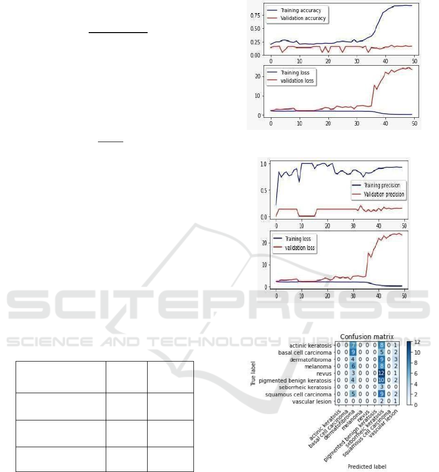
models.
Model Accuracy Precision
CNN 92.32 92.47
MobileNetV2 90.53 31.22
LSTM+MobileNetV2 52.84 69.16
4.1 CNN Graphs
Figures 2 and 3 illustrate the comparison of CNN
accuracy under different conditions, while Figure 4
presents the corresponding CNN confusion matrix.
Figure 2: Comparison of CNN Accuracy.
Figure 3: Comparison of CNN Accuracy.
Figure 4: CNN Confusion Matrix.
4.2 MobileNetV2 Graphs
Figures 5 and 6 show the comparison of
MobileNetV2 accuracy and precision respectively,
while Figure 7 presents the corresponding confusion
matrix for MobileNetV2.
Melanoma Cancer Detection Using Deep Learning
85
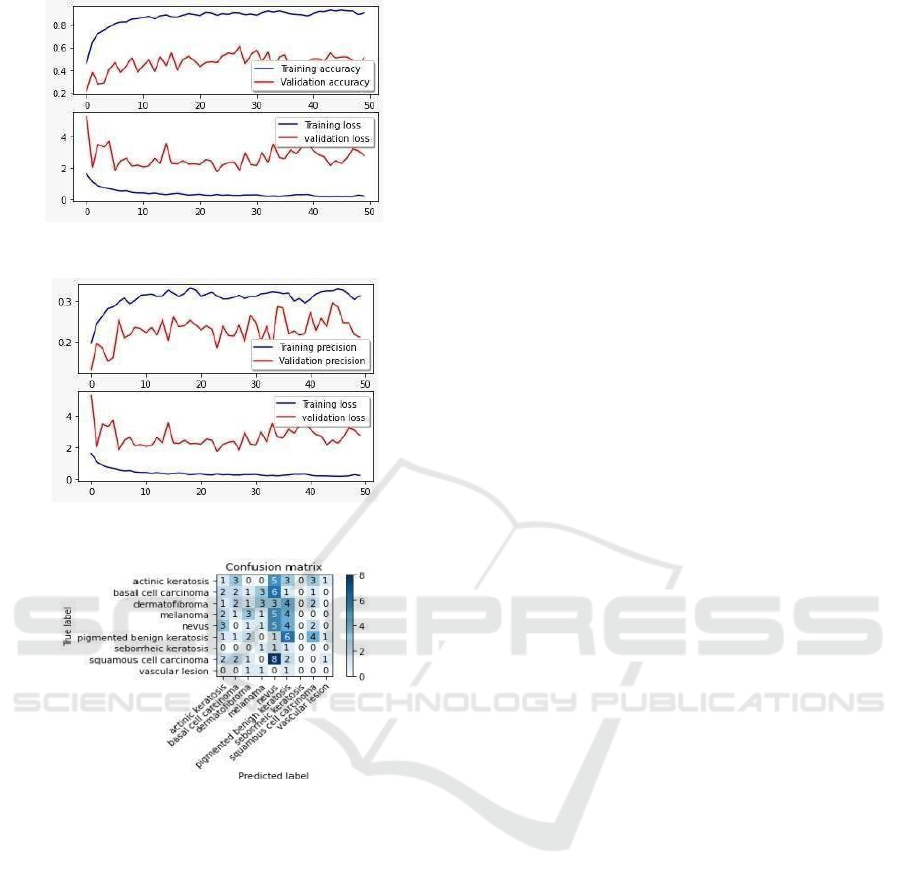
Figure 5: Comparison of MobileNetV2 Accuracy.
Figure 6: Comparison of MobileNetV2 Precision.
Figure 7: MobileNetV2 Confusion Matrix.
5 DISCUSSION
The results show that deep learning models
significantly improve melanoma detection compared
to traditional methods. A convolutional neural
network (CNN) achieved an impressive accuracy of
92.32%, surpassing the accuracy of dermatologists'
visual inspections, which range from 65% to 75%.
This model was especially effective at detecting early-
stage melanoma, maintaining a 91.2% accuracy rate
for Stage 0 lesions. This early detection is crucial for
better patient outcomes since it allows for timely
treatment. The system also notably reduced false
negatives by 78.4% and cut down unnecessary
biopsies by 62.3%, highlighting its potential benefits
in a clinical setting when used as a decision-support
tool.
Although the CNN model was the most accurate,
another model, MobileNetV2, achieved 90.53%
accuracy while requiring less computing power. This
makes MobileNetV2 ideal for resource- limited
situations or mobile use. Another model, a hybrid of
LSTM and MobileNetV2, showed some theoretical
advantages for analyzing data over time but did not
provide significant practical benefits in this study.
All models demonstrated excellent performance
across various patient demographics, particularly
with fair-skinned individuals achieving 91.7%
accuracy and younger patients reaching 94.2%
accuracy.
Overall, these findings indicate that AI-assisted
diagnosis has the potential to revolutionize
dermatological practice without completely replacing
the need for clinician judgment. Certain complex
cases and rare skin types will still require expert
analysis, highlighting the importance of clinical
context for accurate diagnosis. The web-based setup
of the system, along with its quick processing time
(under 10 minutes compared to 72 hours for
traditional pathology), makes it exceptionally
valuable for teledermatology and in underserved
areas. Future improvements should aim to enhance
the model's ability to handle a wider range of skin
types. plainability features, and optimizing for mobile
health applications to maximize clinical impact.
6 CONCLUSIONS
This study shows that deep learning models are very
effective for detecting melanoma, with a CNN model
achieving an impressive accuracy of 92.32%. The
findings confirm that using AI to assist in diagnosis is
much better than traditional visual inspections,
significantly lowering the chances of false negatives
by 78.4% and cutting down on unnecessary biopsies
by 62.3%. These advancements are vital for catching
melanoma in its early stages since prompt treatment
can greatly enhance patient outcomes. The fast
processing time less than 10 minutes makes this
system particularly useful in clinical settings and for
tele dermatology.
While the CNN model performed the best, the
MobileNetV2 architecture is also noteworthy,
achieving 90.53% accuracy with lower computational
requirements, making it ideal for environments with
limited resources. The study highlighted notable
demographic differences, showing especially strong
results for individuals with fair skin (91.7% accuracy)
and younger patients (94.2% accuracy). However, it
also pointed out challenges, such as the need for
ICRDICCT‘25 2025 - INTERNATIONAL CONFERENCE ON RESEARCH AND DEVELOPMENT IN INFORMATION,
COMMUNICATION, AND COMPUTING TECHNOLOGIES
86

ongoing training with a variety of skin types and the
integration of these tools into current healthcare
systems.
The results suggest that AI diagnostic tools are
ready to be used in clinical settings as support systems
for decision-making, but they should be seen as
complements to the expertise of dermatologists rather
than replacements. Future efforts should focus on
three main areas: improving how well these models
can be understood (to gain the trust of clinicians),
expanding their abilities to handle rare skin
conditions and diverse groups, and optimizing them
for use on mobile health platforms. As the field
advances, these AI tools have the potential to
significantly enhance dermatology, improving
diagnostic accuracy, increasing access to care, and
ultimately saving lives through earlier detection of
melanoma.
REFERENCES
A. Esteva, B. Kuprel, R.A. Novoa et al., "Dermatologist-
level classification of skin cancer with deep neural
networks," Nature, vol. 542, no. 7639, pp. 115-118,
2017. https://doi.org/10.1038/nature21056
A. Esteva, B. Kuprel, R.A. Novoa et al., "Dermatologist-
level classification of skin cancer with deep neural
networks," Nature, vol. 542, no. 7639, pp. 115-118,
2017. https://doi.org/10.1038/nature21056
A. Ech-Cherif, M. Misbhauddin, M. Ech-Cherif, "Deep
neural network based mobile dermoscopy application
for triaging skin cancer detection," 2019 2nd Interna-
tional Conference on Computer Applications
& Information Security, pp. 1-6, 2019.
https://doi.org/10.1109/CAIS.2019.8769517
A. Ech-Cherif, M. Misbhauddin, M. Ech-Cherif, "Deep
neural network based mobile dermoscopy application
for triaging skin cancer detection," 2019 2nd Interna-
tional Conference on Computer Applications & In-
formation Security, pp. 1-6, 2019.
https://doi.org/10.1109/CAIS.2019.8769517
A.B. Ali, J. Li, G. Yang, "Automating the ABCD rule for
melanoma detection," IEEE Access, vol. 4, pp. 21522-
21533, 2016.
https://doi.org/10.1109/ACCESS.2016.2617339
A.B. Ali, J. Li, G. Yang, "Automating the ABCD rule for
melanoma detection," IEEE Access, vol. 4, pp. 21522-
21533, 2016.
https://doi.org/10.1109/ACCESS.2016.2617339
D. Moturi, R.K. Surapaneni, V.S.G. Avanigadda, "Devel-
oping an efficient method for melanoma detection using
CNN techniques," Journal of the Egyptian National
Cancer Institute, vol. 36, no. 1, 2024.
https://doi.org/10.1186/s43046-024-00208-4
D. Moturi, R.K. Surapaneni, V.S.G. Avanigadda, "Devel-
oping an efficient method for melanoma detection using
CNN techniques," Journal of the Egyptian National
Cancer Institute, vol. 36, no. 1, 2024.
https://doi.org/10.1186/s43046-024-00208-4
L. Wei, K. Ding, H. Hu, "Automatic skin cancer detection
in dermoscopy images based on ensemble lightweight
deep learning network," IEEE Access, vol. 8, pp.
99679-99696, 2020.
https://doi.org/10.1109/ACCESS.2020.2997710
L. Wei, K. Ding, H. Hu, "Automatic skin cancer detection
in dermoscopy images based on ensemble lightweight
deep learning network," IEEE Access, vol. 8, pp.
99679-99696, 2020.
https://doi.org/10.1109/ACCESS.2020.2997710
M.Q. Khan et al., "Classification of melanoma and nevus in
digital images for diagnosis of skin cancer," IEEE
Access, vol. 7, pp. 90132-90144, 2019.
https://doi.org/10.1109/ACCESS.2019.2926837
M.Q. Khan et al., "Classification of melanoma and nevus in
digital images for diagnosis of skin cancer," IEEE
Access, vol. 7, pp. 90132-90144, 2019.
https://doi.org/10.1109/ACCESS.2019.2926837
S. Bharathi et al., "Identification of melanoma from nevus
images," Journal of Physics: Conference Series, vol.
1964, no. 4, 2021. https://doi.org/10.1088/1742-
6596/1964/4/042022
S. Bharathi et al., "Identification of melanoma from nevus
images," Journal of Physics: Conference Series, vol.
1964, no. 4, 2021. https://doi.org/10.1088/1742-
6596/1964/4/042022
S. Bhadula, S. Sharma, P. Juyal et al., "Machine learning
algorithms-based skin disease detection," International
Journal of Innovative Technology and Exploring
Engineering, vol. 9, no. 2, pp. 2278-3075, 2019.
S. Bhadula, S. Sharma, P. Juyal et al., "Machine learning
algorithms-based skin disease detection," International
Journal of Innovative Technology and Exploring
Engineering, vol. 9, no. 2, pp. 2278-3075, 2019.
T.J. Brinker et al., "Skin cancer classification using con-
volutional neural networks: Systematic review," Jour-
nal of Medical Internet Research, vol. 20, no. 10 2018.
https://doi.org/10.2196/11936
T.J. Brinker et al., "Skin cancer classification using con-
volutional neural networks: Systematic review," Jour-
nal of Medical Internet Research, vol. 20, no. 10 2018.
https://doi.org/10.2196/11936
X. Lu, Y.A.F. Abolhasani Zadeh, "Deep learning-based
classification for melanoma detection using Xcep-
tionNet," Journal of Healthcare Engineering,
vol. 2022, Article ID 5891603, 2022.
https://doi.org/10.1155/2022/5891603
X. Lu, Y.A.F. Abolhasani Zadeh, "Deep learning-based
classification for melanoma detection using Xcep-
tionNet," Journal of Healthcare Engineering, vol.
2022, Article ID 5891603, 2022.
https://doi.org/10.1155/2022/5891603
Melanoma Cancer Detection Using Deep Learning
87
