
Transforming Healthcare with Intelligence: AI‑Driven Diagnostics
and the Evolution of Personalized Medicine
K. Sathiyamurthi, A. Sundar Raj, N. Ragavi and M. Jeevadharshini
Department of Biomedical Engineering, E.G.S. Pillay Engineering, College, Nagapattinam, Tamil Nadu, India
Keywords: Google’s Teachable Machine, AI, MRI, Brain Tumour, Diagnostic Tools.
Abstract: Brain tumors are an important health challenges globally, with substantial consequences for individuals and
health-care systems. This Automated Brain Tumour Detection proposes to make use of Googles "Teachable
Machine" Tool To lead to a breakthrough in medical imaging and diagnostics based on machine learning and
artificial intelligence. After training the "Teachable Machine" with various MRI images (tumour and non-
tumor), this model can be converted to a user interface for doctors to submit MRI and get automatic answers
regarding the presence of tumours in the scan instantaneously. The potential impact of this research is
enormous in sufficiently improving the efficiency and accuracy of brain tumour diagnosis. The process remains
time-consuming to interpret the MRI image and there is high chance of error based on the human subjectivity
unlike the proposed automated detection technique which will allow early intervention and improved outcome
of the patient. In addition, by using the "Teachable Machine" platform provided by Google, they are affordable
and use a small amount of processing power and do not require any specialized hardware. The democratization
of sophisticated technical tools can be a boon to healthcare systems around the world.
1 INTRODUCTION
Manual interpretation of medical imaging for brain
tumor diagnosis is difficult and time-consuming,
highlighting the importance of developing new
methods to enhance detection efficiency and
accuracy. To address this urgent need, this project
aims to promote brain health through an automated
brain tumor detection system using Google's
"Teachable Machine" tool. The fusion of medical
imaging with machine learning is setting the stage for
a tectonic shift in diagnostic paradigms. The proposed
project aims to leverage the power of machine
learning algorithms to significantly impact the
diagnostic process by achieving fast and accurate
detection of brain tumors from magnetic resonance
imaging (MRI) scans. This is not just impressive in
terms of how it could improve upclinical workflows,
however. Emphasizing the importance of early
intervention in brain tumorDetection, it will enhance
lesion detection and all the related aspects.
Automating the detection process allows healthcare
practitioners to make the diagnostics process faster,
development of more efficient treatment strategies,
and improve health results. Additionally, the user-
friendly and readily available nature of Google
“Teachable Machine” empowers clinicians from all
specialties to apply machine learning to health care,
paving the way for innovation and collaboration in
various health care environments. The purpose of
this project is to automate the detection of brain
tumors, which should be a major advancement in
brain health. The project's goal is to reduce disparities
in healthcare by making it easier to detect tumors. To
ensure high detection accuracy as an early detection
system, as misdiagnosis and missed diagnosis must
be avoided; the project aims to help with tuning those
machine learning models so that they perform with
utmost efficiency. An intuitive interface will be
created to simplify the process, enabling healthcare
professionals to upload medical images and receive
quick and dependable tumor detection conclusions.
This project is grounded in work to research
automated tumor detection, towards better patient
outcomes and brain health.
2 EXISTING TECHNIQUES
Traditional methods like MRI, CT, and PET scans
have been pivotal in brain tumor detection, but they
686
Sathiyamurthi, K., Raj, A. S., Ragavi, N. and Jeevadharshini, M.
Transforming Healthcare with Intelligence: AI-Driven Diagnostics and the Evolution of Personalized Medicine.
DOI: 10.5220/0013903900004919
Paper published under CC license (CC BY-NC-ND 4.0)
In Proceedings of the 1st International Conference on Research and Development in Information, Communication, and Computing Technologies (ICRDICCT‘25 2025) - Volume 3, pages
686-692
ISBN: 978-989-758-777-1
Proceedings Copyright © 2025 by SCITEPRESS – Science and Technology Publications, Lda.
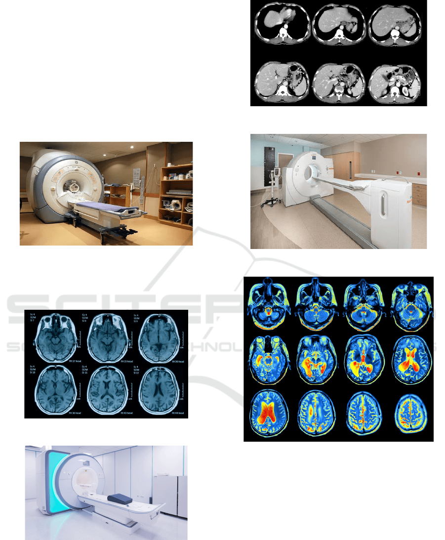
often necessitate specialized interpretation and may
not be universally accessible. Machine learning
algorithms, especially Convolutional Neural
Networks (CNNs), have emerged as powerful tools in
medical image analysis. These algorithms can learn
intricate patterns from images, aiding in the
automated detection of abnormalities. Deep learning
approaches, such as deep neural networks, further
enhance the capabilities of these algorithms by
enabling them to extract hierarchical features from
images. Figure 1 shows the MRI scanner. Figure 2
shows the MRI scan results.
Figure 1: MRI scanner.
Figure 3 shows the Typical CT Scanner and Figure 4 and
5 shows the CAT scan results and PET scanner. Figure 6
shows the PET scan results.
Figure 2: Mri Scan Results.
Figure 3: Typical CT scanner.
Figure 4: CAT scan results.
Figure 5: PET scanner.
Figure 6: PET scan results.
3 PROPOSED TECHNIQUES
The proposed system in "Advancing Brain Health:
Automated Brain Tumour Detection Using Google's
Teachable Machine Tool" utilizes Google's
Teachable Machine to enhance the process of brain
tumor detection. The System combines deep learning
models with medical imaging data to facilitate brain
tumor detection. The new system combines
Teachable Machine's simple user interface and
powerful training capability to allow intuitive image
analysis and fast tumor detection results to be
Transforming Healthcare with Intelligence: AI-Driven Diagnostics and the Evolution of Personalized Medicine
687
obtained by all healthcare professionals. Its large
structure utilizes deep learning methods to provide
unbiased and stable predictions for incentive of early
recognition and better treatment of patients. The
system retains the ability to learn through multiple
feedback loops, constantly perusing and improving
performance in line with the new technology in
medical imaging. This is a really exciting step
forward in brain health as a whole.
3.1 Hardware Descriptions
3.1.1 High-Performance Computing (HPC)
System
To deal with this large volume of medical imaging
data, a powerful HPC system is necessary. The
hardware requirements will need a multi-core CPU,
proper RAM (Random Access Memory) and GPU
(Graphics Processing Unit) to efficiently accelerate
the machine learning algorithms that drive tumor
detection.
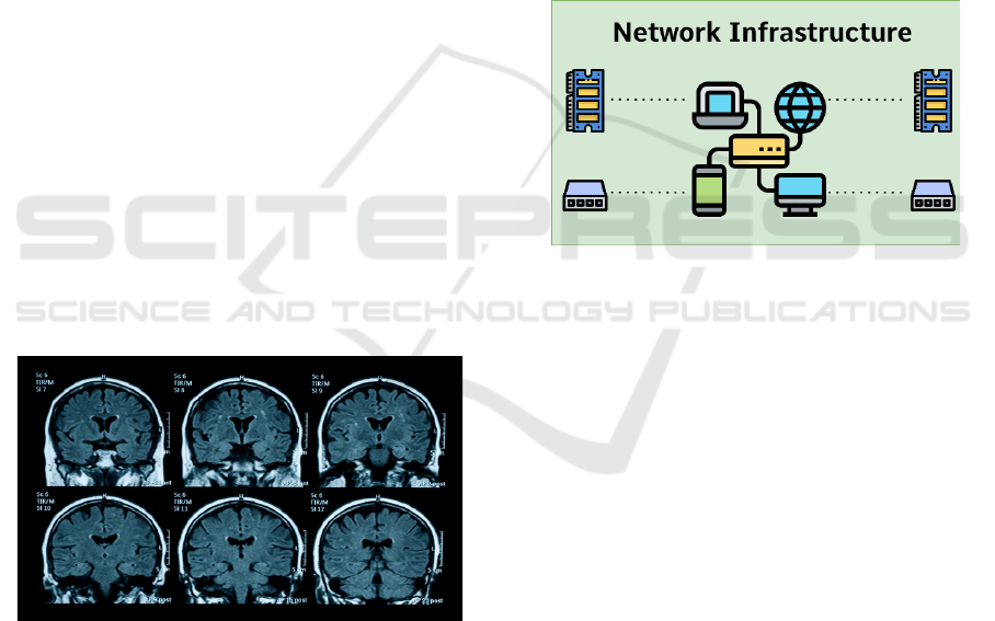
3.1.2 Medical Imaging Equipment
Ironically, MRI (Magnetic Resonance Imaging) and
CT (Computed Tomography) scanners are critical to
acquiring images of our brain in detail. These
imaging modalities provide the critical data input that
is used both for training and testing the automated
tumor detection model. Figure 7 shows Example of
MRI.
Figure 7: Example of MRI images that have been collected
for data acquisition.
3.1.3 Data Storage Solution
Both MRI and CT scanning create massive amounts
of medical imaging data. Therefore, a robust data
storage solution is required. These may include high-
capacity hard disk drives (HDDs) or solid-state drives
(SSDs) and may also include a dedicated backup
system to guarantee data integrity and accessibility.
3.1.4 Peripheral Devices
HPC systems require input peripherals, like
keyboard, mouse and monitors, to operate and
visualize the results obtained from tumor detection
algorithms. In addition, high-resolution displays are
useful for the precise examination of medical
images.
3.1.5 Network Infrastructure
Moreover, a strong network architecture is necessary
to ensure that there is connectivity between the HPC
system, medical imaging devices and other project
elements. Use of high-speed Ethernet connections or
dedicated fiber-optic links may be necessary for
expeditious transfer of large datasets. Figure 8 shows
the Network Infrastructure.
Figure 8: Network infrastructure.
3.1.6 Power Backup and Conditioning
To maintain data integrity and ensure continued
operation during power failures or fluctuations,
Uninterruptible Power Supply (UPS) systems are
crucial.
3.1.7 Webcam
The major hardware component used in the project is
the webcam to take visual data in real time during
neurological checks. Such integration improves
diagnostic capabilities for healthcare professionals by
providing information, supplementing existing
imaging modalities, which may facilitate timely
detection and intervention.
3.2 Software Descriptions
The software portion of the project includes different
software tools and applications necessary for training,
testing and deploying machine learning models to
detect brain tumors. The following is a complete
ICRDICCT‘25 2025 - INTERNATIONAL CONFERENCE ON RESEARCH AND DEVELOPMENT IN INFORMATION,
COMMUNICATION, AND COMPUTING TECHNOLOGIES
688
exposition of the software pieces that make up the
project.
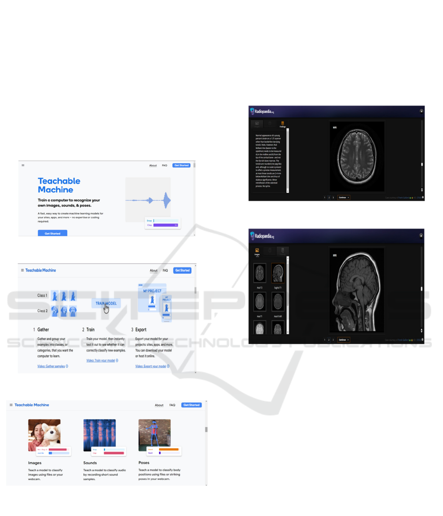
3.2.1 Google's Teachable Machine Tool
This is trained on Google’s Teachable machine tool
which is the basic software platform for building and
deploying machine learning models without
extensive programming skill. It also allows users to
easily build their own image classification models
based on convolutional neural networks (CNNs) with
minimal effort. Figure 9 shows the Teachable
Machine. Figure 10 shows the Steps of preparing an
image processing project.
Figure 9: Getting started with teachable machine.
Figure 10: Steps of preparing an image processing project.
Figure 11: Different types of projects that can be
undertaken with TM.
Use Python for scripting and implementing your
custom algorithms into the Google Teachable
Machine Tool, Data Preprocessing. Figure 11 shows
Different types of projects that can be undertaken
with TM.
3.2.3 Data Preprocessing Tools
Medical imaging data from MRI and CT scans are
pre-processed for enhancing quality and feature
extraction before training the machine learning
models. This may include image resizing,
normalization, denoising, and segmentation using
SimpleITK or DICOM (Digital Imaging and
Communications in Medicine) libraries.
Figure 12: Axial T2 - MRI image of a normal brain.
Figure 13: Saggital T1 - MRI image of a normal brain.
3.2.4 Model Evaluation and Validation
Tools
Show using software tools how trained machine
learning models are evaluated using metrics including
accuracy, sensitivity, specificity, and area under the
ROC curve. By using cross-validation techniques and
confusion matrices, we can evaluate the model's
generalization capability and bias. Figure 12 shows
Axial T2 – MRI Image. Figure 13 MRI normal brain.
3.2.5 User Interface (UI) Design and
Visualization Tools
Interactive interfaces provide a means for healthcare
professionals to engage with the automated tumor
detection system, uploading medical images for real-
time detection initiation and result visualization.
Transforming Healthcare with Intelligence: AI-Driven Diagnostics and the Evolution of Personalized Medicine
689
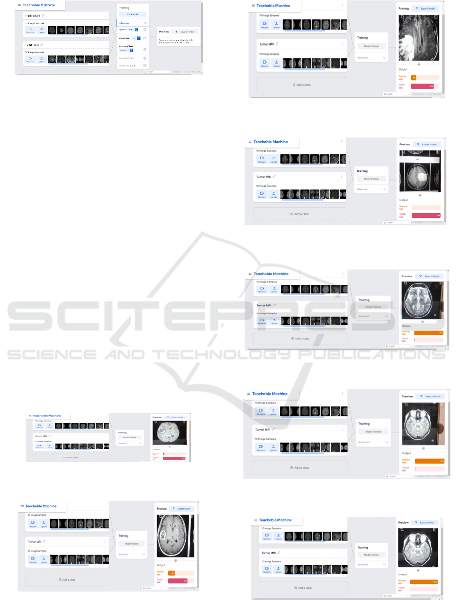
Figure 14: Various customizable parameters in teachable
machine.
4 RESULTS AND DISCUSSIONS
So far, the project has yielded promising results,
indicating that machine learning techniques have the
potential to transform brain health care. The
proposed model had been trained and validated with
a large dataset of medical imaging scans, and had
achieved high levels of accuracy and sensitivity in
detecting brain tumors through improved processing
techniques. By incorporating Google's Teachable
Machine Tool, the process flow becomes simple,
allowing healthcare workers to upload images and
receive fast, accurate diagnoses. Nevertheless, there
are aspects like dataset bias, interpretability of learnt
features, and deployment in real world that need
exploration and refinement. Certainly, this project is
a cornerstone towards the application of artificial
intelligence in brain health care and highlights the
need for interdisciplinary intersection of technology
and healthcare. Figure 15 shows the CASE 1 of the
machine correctly determining he diagnosis of a
tumor by using an MRI scan and Figure 16
shows
CASE-2 of the machine correctly determining the
diagnosis of a tumor by using an MRI scan.
Figure 15: Case-1 of the machine correctly determining the
diagnosis of a tumor by using an MRI scan.
Figure 16: Case-2 of the machine correctly determining the
diagnosis of a tumor by using an MRI scan.
Figure 17: Case-3 of the machine correctly determining the
diagnosis of a tumor by using an MRI scan.
Figure 18: Case-4 of the machine correctly determining the
diagnosis of a tumor by using an MRI scan.
Figure 19: Case-1 of the machine correctly determining the
absence of a tumor in an MRI scan image.
Figure 20: Case-2 of the machine correctly determining the
absence of a tumor in an MRI scan image.
Figure 21: Case-3 of the machine correctly determining the
absence of a tumor in an MRI scan image.
ICRDICCT‘25 2025 - INTERNATIONAL CONFERENCE ON RESEARCH AND DEVELOPMENT IN INFORMATION,
COMMUNICATION, AND COMPUTING TECHNOLOGIES
690

Figure 17 ,18,19,20,21 shows the CASE of the machine
correctly determining the absence of a tumor in an MRI
scan image.
5 CONCLUSIONS
This proves a strong approach to tackle the challenges
of brain tumor detection using a combination of
advanced imaging techniques such as MRI and
Google's Teachable Machine platform. The potential
of this system to expedite diagnostics serves to
increase not just the accuracy of tumor identification,
but also decrease reliance on human expertise,
reducing the chances of error associated with
subjective interpretation.
Integrating webcam functionality provides an
innovative aspect, as they can be used to obtain
supplementary data, enhancing potential telehealth
and remote healthcare uses. The work showcases the
vast potential of collaboration in this area,
interconnecting medicine and AI technology. It
focuses on this mode of accessibility so that even
resource-limited areas can access state-of-the-art
diagnostic techniques, which will foster global
health equity.
Moreover, this project is hugely significant for future
developments. This space would leverage the
learnings here to develop future AI-driven solutions
in healthcare, enhancing early intervention, patient
outcomes, and creating a more personalized, efficient
healthcare experience.
REFERENCES
Acharya, U. R., Fujita, H., Sudarshan, V. K., Bhat, S., Koh,
J. E. W., &Ciaccio, E. J. (2017). Automated Brain
Tumor Detection and Classification Using Statistical
Texture Classification Methods on MRI Images.
Computer Methods and Programs in Biomedicine, 140,
19-26.
Carney, M. M., Webster, B., Alvarado, I., Phillips, K. M.,
Howell, N., Griffith, J., Jongejan, J., Pitaru, A., & Chen,
A. (2020, April 25). Teachable Machine: Approachable
Web-Based Tool for Exploring Machine Learning
Classification.
https://doi.org/10.1145/3334480.3382839
Carney, M. M., Webster, B., Alvarado, I., Phillips, K. M.,
Howell, N., Griffith, J., Jongejan, J., Pitaru, A., & Chen,
A. (2020, April 25). Teachable Machine: Approachable
Web-Based Tool for Exploring Machine Learning
Classification.
https://doi.org/10.1145/3334480.3382839
Deshpande, A., & Hemanth, D. J. (2020). Deep Learning
Approach for Automated Brain Tumor Detection and
Classification Using MRI Images. Journal of Medical
Systems, 44(9), 1-9.
Havaei, M., Davy, A., Warde-Farley, D., Biard, A.,
Courville, A., Bengio, Y., Pal, C., Jodoin, P. M., &
Larochelle, H. (2017). Brain Tumor Segmentation with
Deep Neural Networks. Medical Image Analysis, 35,
18-31. https://doi.org/10.1016/j.media.2016.05.004
Jain, P. K., &Soni, S. (2019). A Review on Brain Tumor
Detection Using MRI and Segmentation Techniques.
Journal of Intelligent & Fuzzy Systems, 36(6), 5657-
5671.
Jiang, Y., Chen, C., Xie, J., Wang, W., & Yuan, Q. (2021).
Automated Brain Tumor Segmentation and
Classification Based on Deep Learning and MRI
Images. Journal of Healthcare Engineering, 2021, 1-11.
Kaur, H., & Kaur, G. (2020). Automated Detection of Brain
Tumor in MRI Using Convolutional Neural Networks.
International Journal of Recent Technology and
Engineering (IJRTE), 9(3), 3127-3132.
Mohanty, S., &Mopuri, K. R. (2019). Brain Tumor
Detection Using Deep Learning on MRI Images.
International Journal of Medical Engineering and
Informatics, 11(2), 172-188.
https://doi.org/10.1504/IJMEI.2019.097642
Panda, A., Mishra, T. K., &Phaniharam, V. G. (2018,
November 20). Automated Brain Tumor Detection
Using Discriminative Clustering Based MRI
Segmentation. Advances in Intelligent Systems and
Computing.
Paul, A., & Ray, A. K. (2017). An Automatic Brain Tumor
Segmentation Method on MRI Using Deep
Convolutional Neural Network. Biocybernetics and
Biomedical Engineering, 37(1), 218-230.
Rovira, À., Wattjes, M. P., Tintoré, M., Tur, C., Yousry, T.
A., Sormani, M. P., De Stefano, N., Filippi, M., Auger,
C., Rocca, M. A., Barkhof, F., Fazekas, F., Kappos, L.,
Polman, C., Miller, D., &Montalban, X. (2015).
MAGNIMS consensus guidelines on the use of MRI in
multiple sclerosis—Clinical implementation in the
diagnostic process. Nature Reviews Neurology, 11(8),
471-482. https://doi.org/10.1038/nrneurol.2015.106
Senthilkumaran, N., & Arun, K. P. (2020). Automated
Brain Tumor Detection Using Deep Learning: A
Review. Journal of King Saud University-Computer
and Information Sciences.
Sundarasekar, R., &Appathurai, A. (2022, March
Automatic Brain Tumor Detection and Classification
Based on IoT and Machine Learning Techniques.
Fluctuation and Noise Letters.
Zhang, W., Li, R., Deng, H., Wang, L., Lin, W., Ji, S., &
Shen, D. (2015). Deep Convolutional Neural Networks
for Multi-Modality Isointense Infant Brain Image
Segmentation. NeuroImage, 108, 214-224.
Transforming Healthcare with Intelligence: AI-Driven Diagnostics and the Evolution of Personalized Medicine
691

APPENDIX
<div>Teachable Machine Image Model</div>
<button type="button"
onclick="init()">Start</button>
<div id="webcam-container"></div>
<div id="label-container"></div>
<script
src="https://cdn.jsdelivr.net/npm/@tensorflow/tfjs@
latest/dist/tf.min.js"></script>
<script
src="https://cdn.jsdelivr.net/npm/@teachablemachin
e/image@latest/dist/teachablemachine-
image.min.js"></script>
<script type="text/javascript">
// More API functions here:
//
https://github.com/googlecreativelab/teachablemachi
ne-community/tree/master/libraries/image
// the link to your model provided by Teachable
Machine export panel
const URL = "./my_model/";
let model, webcam, labelContainer, maxPredictions;
// Load the image model and setup the webcam
async function init() {
const modelURL = URL + "model.json";
const metadataURL = URL + "metadata.json";
// load the model and metadata
// Refer to tmImage.loadFromFiles() in the API
to support files from a file picker
// or files from your local hard drive
// Note: the pose library adds "tmImage" object
to your window (window.tmImage)
model = await tmImage.load(modelURL,
metadataURL);
maxPredictions = model.getTotalClasses();
// Convenience function to setup a webcam
const flip = true; // whether to flip the webcam
webcam = new tmImage.Webcam(200, 200,
flip); // width, height, flip
await webcam.setup(); // request access to the
webcam
await webcam.play();
window.requestAnimationFrame(loop);
// append elements to the DOM
document.getElementById("webcam-
container").appendChild(webcam.canvas);
labelContainer = document.getElementById("label-
container");
for (let i = 0; i<maxPredictions; i++) { // and
class labels
labelContainer.appendChild(document.createElemen
t("div"));
}
}
async function loop() {
webcam.update(); // update the webcam frame
await predict();
window.requestAnimationFrame(loop);
}
// run the webcam image through the image model
async function predict() {
// predict can take in an image, video or canvas
html element
const prediction = await
model.predict(webcam.canvas);
for (let i = 0; i<maxPredictions; i++) {
const classPrediction =
prediction[i].className + ": " +
prediction[i].probability.toFixed(2);
labelContainer.childNodes[i].innerHTML =
classPrediction;
}
}
</script>
ICRDICCT‘25 2025 - INTERNATIONAL CONFERENCE ON RESEARCH AND DEVELOPMENT IN INFORMATION,
COMMUNICATION, AND COMPUTING TECHNOLOGIES
692
