
Enhanced Brain Tumour Detection and Classification through
Sophisticated Machine Learning Approaches
Y. Sujitha, S. Rathnamahi, K. Sheshadri Ramana, N. Divya Sree,
B. Sai Eswara Neha and P. Suniya Begum
Department of Computer Science & Engineering, Ravindra College of Engineering for Women,
Kurnool, Andhra Pradesh, India
Keywords: CNN, VGG16 Model, ResNet50 Model, EfficientNetB0 Model.
Abstract: If not treated, brain tumors pose a significant health risk. Detected and promptly treated. MRI examinations
manual, but improved tumor detection Time-consuming and error-prone diagnosis prone. Deep learning will
be used in this study. Methods, in particular Convolutional Neural Networks (CNNs), to boost precision and
effectiveness in detecting brain tumors. The dataset of 7,023 MRI images is used in the research. From a
variety of sources, such as Figshare, Br35H and SARTAJ. Preprocessing techniques like normalization, image
resizing, and noise cancellation were used to improving the performance of a model. It was made a CNN
model. Using TensorFlow and GPU training acceleration. Data-based additional techniques augmentation,
adjusting the rate of learning, and making use of the Adam optimizer with a beta value made accuracy even
better Callbacks such as Early Stopping and ReduceLR on Plateau were incorporated to prevent overfitting
and ensure a stable training process. The machine learning model successfully divided brain tumors into four
groups, achieving a remarkable accuracy of 99.54 percent. This demonstrates how effective deep learning in
medical imaging and its potential as an accurate diagnostic instrument. The model makes use of important
libraries like TensorFlow, Keras, Pandas and NumPy.
1 INTRODUCTION
Medical-diagnostics, which calls for precise
prediction and treatment. Artificial intelligence (AI)
is needed in medical imaging because traditional
methods like MRI analysis take a long time and
are prone to human error. Brain tumors are
categorized into four categories using in this study:
meningioma, pituitary tumor, no tumor, and glioma.
Meningiomas, on the other hand, are benign but still
require treatment, while gliomas are cancerous and
necessitate immediate medical attention. The
pituitary gland is affected by pituitary tumors, which
can range in severity. Normal brain scans are
represented by the No Tumor category, which serves
as a comparison point. The CNN model's accuracy of
99.54 percent demonstrates its suitability to classify
brain tumors. The dataset consisted of 7,023 MRI
images from various sources. The Adam optimizer,
Early Stopping, ReduceLR on Plateau, and GPU
acceleration were utilized to improve performance.
The study shows that deep learning has the potential
to address real-world medical challenges and pave the
way for future AI-driven healthcare.
2 LITERATURE REVIEW
Manually MRI scans which are time consuming and
error - prone, are the foundation of conventional
diagnostics. Mavrakis et al. (2005) and other early
studies looked at clinical diagnosis without al. Kang et
al. and subsequent methods Tumor features and ML
classifiers face significant difficulties when dealing
with brain tumors classification. Despite its potential,
this was computationally prohibitive and challenging
to make use of big data. Mahobiya and Minz (2017)
also attempted the MRI algorithm AdaBoost.
Classification, which although looked promising had
difficulties with complex feature extraction to current
deep learning.
An XG was described by Mudgal et al. (2017).
optimization required extensive tuning for high-
476
Sujitha, Y., Rathnamahi, S., Ramana, K. S., Sree, N. D., Neha, B. S. E. and Begum, P. S.
Enhanced Brain Tumour Detection and Classification through Sophisticated Machine Learning Approaches.
DOI: 10.5220/0013900200004919
Paper published under CC license (CC BY-NC-ND 4.0)
In Proceedings of the 1st International Conference on Research and Development in Information, Communication, and Computing Technologies (ICRDICCT‘25 2025) - Volume 3, pages
476-482
ISBN: 978-989-758-777-1
Proceedings Copyright © 2025 by SCITEPRESS – Science and Technology Publications, Lda.

dimensional data.
Hemanth et al. (2018) proposed a novel but
imperfectly optimized modified CNN architecture for
large datasets.
Sudharani et al. (2015) used the k-NN algorithm,
which is good for simple classification but bad for
large preprocessing and high-dimensional data.
Togacar et al. (2020) improved accuracy by
optimizing CNN models with hyper columns and
feature selection. However, the increased complexity
raised computational costs and made scaling
challenging.
ResNet-101 was used with squeeze-and-
excitation networks by Ghosal et al. (2019), which
improved classification performance but required a
lot of resources
Szegedy et al. (2015) introduced the Inception
architecture, which enhanced feature extraction in
medical imaging; however, extensive modifications
were required for its application to the classification
of brain tumors.
The increasing use of ML and DL in the diagnosis
of brain tumors was highlighted in systematic reviews
by Khan et al. (2021) and Nadeem et al. (2020).
Although these studies offered insights, they lacked
specifics regarding how they could be put into practice
3 PROPOSED APPROACH
The proposed project eliminates data by employing a
CNN framework that is optimized for processing.
Artifacts solve problems related to the classification
of brain tumors and improve the overall quality of the
images, enhancement and transfer learning. The study
aims to improve brain tumor diagnosis and
classification by combining cutting-edge methods
with real-world solutions, offering a superior
alternative to existing methods.
3.1 Data Collection & Preprocessing
3.1.1 Data Collection
Brain tumors in four categories:
• Glioma: It is a tumor developing in
the spinal cord or brain glial cells and is
benign and malignant with varying growth
rate and severity.
• Meningioma: A benign tumor found in the
brain meninges, which cover the brain and
spinal cord to protect it cord
• No tumor: Normal brain scans that reveal no
tumors
• Pituitary: Tumors of the pituitary gland,
either benign or cancerous.
3.1.2 Preprocessing
When dealing with MRI images that are prone to
variations in resolution, intensity, and noise, it is an
essential phase in preparing the raw data for machine
learning. To address these issues, a methodical
preprocessing pipeline was used.
Image Standardization.
Resizing: The resolution of each MRI image was
resized to 128x128 pixels consistently. This is to keep
important features for classification while making the
images compatible with the CNN architecture.
Gray -Scale conversation: The pictures were
changed to grayscale in order to emphasize the
structural details and simplify computational
processes by removing color variations that aren't
needed.
Noise Reduction: To get rid of the noise, cutting –
edges methods like gaussian method filtering were
used.
The model can now detect more subtle
characteristics of tumors thanks to this improvement.
Image Normalization: The images' pixel
intensities were normalized to the 0-to-1 range. This
step ensures uniformity across the dataset, allowing
the model to learn more quickly during training and
reducing biases caused by variations in image
brightness.
Data Augmentation: To prevent overfitting and
introduce diversity into the dataset, the data
augmentation strategies are rotations, flipping,
zooming, brightness adjustments, sharing and
cropping.
Margin Trimming: Uninformative areas, such as
black margins, were removed to concentrate on the
brain region, raising the signal-to-noise ratio and data
quality in general.
Class Balancing: Targeted data augmentation
compensated for class imbalances to ensure that all
classes were properly represented during training to
avoid model bias.
Label Encoding: Each image received a numerical
label for its class:
• Glioma: 0
• Meningioma: 1
• No Tumor: 2
• Pituitary: 3
Enhanced Brain Tumour Detection and Classification through Sophisticated Machine Learning Approaches
477
The encoding readied the images for the CNN's
classification layer. Because it enables machine
learning algorithms to process categorical labels and
make precise predictions, label encoding is essential
for diagnosing brain tumors Label encoding refers to
the process of changing categorical labels to
numerical labels, which are consumed by machine
learning algorithms.
Label encoding is utilized for numerically catego
rizing different types of brain tumors in the case of
brain tumors However, "glioma," "meningioma," and
"pituitary adenoma” are normally stated while talking
about brain tumors. Label encoding aids in changing
such categorical labels to numerical representations
that machine learning algorithms can handle.
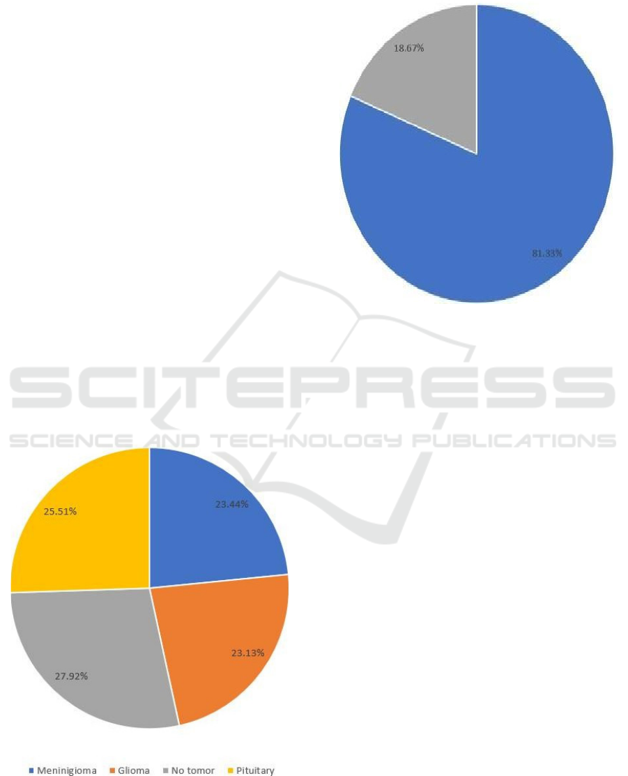
Data Splitting: To work with successful
preparation and impartial assessment, the dataset was
partitioned into three subsets:
•
Training Set: Accustomed to prepare the model
and get familiar with the elements of every
classification (figure 1).
•
Validation Set: Accustomed to tune the model
and assess its presentation during preparing.
•
Test Set: Utilized for definite assessment to
survey the model's speculation capacity (figure
2).
•
Training Set (70%): The biggest piece, used to
prepare the model with expanded information
for speculation.
Figure 1: Training Data.
•
Validation Set (15%): Used for
hyperparameter tuning and to monitor the
performance during training.
Figure 2: Train Test Split.
3.2 Feature Extraction and Transfer
Learning
To further develop order exactness, move learning
was coordinated into the model. Pretrained designs,
like Google Net, were used for include extraction,
giving areas of strength for an in light of information
acquired from huge scope picture datasets. By
utilizing these deeply grounded models, the growing
experience was altogether sped up, permitting the
CNN to rapidly adjust to X-ray cerebrum filters while
keeping up with high exactness.
3.2.1 Dataset
The dataset comprises of X-ray cerebrum pictures
arranged as cancer or non-growth. To address class
irregularity, information expansion was applied to the
minority class. Pictures were resized to 256×256
pixels, changed over completely to grayscale, and
class marks were encoded in double arrangement for
brain network reconciliation. The data were parted
into 70% preparation and 30% test sets with
separation to keep up with class appropriation. This
preprocessing guaranteed a decent, improved dataset
for productive model preparation and assessment.
Table 1 shows the Class Distribution Before and
After Preprocessing. Figure 3 illustrates the several
forms of brain tumor images.
ICRDICCT‘25 2025 - INTERNATIONAL CONFERENCE ON RESEARCH AND DEVELOPMENT IN INFORMATION,
COMMUNICATION, AND COMPUTING TECHNOLOGIES
478
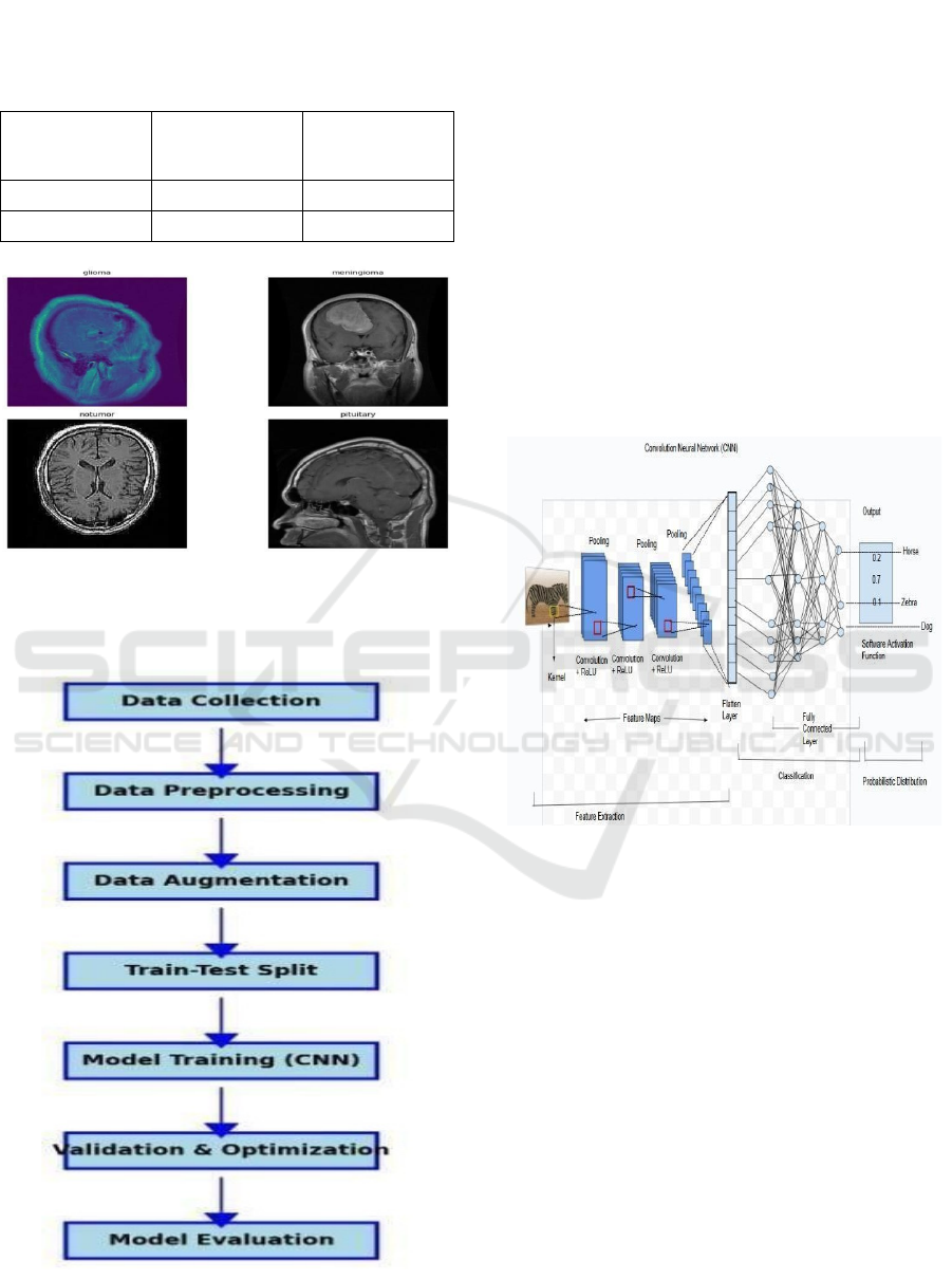
3.2.2 Class Distribution Before and After
Preprocessing
Table 1: Class Distribution Before and After Preprocessing.
Label
Before
Preprocessing
After Preprocessing
Tumor 850 1000
No-Tumor 750 1000
Figure 3: Several Forms of Brain Tumor Images.
4 METHODOLOGY
Figure 4: VGG19 Architecture.
4.1 Model
4.1.1 Convolutional Neural Networks (CNN)
Convolutional Brain Organizations are critical for
mind cancer identification, robotizing highlight
extraction from X-ray and CT filters. Not at all like
conventional strategies, CNNs gain designs from
crude pictures, permitting exact separation among
sound and growth impacted tissues. They are hearty,
precise, and proficient, even with uproarious
information, and perform well with move realizing
when marked information is scant. CNNs diminish
human blunder, give steady judgments, and empower
constant expectations for quicker treatment
arranging. They can likewise arrange growth types,
supporting customized treatment procedures. Figure 5
shows the CNN Architecture.
Figure 5: CNN Architecture.
4.1.2 VGG19 Model
VGG19 is a profound CNN with 19 layers, including
16 convolutional layers, intended to examine picture
grouping execution (figure 4). It utilizes 3×3
convolution channels to catch fine subtleties, making
it powerful for picture acknowledgment errands. By
using pre-prepared loads from datasets like ImageNet
and eliminating all associated layers, VGG19 serves
as an element extractor in brain growth
characterization, reducing preparation time and
further developing execution. VGG19 is able to
successfully separate complex examples for clinical
imaging, despite its computationally demanding
nature. Additionally, it can include multimodal data
for improved indicative precision, enhancing growth
discovery and patient outcomes.
Enhanced Brain Tumour Detection and Classification through Sophisticated Machine Learning Approaches
479
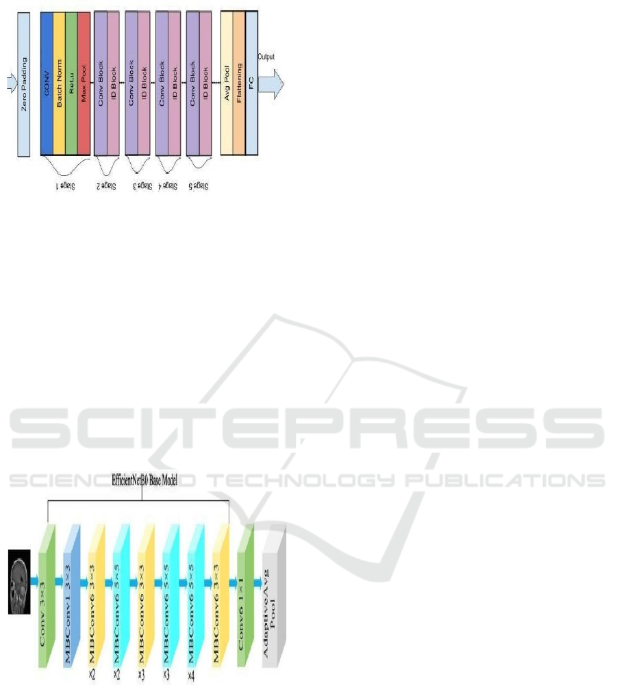
Figure 6: ResNet50 Architecture.
4.1.3 EfficientNet B0 Model
Using a compound scaling approach to change,
EfficientNet B0 is a lightweight CNN that adjusts
accuracy and computational proficiency.
Assignments like characterization of cerebellar cancer
that require very little preparation data. With its solid
execution and low asset use, its proficient plan makes
it ideal for ongoing clinical applications. EfficientNet
B0 is a good choice for resource-constrained
situations because it prioritizes speed and
effectiveness without sacrificing accuracy, making it
less complicated than more advanced models like
ResNet50 (figure 6 and 7).
Figure 7: EfficientNetb0 Architecture.
4.2 Performance Metrics
When determining a model's viability in the mind
cancer grouping, it is important to evaluate its
presentation. Estimating the accuracy, dependability,
and proficiency of the model's expectations is made
easier by the various measurements.
4.2.1 Accuracy
Precision is one of the central measurements used to
assess a grouping model. It addresses the level of
accurately ordered cases out of the all-out
expectations made. A higher exactness shows that the
model performs well across both cancer and non-
growth cases.
𝐴𝑐𝑐𝑢𝑟𝑎𝑐𝑦 = (𝑇𝑃 + 𝑇𝑁)/ 𝑇𝑃 + 𝐹𝑃 + 𝐹𝑁 + 𝑇𝑁) (1)
Where:
•
TP (True Positive) – Correctly predicted tumor
cases
•
TN (True Negative) – Correctly predicted non-
tumor cases
•
FP (False Positive) – Non-tumor cases
incorrectly classified as tumors
•
FN (False Negative) – Tumor cases
incorrectly classified as non-tumor
Classification Report.
4.2.2 Precision
Precision measures how many of the predicted tumor
cases were actually tumors. A greater precision score
demonstrates that the model generates fewer false-
positive errors.
𝑃𝑟𝑒𝑐𝑖𝑠𝑖𝑜𝑛 = 𝑇𝑃/
(
𝑇𝑃 + 𝐹𝑃
)
(2)
4.2.3 Recall (Sensitivity)
Recall, commonly known as sensitivity, evaluates
how many actual tumor cases the model correctly
identifies. A greater recall score ensures that the
model does not miss positive cases.
𝑅𝑒𝑐𝑎𝑙𝑙 = 𝑇𝑃/
(
𝑇𝑃 + 𝐹𝑁
)
(3)
4.2.4 F1-Score
The F1-score is a measure of a model's accuracy,
balancing precision and recall. It is the harmonic
mean of precision and recall, providing a single score
to evaluate performance.
𝐹1𝑠𝑐𝑜𝑟𝑒 =
(
2 × 𝑃𝑟𝑒𝑐𝑖𝑠𝑖𝑜𝑛 × 𝑅𝑒𝑐𝑎𝑙𝑙
)
/
(
𝑃𝑟𝑒𝑐𝑖𝑠𝑖𝑜𝑛 +
𝑅𝑒𝑐𝑎𝑙𝑙
)
(4)
4.3 Loss Function
A loss function quantifies how the model’s predictions
are differ from the actual values. It helps in
optimizing the model by minimizing errors during
ICRDICCT‘25 2025 - INTERNATIONAL CONFERENCE ON RESEARCH AND DEVELOPMENT IN INFORMATION,
COMMUNICATION, AND COMPUTING TECHNOLOGIES
480
training. The loss function used for this classification
task is the Mean Squared Error (MSE):
𝐿𝑜𝑠𝑠 =
(
1/𝑛
)
∗𝛴
(
𝑦ᵢ ȳ
)
(5)
Where:
•
yᵢ indicates actual values
•
ȳ indicates predicted values
•
n is the number of samples
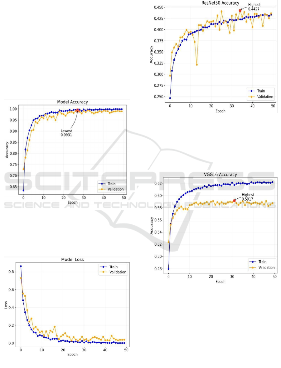
5 RESULTS
5.1 CNN Accuracy and Loss
Figure 8: Progression of Training and Testing Accuracy of
CNN.
Progression of training and testing accuracy of CNN
and Progression of training and testing Loss of CNN
are shown in figures 8 & 9 respectively.
Figure 9: Progression of Training and Testing Loss of CNN.
5.2 Resnet 50 Model Accuracy
Figure 10: Progression of Training and Testing Accuracy of
ResNet50.
Figure 10 depicts the Progression of training and
testing accuracy of Resnet50.
5.3 VGG16 Model Accuracy
Figure 11: Progression of Training and Testing Accuracy of
VGG16 Model.
Figure 11 depicts the Progression of training and
testing Accuracy of VGG16 Model.
5.4 EfficientNet B0 Model Loss and
Accuracy
Figure 12 shows the Progression of training and
testing Accuracy of EfficientNet B0Model.
Enhanced Brain Tumour Detection and Classification through Sophisticated Machine Learning Approaches
481
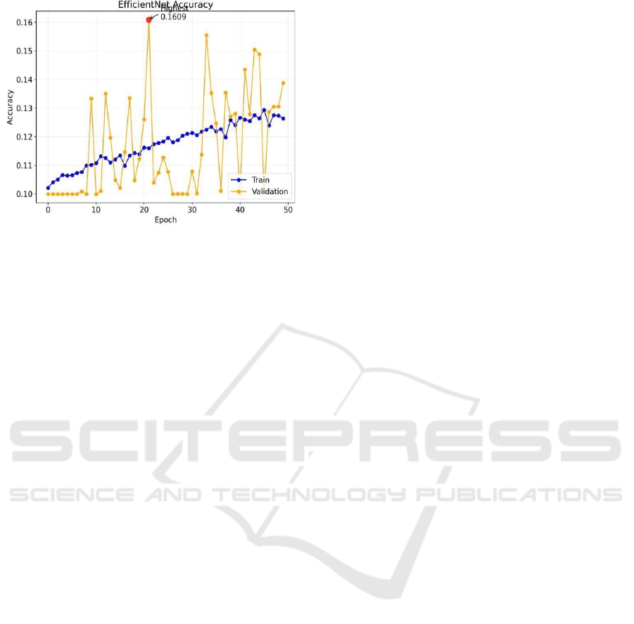
Figure 12: Progression of Training and Testing Accuracy of
EfficientNet B0Model.
6 CONCLUSIONS
This task consolidates computerized picture handling
procedures like division and expansion with profound
learning models (CNNs, VGG16, ResNet50,
EfficientNetB0) to accomplish high precision in mind
cancer discovery and grouping. The model guides
early conclusion by examining X-ray sweeps to
distinguish cancer designs, offering solid outcomes in
regions with restricted admittance to radiologists.
VGG16 played out the best, exhibiting its capacity to
remove complex highlights for exact order. Generally
speaking, this undertaking gives a versatile, effective
answer for cerebrum cancer identification, propelling
clinical diagnostics and further developing medical
care openness.
REFERENCES
4Szegedy C, et al. Going deeper with convolutions.
IEEE Conference on Computer Vision and
Pattern recognition (CVPR). 2015:1–9.
Afshar P, Platanista’s KN, Mohammadi A. Capsule
networks for brain tumor classification based on MRI
images and course tumor boundaries. IEEE
International Conference on Acoustics, Speech and
Signal Processing (ICASSP). 2019:1368–1372.
Brain tumor classification using deep CNN features via
transfer learning. Computers in Biology and Medicine.
2019; 111:103345.
Cinarer G, Emiroglu BG. Classification of brain tumors by
machine learning algorithms. 2019 3rd International
Symposium on Multidisciplinary Studies and
Innovative Technologies (ISMSIT). 2019:1–6
Ghosal P, Nandanwar L, Kanchan S, Bhadra A,
Chakraborty J. Brain tumor classification using
ResNet-101 based squeeze and excitation deep neural
network. Proceedings of the Second International
Conference on Advanced Computational and
Communication Paradigms (ICACCP). 2019:1–6.
Hemanth DJ, Anitha J, Naaji A, et al. A modified deep
convolutional neural network for abnormal brain
classification. IEEE Access. 2018; 7:4275–4283.
K. Intelligent brain tumor lesion classification and
identification from MRI images using k-NN technique.
International Conference on Control, Instrumentation,
Communication and Computational Technologies
(ICCICCT). 2015.
Kang J, Ullah Z, Gwak J. MRI-based brain tumor
classification using ensemble of deep features and
machine learning classifiers. Sensors. 2021; 21:2222.
Khan P, Kader MF, Islam SMR, et al. Machine learning and
deep learning approaches for brain disease diagnosis:
Principles and recent advances. IEEE Access. 2021;
9:37622–37655.
Mavrakis AN, Halpern EF, Baker FG, et al. Diagnostic
evaluation of patients with a brain mass as the
presenting manifestation of cancer. Neurology. 2005;
65:908–911.Deepak S, Ameer PM.
Mehrotra R, Ansari MA, Agrawal R, Anand RS. A transfer
learning approach for AI-based classification of brain
tumors. Machine Learning Applications. 2020; 2:10–
19.
Minz A, Mahobiya C. MR image classification using
AdaBoost for brain tumor type classification. IEEE 7th
International Advance Computing Conference (IACC).
2017:701–705.
Mudgal TK, Gupta A, Jain S, Gusain K. Automated system
for brain tumor detection and classification using
extreme Gradient Boosted decision trees. International
Conference on Soft Computing and Its Engineering
Applications (ICSOFTCOMP). 2017:1–6.
Nadeem MW, Ghamdi MAA, Hussain M, et al. Brain tumor
analysis empowered with deep learning: A review,
taxonomy, and future challenges. Brain Sciences. 2020;
10:118.
Sudharani K, Dr. Sarma TC, Dr. Satya prasad
Togacar M, Comert Z, Ergen B. Classification of brain MRI
using hyper column technique with convolutional
neural network and feature selection method. Expert
Systems with Applications. 2020; 149:113274.
ICRDICCT‘25 2025 - INTERNATIONAL CONFERENCE ON RESEARCH AND DEVELOPMENT IN INFORMATION,
COMMUNICATION, AND COMPUTING TECHNOLOGIES
482
