
Enhancing Lung Cancer Detection Using AI‑Based Deep Learning
Framework
E. S. Vinothkumar
1
, J. Vinoj
2
, M. Nivaashini
3
, A. Ravi Kumar
4
,
Ram Ganesh G. H.
5
and R. Senthilkumar
6
1
Department of CSE, Vel Tech Rangarajan Dr.Sagunthala R&D Institute of Science and Technology, Avadi, Chennai,
Tamil Nadu, India
2
Department of CSE, Vignan’s Foundation for Science, Technology and Research (VFSTR), Guntur, Andhra Pradesh, India
3
Department of AI&DS, Sri Eshwar College of Engineering, Coimbatore, Tamil Nadu, India
4
Department of CSE, Sridevi Women's Engineering College, Hyderabad, Telangana, India
5
Department of IT, Kamaraj College of Engineering and Technology, Viruthunagar, Tamil Nadu, India
6
Department of CSE, Hindusthan Institute of Technology, Coimbatore, Tamil Nadu, India
Keywords: Lung Cancer Detection, Artificial Intelligence (AI), Predictive Analytics, Deep Learning, Deep Convolution
Neural Network (DCNN).
Abstract: Cancer of the lungs is considered the global supreme deadly disease that is life-threatening. However,
premature diagnosis and appropriate cure are crucial for tumbling the transience rate concomitant with this
ailment. Computed tomography scans have emerged as among the most prevalent imagining methods for lung
cancer exposure, particularly when coupled with deep learning models. In this study, propose a deep learning
framework grounded on a Deep Convolutional Neural Network for the timely exposure of lung cancer
expending CT scan images. Additionally, we have analysed the enactment of supplementary models, such as
Inception V3, Xception, and ResNet-50, in comparison to our proposed model. Our comparative analysis
considered various metrics, including accurateness, Area beneath the Curve, recall, and loss. After appraising
the models' presentation, the outcomes show that the DCNN-based approach outperforms the other models
and demonstrates promising potential compared to traditional methods. Specifically, the proposed DCNN
model attained an precision of 98.27%, an Area Under Curve (AUC) of 97.12%, a recall of 98.70%, and a
loss of 0.328.
1 INTRODUCTION
This type of cancer is the deadliest and furthermost
miserable on the planet after all the others. It is
extremely complex in its nature and highly
stimulating to diagnose, as its symptoms are
frequently revealed only during the later and final
stages. However, mortality rates from lung cancer can
definitely be concentrated significantly done
premature recognition and timely therapy. This
disease mainly initiates in lungs but sometimes
completes the entire course with a few minor
noticeable symptoms before it has metastasized to the
other parts of the body. There has been much on-
going research and developments on different
methods, and more of them, in the recent past, have
produced really promising results toward an effective
identification and diagnosis in the case of lung cancer.
One of the best imaging modalities employed here to
assist in diagnosing early medical conditions would
definitely turn out to be CT scan images; however, the
interpretation and detection of such scans from cancer
is a very complicated and challenging practice for
most medical practitioners. Early detection helps the
timely intervention and thus can prove to be highly
crucial for the outcome of patients. Continued
research and innovation in lung cancer screening and
diagnostic methods are very necessary to reduce the
significant impact of this condition on individuals.
2 MATERIAL AND METHODS
Publicly available data set comprising computed
tomography scan images was used in the study, which
went through a whole processing pipeline beginning
Vinothkumar, E. S., Vinoj, J., Nivaashini, M., Kumar, A. R., H., R. G. G. and Senthilkumar, R.
Enhancing Lung Cancer Detection Using AI-Based Deep Learning Framework.
DOI: 10.5220/0013887700004919
Paper published under CC license (CC BY-NC-ND 4.0)
In Proceedings of the 1st International Conference on Research and Development in Information, Communication, and Computing Technologies (ICRDICCT‘25 2025) - Volume 2, pages
643-647
ISBN: 978-989-758-777-1
Proceedings Copyright © 2025 by SCITEPRESS – Science and Technology Publications, Lda.
643
with image resizing, removing artifacts and noise, as
well as advanced image segmentation techniques to
cut out regions of interest. The resulting DCNN
model was used to train, test and validate pre-
processed CT sets with other widely recognized deep
learning architectures such as Inception V3, Xception
and ResNet-50, according to the normal hold-out-
validation method. This performance comparison of
these deep learning models was further evaluated and
analysed to find the best architecture that could
potentially detect different types of cancer. The
DCNN model was custom trained architecture and
ResNet-50, Inception V3, as well as Xception,
compared with pre-trained transmission learning
models that exploited their learned representations
within lung cancer detection capabilities.
2.1 Dataset Collection
The study makes use of a public dataset comprising
computed tomography images which had undergone
an entire processing pipeline from resizing the
pictures to artifact and noise elimination as well as
advanced image segmentation techniques to cut out
the regions of interest. Such that the resulting model
of convolutional neural networks deep was used as
training, testing, and validation in all the pre-
processed CT sets using the mainstream pour-deep
learning architectures such as Inception V3, Xception
and ResNet-50, following the normal threshold
method of comparison. This performance comparison
of such deep learning models was also further
evaluated and analyzed to find the probably best
architecture that might be able to detect cancer types.
Compared to the above models, the DCNN model
was custom trained architecture and pre-trained
transfer learning models Inception V3, Xception and
ResNet-50 were used in their detection capabilities
within lung cancer. As such, the study involved the
consideration of a publicly available computed
tomography scan image database that underwent a
very rigorous pre-processing pipeline that involved
the following strides: the resizing of images, removal
of noise and artifacts, and requiring advanced image
segmentation techniques to isolate areas of interest.
The projected DCCN model was tested, verified, as
well as trained on pre-processed CT scan images by
the regular hold-out-validation technique along with
other known DL architectures. With respect to these
three models of deep learning, thorough evaluation
and analysis were done to find the most suitable
architecture for identifying the three included types of
lung cancers.
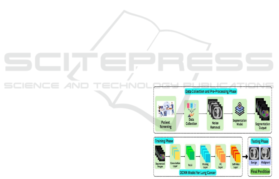
2.2 Dataset Pre-Processing
Feature extraction pipelines a too important pre-
processing step before going for a model analysis
through deep learning. It has different components
which together perform certain important activities
on input data around the needed modelling tasks. Fist,
the raw image data is read, capturing all original pixel
level information. It is here that the rest of this
pipeline begins. The next important pre-processing
activity is resizing the images into a common format.
This is important in ensuring that the deep
learning models will process the inputs. This is
followed by the removal of noise and artefacts from
the images. The model can, otherwise, be affected by
those unwanted characteristics such that, eliminating
them is necessary. Advanced techniques for image
segmentation are applied so that the regions of
interest can be isolated in the images and allow
analysis focusing only on the relevant sections. Then,
further, operations such as dilation and deterioration
are conducted for morphological processes to make
the segmented regions better. It enhances the quality
and integrity of input data for the deep learning model
to be used for classification or pinpointing location
tasks. But by processing data through such an
exhaustive feature extraction pipeline, most
associated deep learning models tend to be more
capable and reliable themselves. Thus, superior
results can be achieved with real-world applications.
Figure 1: Deep Convolutional Neural Network
Architecture.
2.3 Validation Process
These days, with increased availability for datasets of
larger images, looking for an appropriate validation
approach becomes crucial. One of the most used and
effective ways employed for this study is a hold-out
validation scheme, where we have an allocation of a
ICRDICCT‘25 2025 - INTERNATIONAL CONFERENCE ON RESEARCH AND DEVELOPMENT IN INFORMATION,
COMMUNICATION, AND COMPUTING TECHNOLOGIES
644

70:15:15 percent split in data for training, testing, and
validating, respectively. This allows an objective
evaluation of the enactment and generalizability of
DL models. Furthermore, DL models were qualified
for about batch size for 50 epochs is 13. This
configuration was selected keeping convergence and
computation efficiency in a balance. Besides, all the
models were implemented with a random seed of
1000 during the execution so as to give reproducible
results. This step is essential, as it mitigates the
inherent variability that can arise from the random
initialization of model parameters, which could
otherwise lead to inconsistent outputs across different
iterations. By carefully designing and executing the
validation strategy, along with the appropriate
training configurations and seed setting, the study
was able to provide reliable and reproducible insights
into the efficiency of the DL models for initial lung
cancer recognition using CT scan images.
3 PROPOSED DEEP CNN
ARCHITECTURE
The proposed deep CNN has a first convolutional
layer that takes in the 64x64 input image. This layer
has 16 filters and is expected to represent the most
basic features, thus producing 62x62 feature maps.
The convolutional layer served as the primary
fundamental component of the DCNN. Subsequent to
this, the output was conceded over a max pooling
layer, which reduced the longitudinal data size by
half, yielding 31x31 feature maps. Max pooling
chooses the supreme features from the covered
correspond with characteristics region. For further
processing, the output was then fed into a second
convolutional layer with 32 filters and 29x29 feature
atlases. This was trailed by another max pooling
layer, which halved the spatial data size to 14x14
distinctive maps. An additional set of convolutional
and pooling layers was incorporated in the third stage.
The pooling layer in this case contained of 5x5
distinctive maps, while the convolutional layer
utilized 64 filters with 10x10 characteristics atlases.
Lastly, the end results was flattened and passed
through a 260-dimensional dense layer that is
completely interconnected. This was then routed to
the softmax activation function layer, which is
usually employed for multi-class grouping tasks.
Excluding for the end layer, all layers utilized the
ReLU triggering utility without failure. The described
DCNN architecture is represented in Figure 1. The
model was accomplished, authenticated, and verified
using a rate of learning is 0.02, 50 epochs, and a group
size of 13. The Adam optimizer was employed to
compile the model, and a Classification cross-entropy
loss function was utilized, along with other evaluation
indicators like accuracy, recall, and AUC.
Deep CNN Algorithm
Step 1: Convolution layer: The Initial layer serves
as where the input images include are collected.
Step 2: RELU Layer: The picture passes over the
RELU layer, which introduces non-linearity.
Step 3: Pooling Layer: The image is then sent to the
pooling layer, where, if it is too big, the number of
parameters is decreased.
Fully Connected Layer: This layer extracts the sorts
of the pictures with as much as extraordinary
accurateness. It is a vital layer of CNN Split data: Sort
your data with labels into sets for testing, validation,
and training. The model is taught by the training set,
and the validation set monitors its progress, and the
testing set assesses its final performance.
Loss function and back propagation: The predicted
output is associated to the correct label using a loss
function. After that, the error extends reluctant over
and done with the network, correcting the weights and
biases of each layer to minimize future errors.
Step 4: Optimization: Repeat the forward pass and
back propagation for all training images, iteratively
refining the model's parameters using an optimization
algorithm like Adam or SGD.
4 RESULTS AND DISCUSSIONS
The performance results show of four DL
classification models - DCNN, Inception V3,
Xception and ResNet-50 - applied to the Cancer of
the lungs is CT Scan Image collections need
comprehensively summarized in Tables 1, with
comparative insights presented in Figure 2,3,4,5.
These tables provide a detailed breakdown of the
training, validation, and testing show metrics for each
of the respective deep learning models. The inclusion
of these comprehensive performance results allows
for a thorough evaluation and comparison of the
capabilities of the different DL models in the task of
CT scan images to find lung cancer.
Enhancing Lung Cancer Detection Using AI-Based Deep Learning Framework
645
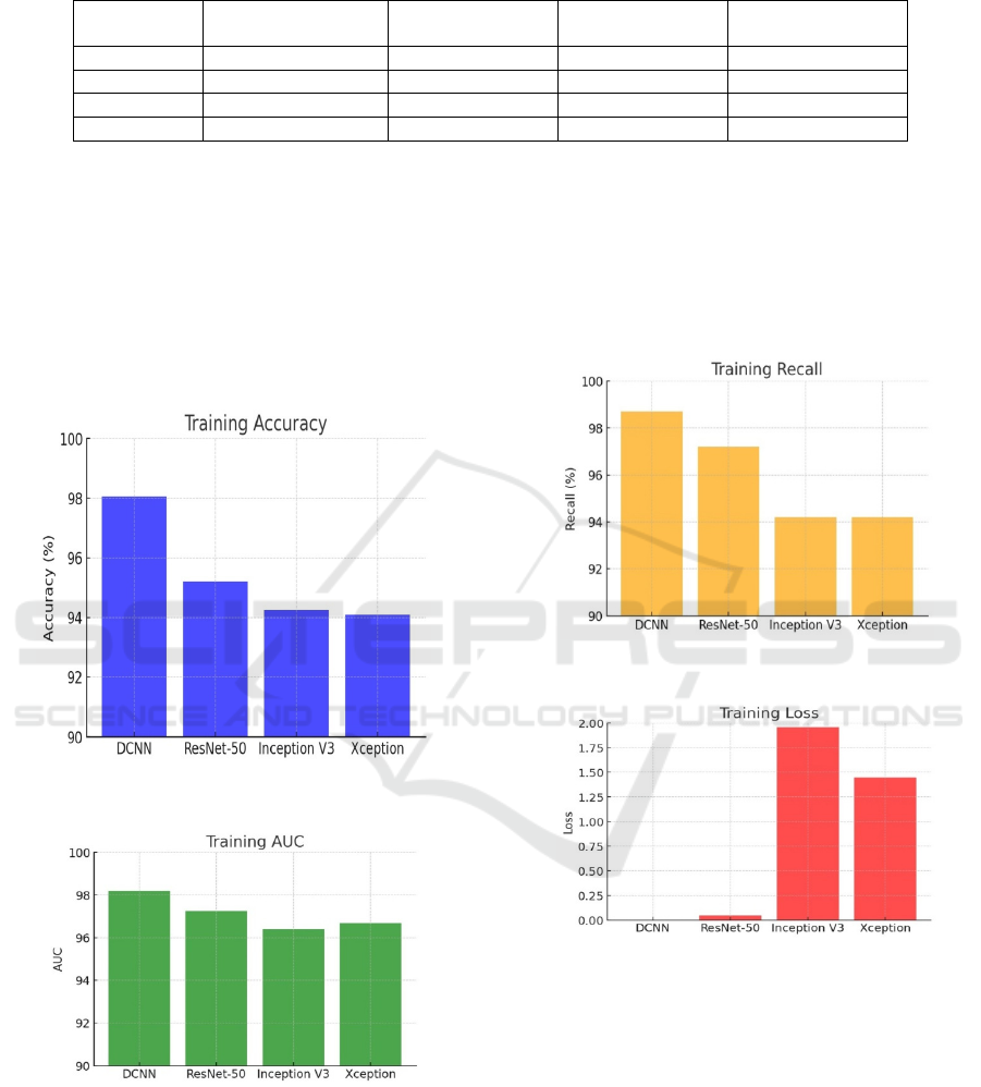
Table 1: Training outcomes for various DL models for lung cancer detection.
Models
Accuracy of
Training
AUC of Training Recall of Training Loss of Training
DCNN 98.27% 97.12 98.70 0.29
ResNet -50 95.20% 97.26 97.20 0.045
Ince
p
tion V3 93.36% 95.3 93.2 1.96
Xce
p
tion 95.24% 95.6 93.2 1.45
The analysis of the methods employed by
the deep convolutional neural network, and other
models reveals that the DCNN model surpasses the
other deep learning approaches, as evidenced by the
comprehensive performance results presented in
Tables 1. The DCNN model was selected as the most
suitable option for the proposed framework aimed at
detecting lung cancer by CT scan images due to its
exceptional performance metrics.
Figure 2: Comparisons for training accuracy levels.
Figure 3: Comparisons for training area under curve.
Specifically, the DCNN model achieved
impressive testing results, including an accurateness
of 98.27%, an area below the curve of 98.21%, a
recall of 98.70%, and a loss of 0.328. These
outstanding performance metrics demonstrate the
DCNN model's effectiveness in accurately
identifying and classifying different kinds of lung
cancer as well as normal cases, from the CT scan data.
The DCNN model's superior performance, compared
to the other DL architectures, for instance Inception
V3, Xception and ResNet-50, makes it a promising
choice for the proposed framework targeting CT
Scans for the preliminary credentials of lung cancer.
Figure 4: Comparisons for training recall.
Figure 5: Training loss function.
5 CONCLUSIONS
Cancer of the lungs is a foremost reason of cancer-
connected humanity global. While it cannot be fully
prevented, early diagnosis and treatment can
significantly improve patient outcomes and survival.
This is a critical priority for healthcare suppliers and
researchers, as lung cancer often goes undetected
until advanced stages. Our research explored a deep
learning framework founded on DCNN for primary
recognition of lung cancer using CT scans. This
ICRDICCT‘25 2025 - INTERNATIONAL CONFERENCE ON RESEARCH AND DEVELOPMENT IN INFORMATION,
COMMUNICATION, AND COMPUTING TECHNOLOGIES
646

DCNN model outperformed other approaches like
ResNet50, Inception V3, and Xception, achieving an
accurateness of 98.05%, AUC of 97.32%, recall of
98.70%, and training loss function of 0.29. To further
enhance early lung cancer diagnosis, we might
incorporate additional datasets and explore other ML
and DL frameworks in the upcoming, aiming to
improve the overall performance and reliability of our
detection methods.
REFERENCES
“Breast cancer statistics of american cancer socity.
[online]. availabe:https://www.cancer.org/cancer.html,
” [Accessed: 13- November-2022].
Abhir Bhandary , G. Ananth Prabhu , V. Rajinikanth , K.
Palani Thanaraj , Suresh Chandra Satapathy , David E.
Robbins , Charles Shasky , YuDong Zhang , Joao
Manuel R.S. Tavares , N. Sri Madhava Raja ,
DeepLearning Framework to Detect Lung ˜
Abnormality – A study with Chest X-Ray and Lung CT
Scan Images, Pattern Recognition Letters (2019), doi:
https://doi.org/10.1016/j.patrec.2019.11.013
Ausawalaithong, W., Thirach, A., Marukatat, S., &
Wilaiprasitporn, T. (2018). Automatic Lung Cancer
Prediction from Chest X-ray Images Using the Deep
Learning Approach. 2018 11th Biomedical Engineerin
g International Conference (BMEiCON).
Da Silva, G.L.F.; da Silva Neto, O.P.; Silva, A.C.; de Paiva,
A.C.; Gattass, M. Lung nodules diagnosis based on
evolutionary convolutional neural network. Multimed.
Tools Appl. 2017, 76, 19039–19055.
Deepa, R., Karthick, R., Velusamy, J., & Senthilkumar, R.
(2025). Performance analysis of multiple-input
multiple output orthogonal frequency division multiple
xing system using arithmetic optimization algorithm.
Computer Standards & Interfaces, 92, 103934.
Hany, M. (2020, August 20). Chest CT-scan images
dataset. Kaggle. Retrieved November 13, 2022, from
https://www.kaggle.com/datasets/mohamedhanyyy/ch
est-ctscan-images
Kaur, S., Hooda, R., Mittal, A., Akashdeep, & Sofat, S.
(2017). Deep CNN-Based Method for Segmenting
Lung Fields in Digital Chest Radiographs. Advanced
Informatics for Computing Research, 185– 194.
doi:10.1007/978-981-10-5780-9_17.
M. Mamun, A. Farjana, M. Al Mamun and M. S.
Ahammed, "Lung cancer prediction model using
ensemble learning techniques and a systematic review
analysis," 2022 IEEE World AI IoT Congress (AIIoT),
2022, pp. 187 193, doi:10.1109/AIIoT54504.2022.981
7326.
M. Mamun, M. I. Mahmud, M. I. Hossain, A. M. Islam, M.
S. Ahammed, M. M. Uddin, "Vocal Feature Guided
Detection of Parkinson's Disease Using Machine
Learning Algorithms", 2022 IEEE 13th Annual
Ubiquitous Computing, Electronics & Mobile
Communication Conference (UEMCON), 2022,
M. Mamun, S. B. Shawkat, M. S. Ahammed, M. M. Uddin,
M. I. Mahmud, A. M. Islam, "Deep Learning Based
Model for Alzheimer's Disease Detection Using Brain
MRI Images", 2022 IEEE 13th Annual Ubiquitous
Computing, Electronics & Mobile Communication
Conference (UEMCON), 2022.
Makaju, Suren, P. W. C. Prasad, Abeer Alsadoon, A. K.
Singh, and A. Elchouemi. "Lung cancer detection using
CT scan images." Procedia Computer Science 125
(2018): 107-114.
Naqi, S.M.; Sharif, M.; Jaffar, A. Lung nodule detection
and classification based on geometric fit in parametric
form and deep learning. Neural Comput. Appl. 2018,
3456789.
Nibali, A.; He, Z.; Wollersheim, D. Pulmonary nodule
classification with deep residual networks. Int. J.
Comput. Assist. Radiol. Surg. 2017, 12, 1799–1808.
Senthilkumar Ramachandraarjunan, Venkatakrishnan
Perumalsamy & Balaji Narayanan 2022, ‘IoT based
artificial intelligence indoor air quality monitoring
system using enabled RNN algorithm techniques’, in
Journal of Intelligent & Fuzzy Systems, vol. 43, no. 3,
pp. 2853-2868
Senthilkumar R, Dr.P.Venkatakrishnan,Dr.N.Balaji, Intelli
gent based novel embedded system based IoT Enabled
air pollution monitoring system, ELSEVIER
Microprocessors and Microsystems Vol.77, June 2020
Shaffie, A.; Soliman, A.; Fraiwan, L.; Ghazal, M.; Taher,
F.; Dunlap, N.; Wang, B.; van Berkel, V.; Keynton, R.;
Elmaghraby, A.; et al. A Generalized Deep
Learning Based Diagnostic System for Early Diagnosi
s of Various Types of Pulmonary Nodules. Technol.
Cancer Res. Treat. 2018, 17
Xie, Y.; Zhang, J.; Xia, Y.; Fulham, M.; Zhang, Y. Fusing
texture, shape and deep model-learned information at
decision level for automated classification of lung
nodules on chest CT. Inf. Fusion 2018, 42, 102–110.
Zhang, T.; Zhao, J.; Luo, J.; Qiang, Y. Deep belief network
for lung nodules diagnosed in CT imaging. Int. J.
Perform. Eng. 2017, 13, 1358– 1370
Enhancing Lung Cancer Detection Using AI-Based Deep Learning Framework
647
