
Classification of Pet Animals Skin Disease Using Vision Transformers
P. Gowsikraja, S. S. Samwilson, S. Joedinesh and K. Saran
Department of Computer Science and Design, Kongu Engineering College (Autonomous) Perundurai,
Erode - 638 060, Tamil Nadu, India
Keywords: Vision Transformers, Pet Skin Diseases, Deep Learning, Image Classification, Veterinary Diagnostics.
Abstract: The classification of pet skin diseases is becoming more and more necessary due to the increasing demand
for early and accurate diagnostics in veterinary healthcare. Complex and diverse skin disease patterns of pets
urge advanced models for reliable recognition and diagnosis. In this paper, we propose an approach based on
Vision Transformers (ViTs) for the classification of skin diseases in pets. The proposed model would be
capable of detecting and categorizing a number of skin diseases efficiently after processing images in such a
way that detailed features crucial for accurate classification are captured. The model has been trained on a
dataset of annotated images to better generalize and perform. These results will be of very important
significance in the diagnosis of different skin diseases found in pets and will contribute a lot to veterinary
dermatology while propagating modern deep learning techniques in improving diagnostic accuracy.
1 INTRODUCTION
It reflects a growing demand for veterinary care,
especially dermatology, due to the skin diseases in
pets that are very common. Therefore, these
conditions should be diagnosed early and correctly
classified so that proper treatment can be initiated,
hence overall health of the animals. However,
traditional methods used for diagnostics present an
unsuitable case as disease manifestation variability is
evident, even in front of seasoned veterinarians.
Techniques based on deep learning are indeed very
promising as they may well automate such diagnoses
and bring improved accuracy into it. A new
architecture in the computer vision domain, known as
Vision Transformers or ViTs, was surprisingly
effective for most classification tasks. This is due to
its ability to model long-range dependencies in
images. The paper is based on the application of ViTs
for the classification of pet skin disease. It elaborates
on how it applies, performs, and compares with other
traditional models, such as CNNs. In this study, the
aim is to design a robust model for distinguishing
various skin diseases in pets and to improve diagnostic
efficiency. Traditional techniques of diagnosis are
mostly based on observation, and hence, vary because
of the asymmetrical presentation of symptoms. This
brings out the emerging importance of AI-based
techniques to aid in support for diagnostic activities,
especially for image classification. Deep learning
architectures, especially of advanced form known as
Vision Transformers, have shown a lot of promise in
image analysis and classification that even includes
medical images. We will apply the application of ViTs
on a pet skin disease classification task and extend
veterinary healthcare diagnostics from a set of images
specially labeled for different kinds of skin diseases.
2 RELATED WORKS
Artificial intelligence, particularly deep learning, has
made significant strides in medical image
classification. Convolutional Neural Networks
(CNNs) have long been the dominant architecture in
image classification tasks, with numerous studies
demonstrating their effectiveness in identifying
human skin diseases such as melanoma and psoriasis.
These models excel at extracting hierarchical features
but can struggle when handling the complex patterns
often present in pet skin diseases.
The Vision Transformer architecture offers an
alternative to CNNs by treating images as a sequence
of patches, which allows the model to retain
information from across the entire image. Unlike
CNNs, which focus on local feature extraction through
convolutional filters, ViTs can process global image
information more effectively. This characteristic
888
Gowsikraja, P., Samwilson, S. S., Joedinesh, S. and Saran, K.
Classification of Pet Animals Skin Disease Using Vision Transformers.
DOI: 10.5220/0013875300004919
Paper published under CC license (CC BY-NC-ND 4.0)
In Proceedings of the 1st International Conference on Research and Development in Information, Communication, and Computing Technologies (ICRDICCT‘25 2025) - Volume 1, pages
888-893
ISBN: 978-989-758-777-1
Proceedings Copyright © 2025 by SCITEPRESS – Science and Technology Publications, Lda.

makes them well-suited for tasks where the
relationships between distant regions of an image are
important, such as in the classification of skin diseases
with varied presentations. While ViTs have shown
promise in fields like object detection and medical
imaging, their application in veterinary diagnostics
remains relatively unexplored.
3 METHODOLOGY
3.1 Dataset Preparation
Table 1: Dataset Distribution by Disease Type.
Disease Type
Number of
Images
Percentage
(%)
Healthy Skin 200 12%
Fungal Infections 350 21%
Bacterial Dermatosis 300 18%
Hypersensitivity
Allergic Dermatosis
350 21%
Other Conditions 100 6%
Total 1300 100
For this table 1, the dataset consists of images of three
primary skin disease categories in dogs namely Fungal
Infections, Hypersensitivity Allergic Dermatosis, and
Bacterial Dermatosis and includes data of healthy skin
to differentiate affected and healthier skins. These
images were collected from veterinary clinics and
medical repositories, ensuring that the dataset covers
a variety of symptoms and conditions for each disease
type.
• Fungal Infections: These include conditions
caused by fungi, such as ringworm,
characterized by scaly patches and hair loss.
• Hypersensitivity Allergic Dermatosis: This
refers to allergic reactions leading to skin
inflammation, itching, and swelling.
• Bacterial Dermatosis: These infections cause
redness, lesions, and, in some cases, pus
formation on the skin.
Each image in the dataset was manually labelled
by veterinary professionals to ensure accurate and
high-quality annotations. To prevent overfitting and
improve model generalization, data augmentation
techniques such as random rotations, scaling, and flips
were applied. This ensured that the model would
perform well across diverse scenarios.
The dataset was divided into three subsets: training
(70%), validation (15%), and testing (15%), with the
image resolution standardized to 224x224 pixels to fit
the input size requirements of the Vision Transformer
model.
3.2 Vision Transformer Model
This architecture based on ViT changes the processing
of an image in comparison with the standard CNNs.
The model does not rely on the usual convolutional
layers of the CNN to extract features but instead
functions by splitting the input image into non-
overlapping small patches. These are usually 16 × 16
pixels in size; then flattened to a one-dimensional
vector. It enables the model to take images as a
sequence, just like the way NLP tasks are addressed in
processing text. This embedding space allows these
vectors to be projected once the image is divided into
patches. In this context, the embedding becomes
crucial for allowing the model to understand
relationships among different patches. The
transformer layers are the backbone of the Vision
Transformer. These employ self-attention
mechanisms, which gives more relevance to some
patches with respect to the other patches, allowing the
model to capture more patterns and the correlations
between the local and the global features that exist in
the images.
The multi-head attention mechanism in the
transformer layers gives a deep, nuanced
understanding of content through focusing differently
at various places of the images at once. This is
especially the case for disease classification problems,
where sometimes slight differences in texture and
structure can signify different skin diseases. The
output of the transformer layers proceeds to a
classification head, often a Multi-Layer Perceptron
(MLP), to produce the final predictions for the
categories for the skin disease. See the architecture of
the vision transformer in Figure 1.
Classification of Pet Animals Skin Disease Using Vision Transformers
889
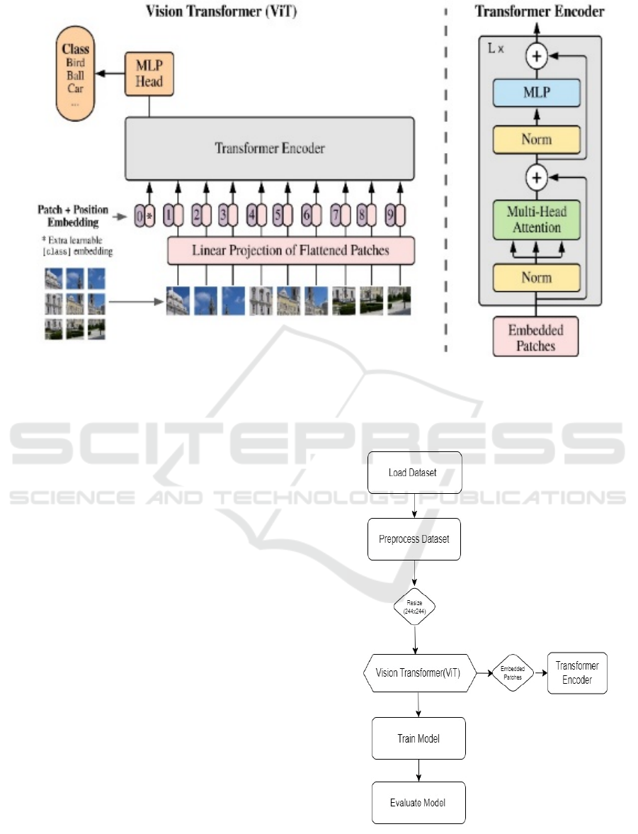
Figure 1: Vision Transformer Architecture.
3.3 Model Specifications
The Vision Transformer model for this study was
configured with the following specifications:
• Input Image Size: 224×224 pixels. This size
is commonly used to maintain a balance
between computational efficiency and
sufficient detail for accurate classification.
• Patch Size: 16×16 pixels. This patch size
allows for capturing localized features while
maintaining a manageable number of patches
for processing.
• Number of Layers: 12 transformer layers.
The depth of the model helps it learn
complex representations.
• Embedding Dimension: 768. This high-
dimensional space allows for a more
expressive representation of the input data.
• Attention Heads: 12. Multiple attention
heads enable the model to attend to different
parts of the image, capturing various features
effectively.
• Classification Head: Multi-Layer
Perceptron (MLP), which processes the
aggregated information from the transformer
layers and outputs probabilities for each class
of skin disease.
3.4 Flow Chart
Figure 2: Flow Chart.
ICRDICCT‘25 2025 - INTERNATIONAL CONFERENCE ON RESEARCH AND DEVELOPMENT IN INFORMATION,
COMMUNICATION, AND COMPUTING TECHNOLOGIES
890
3.5 Training Process
The training process followed a stepwise procedure to
the Vision Transformer, thus ensuring correct learning
and proper generalization for unseen data. Using the
Adam optimizer was pertinent, considering that it
efficiently takes care of sparse gradients and adjusts
learning rates in real time. The chosen learning rate
was 0.0001, balanced to provide maximum
convergence speed within stable training processes.
Training has been performed in batches of 32 images;
this is ensured to provide sufficient computation with
regard to memory use. Batch size influences the
generalization capability of the model; too large a
batch size tends to generalize poorly while the
opposite may lead to noisy updates. The loss function
is cross-entropy that is well suited for multi-class
classification problems. This function calculates the
difference of class probabilities, which the model has
predicted with the actual class labels, and these
differences guide the model to reduce its prediction
errors. It is trained for 50 epochs with the early
stopping mechanisms, which mean that the training is
stopped if the model fails to improve for a specified
number of epochs on the validation set. This can
prevent overfitting in the models, a typical problem
that often arises in deep learning models when the
dataset is relatively small.
The above random rotations, scaling, flipping, and
colour augmentation are then used to build the model
as robust. The model learns it to be exposed to a lot
more scenarios than it would otherwise have been
under if all of these were to have been learned on live
data. Finally, there was transfer learning. Here the
initialization is with the weights attained by
pretraining on the ImageNet dataset. This approach
now puts the knowledge learned from a large-scale
dataset to good use: it now constitutes an extremely
strong initialization for the model. It would allow the
ViT model to adapt even better to the particular task
of classification of skin disease, even by being
compatible with a relatively small target dataset.
4 RESULT AND DISCUSSIONS
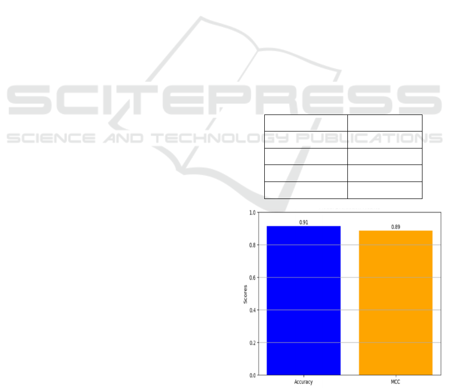
4.1 Performance Evaluation
Test set performance was conducted on the ViT model
by focusing considerably on major three key
performance metrics of evaluation-precision, recall,
F1-score, and accuracy. The overall accuracy rate
returned was 90.5%, showing how the model can
classify images very well according to different
categories of skin diseases. Besides this, all precision,
recall, and F1-score of the model were more than 90%
with respect to diagnostic accuracy, as well as
consistency in finding accuracy with the right disease.
The precision value refers to the accuracy with which
the model will classify its positive instances without
producing excessive false positives. It is very reliable
for diagnostics. The recall score shows how good the
model is in classifying true positives and, thus,
avoiding a possible misclassification. The high F1-
score combines precision and recall, indicating that
the model has the strength to handle the complexity
associated with variable presentations of the disease.
These results confirm reliability for models in real-
veterinary and actual medical application fields where
diagnoses are important as they have to be both precise
yet speedy. The ViT Vision transformer can view full
images and identify between two very similar
conditions so that such specifics appear well distinct.
In this success, therefore, deep learning models like
ViT are most likely to change practice in the sense that
they are going to make such a diagnosis. This is
concerning areas such as dermatology. Table 2
represents the model performance metrics and figure
3 and 4 shows the model evaluation metrices and
confusion matrix.
Table 2: Model Performance Metrics.
Metric Value
Accuracy 90.5%
Precision 91.2%
Recall 90.8%
F1- Score 91.0%
Figure 3: Model Evaluation Metrices.
Classification of Pet Animals Skin Disease Using Vision Transformers
891
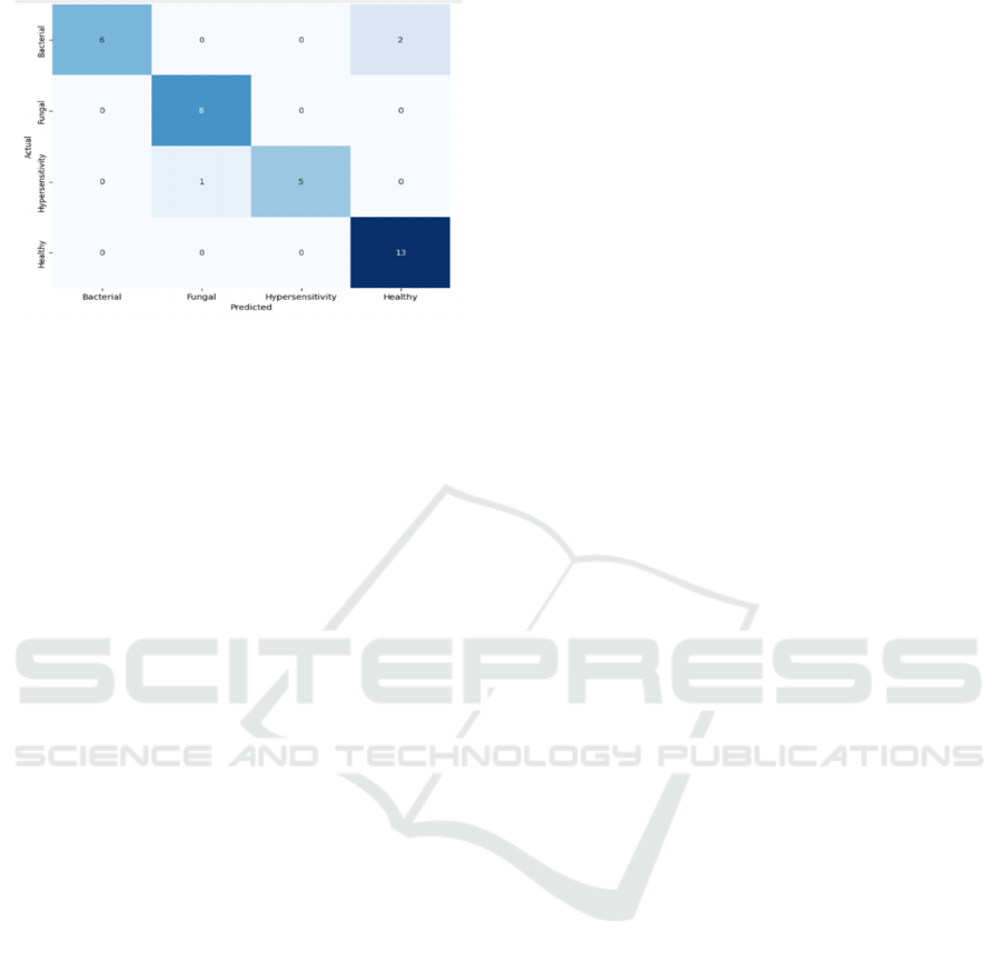
Figure 4: Confusion Matrix.
4.2 Discussion
The model proved to outperform on the different
classes of disease. It can extract a very wide-ranging
pattern in the entire image and proved to be way better
than approaches that highly depend on these local
features. Actually, it showed a really impressive
performance between different skin conditions which
look pretty similar, such as fungal infections versus
bacterial infections. Still, the scope to enhance the
performance further is still in place. Generalizing
capabilities of this model will also be remarkably high
if diversity, such as greater instances of uncommon
skin diseases or even broader species than cats and
dogs, of the dataset will be enhanced. This would
ensure that more precise diagnoses occur in real
veterinary practice as its capability to robustly deal
with different scenarios it might face would be
increased.
5 CONCLUSIONS
This research demonstrates the promising capabilities
of the ViTs in accomplishing the task of skin disease
classification in pets. The results show that it was
found to be accurate under different conditions, hence
really applicable to help veterinary diagnostics get
better. Application of ViTs would therefore greatly
enhance the accuracy as well as the speed of diagnosis,
which, in a way, assists in better planning of treatment
for pets, indirectly enhancing their quality of life. In
particular, the ability to pull complex patterns and
global features from images will be helpful in
differentiating presentations of skin disease that may
look very much alike. This feature of the ViTs will
prove to be of utmost value in veterinary practice,
thanks to the ability to make a diagnosis rapidly and
precisely, therefore arresting the course of disease and
allowing intervention sooner rather than later.
Add new data modalities, for example, clinical notes
or owner-reported symptoms and treatment history, in
order to have a holistic diagnostics approach that is
going to stretch the model very far. A multimodal
approach like this one would add visual context,
beside the mere analysis of vision; it increases the
accuracy of the classification more than just purely
visual analysis of the model above. It will also require
a dataset large and varied enough to cover different
skin conditions and species of pets so that the model
would be robust and generalizable in real-world
veterinary settings.
In summary, the successful application of Vision
Transformers in the present study suggests that these
have the potential to revolutionize the face of
veterinary diagnostics. With development in the
future, technologies of AI are bound to change how
veterinarians will handle diagnostics and treatment on
balance, better outcomes for the pets and less stress
among the owners.
REFERENCES
Bhavsar, S., & Mehendale, N. (2022). Deep Learning-
Based Automatic System for Diagnosis and
Classification of
Skin Dermatoses. https://doi.org/10.21203/rs.3.rs- 236
0579/v1
Gupta, P., & Gupta, S. (2022). Deep learning in medical
image classification and object detection: A survey.
International Journal of Image Processing and Pattern
Recognition. https://doi.org/10.37628/ijippr.v8i2.846
Himel, G. M., Islam, Md. M., Al-Aff, Kh. A., Karim, S. I.,
& Sikder, Md. K. (2024). Skin cancer segmentation and
classification using vision transformer for automatic
analysis in dermatoscopy-based noninvasive digital
system. International Journal of Biomedical Imaging,
2024, 1–18. https://doi.org/10.1155/2024/3022192
Hwang, S., Shin, H. K., Park, J. M., Kwon, B., & Kang, M.-
G. (2022). Classification of dog skin diseases using
deep learning with images captured from Multispectral
Imaging device. Molecular & Cellular
Toxicology, 18(3), 299–309.
https://doi.org/10.1007/s13273-022-00249-7
Hyeon Ki Jeong 1 2, 1, 2, & Artificial intelligence (AI) has
recently made great advances in image classification
and malignancy prediction in the field of dermatology.
However. (2022, August 23). Deep learning in
dermatology: A systematic review of current approach
-es, outcomes, and limitations. JID Innovations.
https://www.sciencedirect.com/science/article/pii/S26
67026722000583
Jiang, Z., Gu, X., Chen, D., Zhang, M., & Xu, C. (2024).
Deep learning-assisted multispectral imaging for early
ICRDICCT‘25 2025 - INTERNATIONAL CONFERENCE ON RESEARCH AND DEVELOPMENT IN INFORMATION,
COMMUNICATION, AND COMPUTING TECHNOLOGIES
892

screening of skin diseases. Photodiagnosis and
Photodynamic Therapy, 48, 104292. https://doi.org/10
.1016/j.pdpdt.2024.104292
Mohsin Ali, M., Chandra Joshi, R., & Kishore Dutta, M.
(2022). An automated and efficient deep learning-based
classification of multiple skin disorders from skin
lesion images. 2022 International Conference on Edge
Computing and Applications (ICECAA), 1156–
1161.https://doi.org/10.1109/icecaa55415.2022.99360
97
Rathnayaka, R. M. N. A., Anuththara, K. G. S. N.,
Wickramasinghe, R. J. P., Gimhana, P. S.,
Weerasinghe, L., & Wimalaratne, G. (2022). Intelligent
system for skin disease detection of dogs with ontology
based clinical information extraction. 2022 IEEE 13th
Annual Ubiquitous Computing, Electronics &
Mobile Communication Conference (UEMCON),
0059–0066.
https://doi.org/10.1109/uemcon54665.2022.9965696
S. P. R. R. Raj and P. K. Gupta, "Vision Transformers in
Medical Image Analysis: A Review," IEEE
Transactions on Medical Imaging, vol. 41, no. 9, pp.
2161- 2175, Sep. 2022. https://www.semanticscholar.
org/paper/Transformers-in-Medical-Image-Analysis:-
A-Review-He-
Gan/42bad1b72259aa1ff70d7ce2539220a83f1af9a4
Saraf, P., Tharaniesh, P. R., & Singh, S. (2024). Skin
disease detection using convolutional neural network.
2024 Second International Conference on Networks,
Multimedia and Information Technology (NMITCON),
1– 6. https://doi.org/10.1109/nmitcon62075.2024.106
99196
Upadhyay, A., Singh, G., Mhatre, S., & Nadar, P. (2023).
Dog skin diseases detection and identification using
convolutional neural networks. SN Computer Science,
4(3). https://doi.org/10.1007/s42979-022-01645-5
Classification of Pet Animals Skin Disease Using Vision Transformers
893
