Real-Time Monitoring Technology for Physiological States in Brain
Computer Interface Systems
Chenxi Hu
Department of Physics, King’s College London, U.K.
Keywords: Real-Time Monitoring Technology, BCI, fNIRs.
Abstract: Since the early 2000s, Brain-Computer Interface (BCI) systems have emerged as a transforma-tive technology
in neuroscience, enabling direct communication between the brain and exter-nal devices. Originally
developed to restore motor functions and control external systems, BCIs now extends to real-time
physiological state monitoring. This paper explores the evolu-tion and methodologies of BCIs, focusing on
signal detection techniques. Non-invasive meth-ods, such as Electroencephalography (EEG) and Functional
Near-Infrared Spectroscopy (fNIRS), provide safe and accessible options, while invasive techniques like
Electrocorticogra-phy (ECoG) offer superior precision. Hybrid BCIs, integrating modalities such as EEG-
fNIRS, enhance performance by combining the strengths of individual technologies. The applications of BCIs
span clinical and non-clinical domains, including stroke rehabilitation, communica-tion for individuals with
severe impairments, brain-controlled gaming, and artistic creation. Recent advancements in signal acquisition,
processing, and integration, such as improved electrode designs and real-time signal processing algorithms,
have established BCIs as a criti-cal tool for neurotechnological innovation, with immense potential to
transform healthcare and human-computer interaction.
1 INTRODUCTION
Brain-Computer Interface (BCI) systems have
become a key focus of research, offering direct
communication pathways between the brain and
external devices. Early BCI studies focused on
restoring motor functions and controlling external
systems, and this research has expanded to include
real-time monitoring of physiological states. The
foundation of BCI research was laid in the early 20th
century with Hans Berger’s discovery of
electroencephalography (EEG). This breakthrough
demonstrated that neural activity could be measured
and analysed, forming the basis for the modern
exploration of direct brain-to-machine
communication. From this foundational work, the
field of BCIs has advanced significantly, with a
particular focus on the development of technologies
capable of accurately detecting and interpreting
neural signals. These signals serve as the fundamental
medium through which brain activity is translated
into actionable commands for controlling external
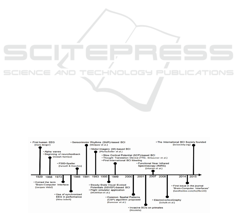
devices. The brief history of BCIs evolution is shown
in Figure 1.
Figure 1. This diagram shown a span of evolution of BCIs range from 1929 to 2015 (Fabien et. al. 2018).
Hu, C.
Real-Time Monitoring Technology for Physiological States in Brain Computer Interface Systems.
DOI: 10.5220/0013851800004708
Paper published under CC license (CC BY-NC-ND 4.0)
In Proceedings of the 2nd International Conference on Innovations in Applied Mathematics, Physics, and Astronomy (IAMPA 2025), pages 649-654
ISBN: 978-989-758-774-0
Proceedings Copyright © 2025 by SCITEPRESS – Science and Technology Publications, Lda.
649
Alt Text for the figure: the timeline labelled with various important events of BCI evolution from 1929 to 2015.
The brackets conclude the names of scientists who discovered or invented this specific event. For example, Hans
Berger recorded the first human EEG in the 1920s.
Following the historical development, it is
essential to understand how BCIs function. The
methodology of BCI involves several steps that
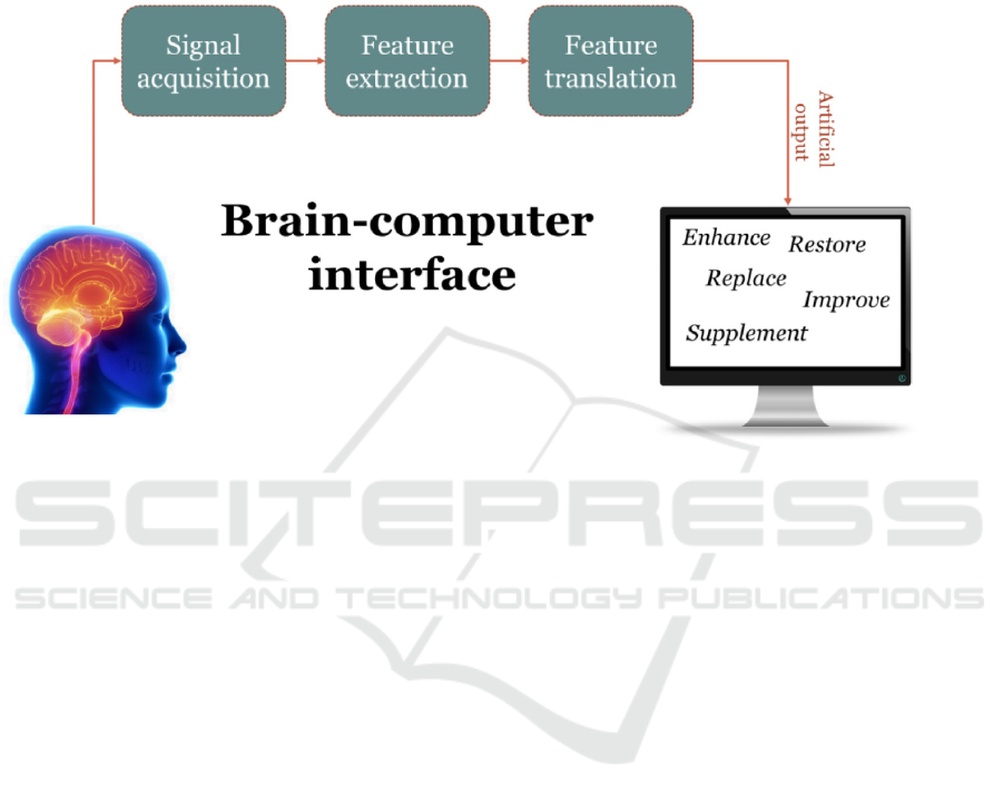
enable direct communication between the brain and
external devices. Figure 2 shows the basic processing
of BCI system.
Figure 2. A schematic representation of the basic processing of BCI systems (Aricò et. al. 2018).
Alt Text for the figure: the Brain-computer interface starts at brain to signal acquisition, then feature extraction,
and feature translation. The artificial output from the translation will then enhance the brain instruction to the
computer device.
The first step in any Brain-Computer Interface
(BCI) system is signal acquisition, where brain
signals is captured using various techniques. While
all these detection methods aim to capture brain
signals accurately, they can be broadly categorized
into non-invasive and inva-sive approaches based on
their implementation. Non-invasive techniques, such
as Electroen-cephalography (EEG) and functional
near-infrared spectroscopy (fNIRS), are widely used
due to their safety, ease of use, and accessibility.
These methods measure neural activity indirectly
through the skull and scalp, making them particularly
well-suited for real-world applications due to their
non-invasive nature and minimal risk to users. In
contrast, invasive methods like Electrocorticography
(ECoG) directly record electrical activity from the
cortical surface, offer-ing superior spatial and
temporal resolution, which is ideal for specific
applications requiring high precision. Recently,
hybrid systems that integrate multiple modalities
have emerged, combining the strengths of each
technology to overcome individual limitations and
enhance overall performance. However, raw brain
signals are often noisy and require preprocessing to
remove artifacts. One of the most common artifacts,
especially in EEG signals, is eye move-ment. To
address this, specific preprocessing techniques are
employed, such as digital filtering for noise removal
and Independent Component Analysis (ICA) for
artifact correction.
Next, the feature extraction phase begins, where
relevant features are extracted from the pre-processed
signals to identify specific patterns in brain activity.
Common techniques in-clude time-domain features
(such as signal amplitude, variance, and peak values)
and frequen-cy-domain features (such as power
spectral density and wavelet transforms), which
together provide a comprehensive description of
signal characteristics. Effective feature extraction is
critical for improving the accuracy and robustness of
BCI systems, as it directly influences the performance
of the subsequent classification algorithms. There are
three main types of classi-fication algorithms used in
the BCI field: Machine Learning (ML), Deep
Learning (DL), and Transformer-based models.
These algorithms translate brain activity into
actionable outputs, which can then be used to control
external devices such as robotic arms, prosthetics, or
speech synthesis systems. This paper primarily
focuses on the signal detection aspect of BCI systems,
IAMPA 2025 - The International Conference on Innovations in Applied Mathematics, Physics, and Astronomy
650

exploring various techniques and their role in
enhancing the quality and reliability of brain signal
acquisition.
2 TECHNOLOGY OF SIGNAL
DETECTION
Neural signals are essential in brain-computer
interfaces (BCIs) for translating brain activity into
device control commands. These signals reflect the
electrical and physiological activity of the brain,
enabling direct interaction between neural functions
and external systems. BCIs primarily rely on
accurately capturing these signals, thereby enabling
applications such as prosthetic control and
communication aids. To achieve this, various
techniques are employed to measure brain activity,
with non-invasive methods being the most widely
used due to their safety, ease of application, and
accessibility.
2.1 Non-Invasive Signal Detection
Currently, among the non-invasive approaches,
Electroencephalography (EEG) and Functional Near-
Infrared Spectroscopy (fNIRS) are two prominent
technologies for recording brain activity.
2.1.1 Electroencephalography (EEG)
Electroencephalography (EEG) is a widely adopted
non-invasive technique for monitoring electrical
activity in the brain. By placing electrodes on the
scalp, EEG captures voltage fluc-tuations arising
from current flows within neuronal networks (Finnis
et. al. 2024). Since its introduction nearly a century
ago, EEG has been a foundational tool in both clinical
diagnos-tics and neuroscience research. Rather than
detecting action potential, EEG measures
postsynaptic potentials generated by neurotransmitter
activity. These signals originate from cortical
pyramidal neurons, which are aligned in a way that
makes their synchronized activity more detectable.
However, the signal is often modulated by factors
such as cerebrospinal fluid and the skull, which can
distort or attenuate its propagation (Andrea et. al.
2019).
EEG plays a pivotal role in diagnosing
neurological disorders, including epilepsy, sleep dys-
functions, and other conditions (Andrea et. al. 2019).
In recent years, its integration with brain-computer
interface (BCI) systems has expanded its
applications, enabling innovations such as mind-
controlled prosthetics and rehabilitation devices (Lee
et. al. 2017). A major ad-vantage of EEG lies in its
exceptional temporal resolution, which allows it to
track rapid neu-ral changes in real-time (Mathewson
et. al. 2017). Modern EEG systems, which can
support over 128 recording channels and achieve
sampling rates exceeding 10 kHz, are lightweight,
portable, and cost-efficient (Andrea et. al. 2019).
These features make EEG suitable for both controlled
laboratory environments and real-world applications,
such as classrooms and athlet-ic training.
However, EEG systems are not without
limitations. They are highly sensitive to noise, in-
cluding electrical interference and movement
artifacts, such as eye blinks or head motion
(Mathewson et. al. 2017). Moreover, variations in
signal preprocessing methods and referenc-ing
techniques between different research studies can
reduce result reproducibility and limit their broader
application. Despite these challenges, continued
advancements in signal pro-cessing and
computational analysis ensure that EEG remains a
critical tool for exploring brain function and
developing neurotechnological innovations (Pfeffer
et. al. 2024).
2.1.2 Functional Near-Infrared
Spectroscopy (fNIRS)
Functional Near-Infrared Spectroscopy (fNIRS) is an
emerging non-invasive imaging technol-ogy that
monitors brain activity by measuring changes in
blood oxygenation (Finnis et. al. 2024). Compared to
EEG which directly records electrical activity, fNIRS
indirectly tracks neural processes by capturing
fluctuations in oxyhaemoglobin (HbO2) and
deoxyhaemoglobin (HbR) levels. These changes are
indicative of hemodynamic responses to brain
activation. Employing near-infrared light, fNIRS
detects these variations with greater spatial resolution
(approximately 1 cm) than EEG (roughly 3 cm)
(Borgheai et. al. 2020). Furthermore, fNIRS is less
susceptible to artifacts caused by muscle activity or
motion, making it an advantageous choice in many
scenarios (Finnis et. al. 2024). Unlike EEG, one of
fNIRS’s most notable ben-efits is its immunity to
electromagnetic interference, which is particularly
valuable in envi-ronments where electrical noise
poses a challenge. This characteristic has made
fNIRS a pre-ferred tool in applications such as
controlling prosthetic devices and studying brain
activity under real-world conditions. In the context of
BCIs, fNIRS has shown great promise for assist-ing
individuals with severe motor impairments, such as
Real-Time Monitoring Technology for Physiological States in Brain Computer Interface Systems
651

late-stage amyotrophic lateral sclerosis (ALS)
patients (Borgheai et. al. 2020). It can translate
haemodynamic changes into actionable control
signals during cognitive tasks, such as mental
arithmetic or imagery.
Recent innovations include the development of
hybrid EEG-fNIRS systems, which combine the
temporal resolution of EEG with the spatial precision
of fNIRS (Liu et. al. 2021). Fur-thermore, advanced
paradigms such as the Visuo-Mental (VM) task
combine visual stimuli and mental calculations to
generate distinctive hemodynamic patterns in single
trials (Bor-gheai et. al. 2020). These advances reduce
response times and enhance usability, particularly in
spelling systems for communication. Unlike
traditional methods requiring multiple trials, fNIRS-
based systems can identify target responses rapidly,
often achieving classification accu-racies above 80%
(Liu et. al. 2021). The robustness of fNIRS against
motion artifacts and its compatibility with bedside
setups highlight its transformative potential for
neurotechnological applications (Cutini et. al. 2012).
As research continues, refinements in algorithms,
real-time processing, and system integration are
expected to further enhance its effectiveness, particu-
larly in personalized and clinical settings (Yücel et.
al. 2017).
2.2 Invasive Signal Detection
ECoG is a neurophysiological method used to record
electrical activity directly from the sur-face of the
brain. It involves placing electrode grids on the
exposed cerebral cortex, typically during a surgical
procedure. It is considered a minimally invasive
technique compared to fully invasive methods like
intracortical recordings, as the electrodes rest on the
brain surface rather than penetrating it (Wilson et. al.
2006).
ECoG based BCIs leverage several key
advantages over non-invasive alternatives. The signal
quality is enhanced in ECoG as the electrodes are
closer to the neural sources, yielding signals with
higher amplitude compared with EEG (Wilson et. al.
2006). This reduces signal noise and allows for better
artifact rejection. It also has higher spatial and
temporal resolution. The millimetre-scale spatial
resolution achievable with ECoG enables
discrimination of fine neural patterns. This contrasts
with the centimetre-scale resolution of EEG, which
often leads to sig-nal overlap. The applications of
ECoG have proven effective for both motor and
sensory im-agery-based control tasks, particularly in
tasks like imagining limb movements which activate
distinct sensorimotor rhythms. ECoG’s precision
allows mapping these activities across adja-cent
cortical areas. Despite its advantages, ECoG-based
systems face challenges including sur-gical risks,
chronic viability, and signal interpretation. The
implantation of ECoG grids re-quires craniotomy,
carrying inherent risks such as infection and
inflammation. In the long-term, it raises concerns
about electro stability and biocompatibility.
2.3 Hybrid BCI (hBCI)
To enhance BCI performance, BCI systems are
increasingly being incorporated with other
physiological signals. The EEG-fNIRS mentioned in
the fNIRS technology part is one of the most
promising hybrid BCI systems. It combines the high
temporal resolution of EEG and the spatial resolution
of fNIRS, which provides a complementary insight
into brain dynamics.
Electrocardiography (ECG) and heart rate
variability (HRV) are also gaining attention in BCIs
for detecting emotional and autonomic responses.
The study in states that the fusion of ECG and EEG
features for hBCI enhances the average imagery
classification accuracy in training and evaluation
stages (Shahid et. al. 2011). However, more recent
studies have pre-dominantly focused on combining
EEG with other modalities such as electromyography
(EMG) and functional near-infrared spectroscopy
(fNIRS). For example, a 2024 study intro-duced a
motor imagery classification model based on a hybrid
BCI that integrates EEG and EMG signals,
demonstrating improved classification accuracy.
Another study in 2020 eval-uated the performance of
a compact hybrid BCI combining EEG and fNIRS,
achieving high classification accuracy with a reduced
number of channels (Choi et. al. 2017). These
develop-ments suggest that while the fusion of ECG
and EEG in hybrid BCIs has been explored, re-cent
research trends have shifted towards other
combinations of physiological signals to en-hance
BCI performance and practicality.
3 APPLICATIONS OF BCI
The BCI has significant potential in both clinical
and non-clinical fields, with different
applications tailored to distinct purposes.
IAMPA 2025 - The International Conference on Innovations in Applied Mathematics, Physics, and Astronomy
652

3.1 Clinical Applications
Brain-Computer Interfaces (BCIs) have
revolutionized clinical rehabilitation and
assistive technologies. These systems offer
transformative solutions for patients with severe
motor or communication impairments. In stroke
rehabilitation, BCIs leverage motor imagery and
real-time feedback to activate neural pathways,
promoting neuroplasticity and aiding motor
recovery, especially when combined with
robotic devices or functional electrical
stimulation (Ang et. al. 2015). For individuals
with amyotrophic lateral sclerosis (ALS), BCIs
provide an essential communication channel by
detecting brain signals like P300 or steady-state
visual evoked potentials (SSVEP), enabling
word spelling or device control even in advanced
disease stages (Vansteensel et. al. 2016).
Furthermore, BCIs enable intuitive control of
prosthetic limbs and wheelchairs by translating
electroencephalography (EEG) signals into
commands, empowering individuals with severe
motor impairments to regain mobility and
independence. Additionally, combining BCI
with machine learning has led to significant
advancements in natural language processing
(NLP), allowing real-time text generation or
speech synthesis through neural decoding, which
is especially beneficial for patients with locked-
in syndrome (Moses 2021). These clinical
applications highlight the profound impact of
BCIs on improving patient quality of life and
enabling greater independence.
3.2 Non-Clinical Applications
In non-clinical fields, Brain-Computer Interfaces
(BCIs) have demonstrated transformative
potential across diverse fields such as gaming
and creative arts. In gaming, BCIs enable brain-
controlled experiences that allow players to
interact with games through their thoughts,
which enhance engagement and creates
innovative design possibilities. This
advancement highlights the potential of BCIs to
revolutionize entertainment and education by
driving the development of more intuitive
human-computer interfaces (Nijholt et. al.
2015). Similarly, in the creative arts, BCIs allow
users to create music, paintings, or digital art
through neural activity, providing a unique
platform for self-expression and creativity. This
is particularly impactful for individuals with
physical disabilities, as it broadens access to
artistic creation while pushing the boundaries of
traditional art production and experience
(Miranda et. al. 2011). These applications
underscore the versatility of BCIs in shaping
interactions with technology beyond clinical use.
4 CONCLUSIONS
Brain-Computer Interface (BCI) systems have
emerged as one of the most transformative
technologies in modern science, bridging the gap
between neural activity and external device control.
BCIs have come a long way since their foundational
discovery with EEG in the early 20th century. Today's
advanced hybrid systems have demonstrated
remarkable potential in both clinical and non-clinical
domains. Central to the effectiveness of these systems
is the methodology of signal detection, which
encompasses non-invasive techniques like EEG and
fNIRS, invasive methods such as ECoG, and hybrid
BCIs that combine multiple modalities for enhanced
performance. Each of these approaches offers unique
advantages: EEG provides ex-ceptional temporal
resolution, fNIRS delivers superior spatial resolution,
and ECoG achieves unmatched precision through
direct cortical contact.
The clinical applications of BCIs are diverse,
including stroke rehabilitation, assistive tech-
nologies for individuals with ALS, and
communication solutions for locked-in syndrome.
These applications demonstrate BCIs' capacity to
significantly improve quality of life. These systems
leverage advanced signal processing and machine
learning to translate neural activity into actionable
outputs, facilitating motor recovery, communication,
and mobility. Non-clinical applications, such as
brain-controlled gaming and artistic creation,
demonstrate the versatility of BCIs beyond
healthcare, offering new platforms for self-
expression, creativity, and intuitive interaction with
technology. Despite these advancements, several
challenges need to be addressed through ongoing
research, including signal noise reduction, movement
artifact compensation, and minimizing risks
associated with invasive methods. Variability in
preprocessing techniques and the complexity of
integrating multimodal systems also present obstacles
to widespread adoption. However, ongoing research
Real-Time Monitoring Technology for Physiological States in Brain Computer Interface Systems
653

in computational algorithms, re-al-time signal
processing, and system miniaturization continues to
address these limitations, paving the way for broader
usability in both laboratory and real-world settings.
Looking ahead, BCIs are positioned to
revolutionize human-computer interaction, enabling
seamless integration between neural processes and
external systems. Emerging hybrid sys-tems, such as
EEG-fNIRS combinations, highlight the potential to
enhance classification ac-curacy and usability,
particularly for personalized and clinical applications.
The fusion of BCIs with fields like artificial
intelligence, natural language processing, and
robotics is creating synergistic effects. These
combinations are accelerating innovation by enabling
more sophisti-cated interpretation of neural signals,
thus opening doors to new possibilities in
communica-tion, rehabilitation, and entertainment. In
conclusion, BCIs have the potential to redefine the
relationship between humans and technology,
transforming how humans interact with ma-chines
and the environment. While significant challenges
remain, continued advancements in signal
acquisition, processing techniques, and system
integration ensure that BCIs will play an increasingly
vital role in addressing societal needs, improving
accessibility, and enhancing the overall quality of life
for individuals across the globe.
REFERENCES
Ang, K. K., Guan, C., Chua, K. S. G., et al. 2015. A large
clinical study on the ability of motor imagery BCI to
facilitate post-stroke upper-limb recovery.
Neurorehabilitation and Neural Repair 29(5): 433-442.
Aricò, P., Aloise, F., Schettini, F., et al. 2018. Passive BCI
beyond the lab: current trends and future directions.
Measurement Science and Technology 29(8): 08TR02.
Borgheai, S. B., Khalilzadeh, M., Nazerfard, E., et al. 2020.
Enhancing communication for people in late-stage ALS
using an fNIRS-based BCI system. IEEE Transactions
on Neural Systems and Rehabilitation Engineering
28(5): 1198-1207.
Choi, I., Rhiu, I., Lee, Y., Yun, M. H., Nam, C. S. 2017. A
systematic review of hybrid brain-computer interfaces:
taxonomy and usability perspectives. PLoS ONE 12(4):
e0176674.
Cutini, S., Moro, S. B., Bisconti, S. 2012. Functional near-
infrared optical imaging in cognitive neuroscience: an
introductory review. Frontiers in Human Neuroscience
6:117.
Fabien, L., Chang, S., Nijholt, A. 2018. Introduction:
Evolution of brain-computer interfaces. Brain-
Computer Interfaces Handbook: Technological and
Theoretical Advance. Taylor & Francis (CRC Press) 1-
11.
Finnis, R., Mehmood, A., Iqbal, J. 2024. fNIRS vs. EEG-
based prosthetic limb control: a systematic literature
review. Yorkshire Innovation in Science and
Engineering Conference (YISEC) 2024, Hull, UK.
Lee, J., Kim, D., Jeong, J., et al. 2017. A multichannel-near-
infrared-spectroscopy-triggered robotic hand
rehabilitation system for stroke patients. IEEE
International Conference on Rehabilitation Robotics
(ICORR), 17: 158-163.
Liu, Z., Shore, J., Wang, M., Yuan, F., Buss, A., Zhao, X.
2021. A systematic review on hybrid EEG/fNIRS in
brain-computer interface. Biomedical Signal
Processing and Control 68:102595.
Mathewson, K. E., Harrison, T. J., Kizuk, S. A. 2017. High
and dry? Comparing active dry EEG electrodes to
active and passive wet electrodes. Psychophysiology
54: 74-82.
Miranda, E. R., Durrant, S., Anders, T. 2011. Brain-
computer music interfacing: interdisciplinary research
at the crossroads of music, science, and technology.
Music and Medicine 3(3): 150-160.
Moses, D. A., Leonard, M. K., Makin, J. G., Chang, E. F.
2021. Real-time decoding of question-and-answer
speech dialogue using human cortical activity. Nature
Communications 12: 363.
Nijholt, A. 2015. Brain-computer interfaces for games and
entertainment. Springer.
Pfeffer, M. A., Ling, S. S. H., Wong, J. K. W. 2024.
Exploring the frontier: transformer-based models in
EEG signal analysis for brain-computer interfaces.
Computers in Biology and Medicine 178: 108705.
Shahid, S., Prasad, G., Sinha, R. K. 2011. On fusion of heart
and brain signals for hybrid BCI. 2011 5th International
IEEE/EMBS Conference on Neural Engineering,
Cancun, Mexico, 48-52.
Vansteensel, M. J., Pels, E. G., Bleichner, M. G., et al. 2016.
Fully implanted brain-computer interface in a locked-in
patient with ALS. New England Journal of Medicine
375(21): 2060-2066.
Wilson, J. A., Felton, E. A., Garell, P. C., Schalk, G.,
Williams, J. C. 2006. ECoG factors underlying
multimodal control of a brain-computer interface. IEEE
Transactions on Neural Systems and Rehabilitation
Engineering 14(2): 246-250.
Yücel, M. A., Selb, J., Huppert, T. J., Franceschini, M. A.,
Boas, D. A. 2017. Functional near-infrared
spectroscopy: enabling routine functional brain
imaging. Current Opinion in Biomedical Engineering
4: 78-86.
IAMPA 2025 - The International Conference on Innovations in Applied Mathematics, Physics, and Astronomy
654
