
Super-Resolution Image Generation for Diabetic Retinopathy
Detection by SRGAN
Yi Zhao
a
Data Science, Beijing Normal University-Hong Kong Baptist University United International College, Zhuhai, China
Keywords: Super-Resolution Generative Adversarial Network, Diabetic Retinopathy Images, Medical Image
Augmentation, Super-Resolution.
Abstract: As computer vision technology progresses, the Super-Resolution method is essential in medical image
enhancement. In this article, Super-Resolution Generative Adversarial Network (SRGAN) is trained to
produce high-resolution diabetic retinopathy images, aiming to assist numerous model training processes such
as U-net and ResU-net. As a result of improving the original SRGAN framework, the resolution and quality
of images reach a higher level, capturing more detailed information. Through this way, segmentation models
can more accurately determine the location of lesions and tumor nodules, which enables early disease
prediction and precise localization. Nowadays, many advanced segmentation studies work with high-
resolution processing of medical images. The experiment results indicate that SRGAN has commendable
efficacy in the APTOS-2019 dataset, achieving PSNR-43 SSIM-0.93, Precision-0.965, Recall-0.913, F1-
Score-0.937, which demonstrates its superiority in detail restoration. SRGAN provides strong support for
subsequent disease detection tasks, definitely facilitating more accurate diagnostic outcomes and ascending
the reliability of medical image analysis.
1 INTRODUCTION
Super-resolution technology is capable of creating
high-resolution pictures from low-resolution ones,
tremendously improving the details of medical
images. This technology provides more precise visual
information for clinical analysis and diagnosis,
especially diabetic retinopathy (DR) which requires
clearer images to train the segment model. However,
it is challenging for traditional methods to recover
information from complex pictures, because of the
limits of resolution increase and loss of detail.
The SRGAN was first presented in 2017. It
combined traditional pixel-level loss and perceptual
loss, utilizing a generative adversarial network
framework. This model successfully enhances single-
image super-resolution (Ledig, et al, 2017). To make
the medical images look more realistic, SRGAN
ascends the perceptual quality of generated images
through adversarial training, meanwhile, maintaining
high fidelity of detail and structure. Researchers
reviewed the research on GANs and pointed out the
pattern collapse problems, a kind of training
a
https://orcid.org/0009-0000-6769-464X
instability that may occur during the training process
of GANs (Gonog and Zhou, 2019). After that, many
schemes were proposed to enhance the performance
of SRGAN. For instance, scientists constructed the
super-resolution algorithm that is based on the
attention mechanism to better focus on key
information areas of the image (Liu and Chen, 2021).
This method achieved excellent results in MRI, CT,
and retinal imaging. A more advanced SRGAN-
based super-resolution method was created for CT
images, optimizing network architecture to create
certain medical photos (Jiang et al, 2020). Super-
resolution residual network model can assist
researchers in recovering high-level image semantic
information (Abbas and Gu, 2023). In addition, two
researchers discussed the challenges of adversarial
training, suggesting some possible solutions, such as
utilizing techniques for multimodal generation and
enhancing the robustness of the model (Sajeeda and
Hossain, 2022).
The core of this research is how to enhance the
model quality based on SRGAN to provide more
efficient and reliable data support for subsequent
model training and clinical applications. In this
400
Zhao, Y.
Super-Resolution Image Generation for Diabetic Retinopathy Detection by SRGAN.
DOI: 10.5220/0013698200004670
Paper published under CC license (CC BY-NC-ND 4.0)
In Proceedings of the 2nd International Conference on Data Science and Engineering (ICDSE 2025), pages 400-403
ISBN: 978-989-758-765-8
Proceedings Copyright © 2025 by SCITEPRESS – Science and Technology Publications, Lda.
research, the powerful VGG-19 is used as the
discriminator to better recognize the image realism.
In the training process, the generator is trained for 50
epochs first, avoiding the mismatch problem between
the generator and the discriminator. The code was
uploaded to GitHub: https://github.com/ZhaoYi-10-
13/Super-Resolution-Image-Generation-for-APTOS-
2019-Dataset-Based-on-SRGAN
2 METHODOLOGIES
This section illustrates the SRGAN framework
utilized to enhance diabetic retinopathy fundus
images. This approach uses residual learning, a
modified discriminator built upon a pre-trained VGG-
19 network, and comprehensive data augmentation
techniques to generate high-resolution images. These
SR pictures will serve for clinical image analysis and
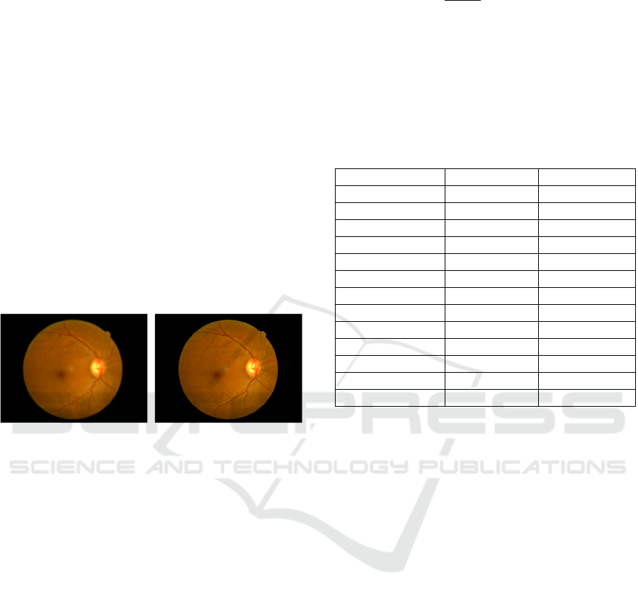
diagnosis. In Figure 1, SRGAN dramatically
increases the quality of fundus images.
Figure 1: Comparison of fundus images before and after
SRGAN processing. (Picture credit: Original)
2.1 Overview of SRGAN
The proposed SRGAN framework contains a
generator and a discriminator. Generator G
transforms a LR diabetic retinopathy image into SR
images:
𝐼
=𝐺
𝐼
(1)
In this model, discriminator D is replaced by pre-
trained VGG-19, which helps to capture subtle
textural and structural features in medical images,
meanwhile, the adversarial training framework is
formulated in a minimax method:
min
max
𝐸
~
log𝐷
𝐼
𝐸
~
log
1 𝐷𝐺
𝐼
(2)
To assist SR images in maintaining important
perceptual details, we incorporate a perceptual
content loss computed using feature maps from the
VGG-19 network:
𝐿
=
,
,
∑∑
𝜙
,
𝐼
,
,
,
𝜙
,
𝐺𝐼
(3)
where Φ
,
represents the feature map which is
extracted from the j-th convolution layer before the i-
th pooling layer in the VGG-19 model. 𝑊
,
, 𝐻
,
are
the feature map dimensions. Table 1 shows the
structure of VGG-19.
Table 1: Structure of VGG-19
La
y
e
r
Output Shape Parameters
Conv
_
2D
(224 224 64)
1792
Conv
_
2D
(224 224 64)
36928
MaxPoolin
g
_
2D
(112 112 64)
0
Conv
_
2D
(112 112 128)
73856
Conv
_
2D
(112 112 128)
147584
MaxPoolin
g
_
2D
(56 56 128)
0
Conv
_
2D
(56 56 256)
295168
Conv
_
2D
(56 56 256)
590080
Conv
_
2D
(56 56 256)
590080
…… …… ……
Flatten
(25088)
0
Dense
_
1
(4096)
16781312
Dense
_
2
(1000)
4097000
The whole generator loss is a weighted sum of the
content and adversarial losses:
𝐿
= 𝐿
𝜆𝐿
(4)
And the adversarial loss defined as:
𝐿
= log
𝐷𝐺
𝐼
(5)
where 𝜆 serves as a hyperparameter balancing the
two terms.
2.2 Key Improvements
The improvements used in this SRGAN framework
for enhancing diabetic retinopathy images are shown
here:
1. Enhanced Residual Learning: The generator G
contains numerous deep residual blocks so that it can
effectively learn the mapping from LR to HR images.
Each residual block is formulated as:
𝐹
𝑥
=𝑥𝐻
𝑥
(6)
where 𝐻
𝑥
is the residual function of the block.
Super-Resolution Image Generation for Diabetic Retinopathy Detection by SRGAN
401
2. Modified Discriminator Architecture: This
SRGAN model utilizes VGG-19 network with
additional convolutional layers, and its architecture
improves the network's capture of richer semantic
features in diabetic retinopathy images.
3. Perceptual Loss Optimization: By computing
the loss in the deep feature space of the VGG network,
the images maintain more high-frequency details and
realistic textures.
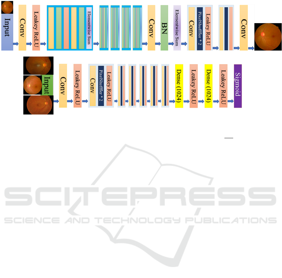
2.3 Model Structure
In Figure 2, the generator consists of multiple
components, including convolutional layers, Leaky
ReLU activations, element-wise summation, and
batch normalization. Every layer inherits the
parameter from previous parts and outputs the final
SR images.
In Figure 3, the discriminator receives various
images and processes them through a series of blocks,
which were composed of two PixelShuffler layers,
convolutional layers, and Leaky ReLU activations.
After that, the extracted features are passed to a dense
layer. Then, a sigmoid activation outputs the final
probability to determine whether the image is real or
generated.
3 EXPERIMENTS AND RESULTS
3.1 Data Preprocessing and
Augmentation
Robust data preprocessing and augmentation are
essential in model training, so each input image 𝐼 is
normalized as follows:
𝐼
=
(7)
where 𝜇 and 𝜎 denote the mean and standard
deviation of the training set, respectively.
Data augmentation is performed via a set of
transformations 𝑇, to further enhance the diversity of
the dataset and improve model robustness:
𝐼
=𝑇
𝐼
,𝐼
=𝑇
𝐼
(8)
with the augmentation set defined as:
random crop,horizontal flip,vertical flip,rotation
3.2 Training Process
3.2.1 Stage 1: Pre-training the Generator
To mitigate the mismatch between the generator and
discriminator, the generator is pre-trained for 50
epochs using only the content loss.
This stage allows 𝐺 to learn an initial mapping
from LR to HR images perceptually and meaningfully.
3.2.2 Stage 2: Adversarial Training
Following pre-training, adversarial training is
initiated. In each training iteration, the parameters of
both the generator 𝐺 and the modified VGG-19-
based discriminator are updated using gradient
descent:
𝜃 ← 𝜃 𝜂∇
𝐿 (9)
where 𝜂 is the learning rate, and the discriminator
loss is defined as:
𝐿
=𝐸
~
log𝐷
𝐼
𝐸
~
log
1 𝐷𝐺
𝐼
(10)
Figure 2: Structure of Generator (Picture credit: Original)
Figure 3: Structure of Discriminator (Picture credit: Original)
ICDSE 2025 - The International Conference on Data Science and Engineering
402

3.3 Evaluation Metrics
The performance of our SRGAN framework is
evaluated using both image quality metrics and
downstream task metrics relevant to diabetic
retinopathy analysis.
(1) Structural Similarity Index:
𝑆𝑆𝐼𝑀
𝐼
,𝐼
=
(11)
Where 𝜇 and 𝜎 , 𝜎
denote the means,
variances, and covariance of the images, and 𝐶
,
𝐶
are constants for stability.
(2) Peak Signal to Noise Ratio
𝑃𝑆𝑁𝑅 = 10 ∙ log
(12)
Where 𝑀𝐴𝑋 is the maximum pixel value.
Table 2: The metrics of the model
Metric Value
PSNR
_
dB 43.00
SSIM 0.93
Precision 0.965
Recall 0.913
F1-Score 0.937
Table 2 indicates that SRGAN performed
exceptionally well in APTOS-2019, because of the
outstanding PSNR_dB, SSIM and Precision Recall
F1-score.
4 CONCLUSION
APTOS-2019 dataset includes 3662 diabetic
retinopathy fundus images, which are used to
downsample the LR images for model training. This
method simulates the clinical image-generating
challenges. The generator was first trained for 50
epochs to prevent the model from collapsing, after
that, both G and D performed the adversarial training
with a learning rate of 0.0001 and 500 epochs.
Aiming to increase the model robustness, data
augmentation is utilized at the beginning of training,
such as random cropping, horizontal and vertical
flipping, and rotation (every image is coped with the
normalization process)
SRGAN is specially designed for generating SR
retinopathy fundus images, because of the VGG-19-
based discriminator. It greatly enhances the resolution
of images and recreates the details. From the
experimental results, SRGAN has a potential clinical
image analysis application value, especially in
segment.
Although SRGAN performs well in this case,
there are still some challenges: certain areas in other
pictures may appear blurred, and low-level detail
reconstruction; Significant computational resources
are used in training that obstacles to wider application;
This dataset has limited variability of diabetic
retinopathy images, which may limit the
generalization ability of the model.
That is the reason why future research will focus
on improving the model generalization ability and
making it easier to be trained. In the end, integrating
SRGAN will aid in early detection and intervention,
aiming to develop medical technology.
REFERENCES
Abbas, R., Gu, N., 2023. Improving deep learning-based
image super-resolution with residual learning and
perceptual loss using SRGAN model. Soft Computing,
27(21), 16041-16057.
Deng, Z., Zhang, H., Liang, X., Yang, L., Xu, S., Zhu, J., &
Xing, E. P., 2017. Structured generative adversarial
networks. In Advances in neural information
processing systems, 30.
Elanwar, R., Betke, M., 2024. Generative adversarial
networks for handwriting image generation: a review.
The Visual Computer, 1-24.
Goodfellow, I., Pouget-Abadie, J., Mirza, M., Xu, B.,
Warde-Farley, D., Ozair, S., & Bengio, Y., 2020.
Generative adversarial networks. Communications of
the ACM, 63(11), 139-144.
Gonog, L., & Zhou, Y., 2019. A review: generative
adversarial networks. In 2019 14th IEEE Conference on
Industrial Electronics and Applications (ICTEA) (pp.
505-510). IEEE.
Jiang, X., Xu, Y., Wei, P., & Zhou, Z., 2020. CT image
super resolution based on improved SRGAN. In 2020
5th International Conference on Computer and
Communication Systems (ICCCS) (pp. 363-367). IEEE.
Ledig, C., Theis, L., Huszar, F., Caballero, J., Cunningham,
A., Acosta, A., & Shi, W., 2017. Photorealistic single
image super-resolution using a generative adversarial
network. In Proceedings of the IEEE conference on
computer vision and pattern recognition (pp. 4681-
4690).
Liu, B., & Chen, J., 2021. A super resolution algorithm
based on attention mechanism and SRGAN network.
IEEE Access, 9, 139138-139145.
Sajeeda, A., & Hossain, B. M., 2022. Exploring generative
adversarial networks and adversarial training.
International Journal of Cognitive Computing in
Engineering, 3, 78-89.
Wang, K., Gou, C., Duan, Y., Lin, Y., Zheng, X., & Wang,
F. Y., 2017. Generative adversarial networks:
introduction and outlook. IEEE/CAA Journal of
Automatica Sinica, 4(4), 588-598.
Super-Resolution Image Generation for Diabetic Retinopathy Detection by SRGAN
403
