
Towards Transparent AI in Medical Imaging: Fracture Detection in
Hand Radiographs with Grad-CAM Insights
Mustafa Juzer Fatehi, Siddharath Malavalli Nagesh, Mandalam Akshit Rao, Stellin John George,
J. Angel Arul Jothi
a
and Elakkiya Rajasekar
b
Department of Computer Science, Birla Institute of Technology and Science, Pilani, Dubai Campus,
Dubai International Academic City, Dubai, U.A.E.
Keywords:
Deep Learning (DL), YOLO, Faster R-CNN, Explainable Artificial Intelligence (XAI), Grad-CAM, Fracture
Detection.
Abstract:
Timely and accurate detection of bone fractures in hand radiographs, particularly in fingers and wrists re-
mains a critical challenge in clinical diagnostics due to anatomical complexity and subtle fracture patterns.
This study presents an explainable AI framework for automatic fracture detection using a single-shot detec-
tion framework-YOLOv5 Medium (YOLOv5m) model, optimized through targeted preprocessing and inter-
pretability techniques. A dedicated preprocessing pipeline is used to enhance fracture visibility and reduce
irrelevant noise. This includes key steps like histogram equalization, Gaussian filtering, Laplacian filtering,
and intensity normalization. To foster clinical trust and transparency, we integrate Gradient-weighted Class
Activation Mapping (Grad-CAM) to visualize regions of interest influencing the model’s predictions. Trained
on a curated dataset of over 9,000 annotated X-ray images, YOLOv5m achieved outstanding performance,
with a mean Average Precision mAP@50 of 95.87% and an inference speed of 690 ms, making it suitable for
real-time diagnostic support. This work demonstrates the potential of AI-assisted systems not only to improve
fracture diagnosis but also to bridge the trust gap in clinical deployment through transparent decision-making
support.
1 INTRODUCTION
Finger and wrist fractures represent a significant por-
tion of the 178 million global bone fractures annually,
posing diagnostic challenges due to their subtle and
varied presentation in X-ray imaging Wu et al. (2021).
Undetected or misdiagnosed fractures can lead to
long-term complications, underscoring the need for
accurate and timely diagnosis. While X-ray imag-
ing remains the gold standard for fracture detection,
manual interpretation is time-intensive and prone to
variability among clinicians Valali et al. (2023). This
work aims to address these limitations by leveraging
artificial intelligence (AI) to improve fracture detec-
tion and localization, ultimately reducing diagnostic
time, easing the burden on medical staff, and improv-
ing patient outcomes.
There are various difficulties in using AI to de-
tect fractures. The scarcity of high-quality annotated
a
https://orcid.org/0000-0002-1773-8779
b
https://orcid.org/0000-0002-2257-0640
data, which is necessary for building reliable mod-
els, is one of the main challenges. Furthermore, frac-
tures can happen in a variety of places along the bone
and have a wide range of orientations, which makes
it challenging to create a universal detection model.
Overlapping structures in X-ray images add to the
complexity and can make fractures harder to see and
make detection less accurate. Moreover, different pa-
tients present unique anatomical variations, adding to
the difficulty of creating models that can generalize
across diverse populations. Addressing these chal-
lenges is crucial for developing reliable AI models
capable of improving diagnostic accuracy and consis-
tency in clinical settings.
This study explores the use of deep learning (DL)
techniques for detecting and localizing bone frac-
tures in X-ray images, with a focus on finger joint
and wrist fractures captured from diverse perspec-
tives. By leveraging a dataset of over 9,000 im-
ages, significantly larger and more comprehensive
than those used in prior studies, we address critical
Fatehi, M. J., Nagesh, S. M., Rao, M. A., George, S. J., Jothi, J. A. A. and Rajasekar, E.
Towards Transparent AI in Medical Imaging: Fracture Detection in Hand Radiographs with Grad-CAM Insights.
DOI: 10.5220/0013676200004000
Paper published under CC license (CC BY-NC-ND 4.0)
In Proceedings of the 17th International Joint Conference on Knowledge Discovery, Knowledge Engineering and Knowledge Management (IC3K 2025) - Volume 1: KDIR, pages 39-48
Proceedings Copyright © 2025 by SCITEPRESS – Science and Technology Publications, Lda.
39

limitations in existing research related to dataset di-
versity and size. We finetuned the YOLOv5 Medium
(YOLOv5m) model. It is a single stage object detec-
tion model known for its balance of speed, accuracy,
and lightweight architecture-on this dataset and ex-
tended the detection pipeline to incorporate explain-
ability via Grad-CAM, modifying the model’s final
layers to enable attention-based visualization of pre-
dicted regions.
Our evaluation examines the model’s perfor-
mance across multiple fracture types within the same
anatomical region, assessing both detection accuracy
and generalizability. By offering a robust evaluation
framework, our research contributes to the develop-
ment of more reliable, effective and transparent auto-
mated diagnostic systems, paving the way for future
advancements in fracture detection and medical imag-
ing.
This paper is organized into seven sections. Sec-
tion 1 provides an overview of the research problem
and objectives. Section 2 situates the study within ex-
isting literature. The Section 3 outlines the data used,
followed by the Methodology in Section 4 which de-
tails the proposed approach. System Requirements
and Evaluation Metrics are specified in Section 5. The
Section 6 discusses the findings, and the Conclusion
section highlights key insights and future directions.
2 RELATED WORK
In recent years, the application of machine learn-
ing (ML) and DL techniques have significantly ad-
vanced the field of automated bone fracture detec-
tion (Ahmed and Hawezi, 2023). Zhang et al.
(2021) proposed a traditional ML pipeline involving
grayscale conversion, Gaussian filtering, adaptive his-
togram equalization, Canny edge detection, and Gray-
Level Co-occurrence Matrix (GLCM)-based feature
extraction, with classification using models like Sup-
port Vector Machine (SVM) achieving up to 92% ac-
curacy. Addressing annotation ambiguity, a point-
based annotation with “Window Loss,” was intro-
duced achieving an Area Under the Receiver Operat-
ing Characteristic curve (AUROC) of 0.983 and Free-
Response Receiver Operating Characteristic (FROC)
of 89.6%, outperforming standard detectors.
Building on traditional ML, several studies
demonstrated the superior capability of DL models
in capturing complex patterns. Karanam et al. (2021)
emphasized the effectiveness of Convolutional Neural
Networks (CNN) for hierarchical feature learning, es-
pecially in large datasets. Ghosh et al. (2024) further
improved accuracy (97%) by applying anatomical
feature enhancement before feeding the images into
CNNs. Lee et al. (2020) proposed a meta-learning-
based encoder-decoder using GoogLeNet, utilizing
shared latent representation for improved classifica-
tion across modalities.
Hybrid and transfer learning strategies have also
shown significant promise. Khatik and Kadam (2022)
and Warin et al. (2023) explored the use of pretrained
models such as ResNet and Faster R-CNN, integrat-
ing transfer learning and data augmentation to en-
hance performance. Meena and Roy (2022) demon-
strated the integration of real-time DL models like
ResNet, VGGNet, and U-Net, achieving high accu-
racy for wrist and hip fractures while highlighting
challenges such as class imbalance and rare case de-
tection.
Fracture localization has become increasingly im-
portant. Ma (2021) proposed a two-stage framework
combining Faster R-CNN with a Crack-Sensitive
CNN (CrackNet) for detecting and classifying spe-
cific bone regions. Similar detection-refinement
pipelines were explored by Abbas et al. (2020) and
Su et al. (2023), reporting mAP scores around 60%.
One-shot detectors such as the YOLO fam-
ily have gained substantial traction for their speed
and efficiency. Zou and Arshad (2024) demon-
strated YOLO’s effectiveness over two-stage detec-
tors. Ju and Cai (2023) showcased YOLOv8’s per-
formance, achieving a mAP of 0.638 using multi-
scale feature fusion.Morita et al. (2024) confirmed
YOLOv8’s superiority over SSD after extended train-
ing. The YOLOv7-ATT model by Zou and Arshad
(2024), with an attention mechanism, achieved 86.2%
mAP on the FracAtlas dataset by focusing on sub-
tle fracture-specific cues. Moon et al. (2022) used
YOLOX-S for nasal bone fractures, achieving 100%
sensitivity and 69.8% precision, thereby easing diag-
nostic burden for specialists.
Beyond YOLO, other architectures have been
tested. AFFNet, as proposed by Nguyen et al. (2024),
improved upon ResNet-50 while integrating activa-
tion maps to visualize important regions. While Reti-
naNet lagged behind with approximately 76% accu-
racy, Yadav et al. (2022) introduced SFNet—using
multi-scale fusion and edge detection—to achieve
99.12% accuracy, 100% precision, and 98% recall,
outperforming U-Net, YOLOv4, and R-CNN. In ad-
dition, Beyraghi et al. (2023) explored microwave
imaging as a novel, radiation-free method for fracture
detection using S-parameter data and deep neural net-
works, achieving high classification accuracy and low
regression error.
Parallel to the advancements in detection architec-
tures, the role of XAI has grown critical in ensuring
KDIR 2025 - 17th International Conference on Knowledge Discovery and Information Retrieval
40
model transparency and trust. Borys et al. (2023) cat-
egorized saliency-based XAI methods such as Grad-
CAM, LIME, SHAP, and Occlusion into perturbation-
based and backpropagation-based techniques, outlin-
ing their respective strengths and limitations in medi-
cal image analysis. They also addressed the practical
challenges in deploying XAI, emphasizing variabil-
ity in heatmap-based visual attributions across meth-
ods. Their study called for a standardized, multi-
dimensional evaluation framework to assess XAI reli-
ability and alignment with clinical decision-making,
reinforcing the synergy between accurate detection
and explainable outputs. Volkov and Averkin (2023)
further examined XAI’s domain-specific applications,
noting Grad-CAM’s success in radiology, derma-
tology, and histopathology, particularly in detecting
COVID-19 pneumonia, brain tumors, and skin le-
sions. They advocated for clinician-centered design
and proposed integrating XAI with fuzzy logic to en-
hance diagnostic support in real-world clinical work-
flows.
3 DATASET DESCRIPTION
The Bone Fracture Detection Dataset (Phanan, 2024)
used in this work contains about 9,585 X-ray images
of finger and wrist fractures, encompassing diverse
orientations and imaging conditions, including top-
down and lateral views. The dataset is split into train-
ing (70%), validation (20%), and testing (10%) sub-
sets.
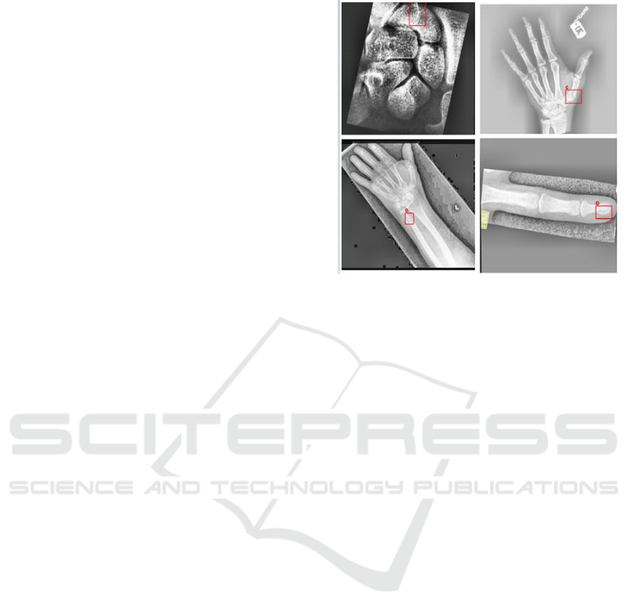
Images of fractured bones are annotated using
bounding boxes to highlight the fracture areas as ob-
served in Figure 1. These annotations follow the
YOLO format, which includes the class label and
normalized coordinates of the bounding boxes (cen-
ter, width, and height) relative to the image size.
The images, provided as 640 × 640 pixel JPEGs,
are readily compatible with standard computer vi-
sion frameworks and preprocessing pipelines. The
dataset also includes images featuring multiple frac-
tures within the same frame, enabling the detection
of cases with more than one fracture simultaneously.
Focused specifically on finger and wrist fractures, this
dataset offers a rich collection of clinically relevant
images that enable rigorous evaluation and compari-
son of automated fracture detection approaches.
4 METHODOLOGY
The methodology for bone fracture detection in this
paper is structured into five key stages: data collec-
Figure 1: X ray images with ground truth boxes.
tion, preprocessing, model implementation, training,
evaluation, and testing. These stages are designed to
comprehensively address the challenges of accurate
fracture detection, from processing the dataset to as-
sessing the performance of fine-tuned models.
The core of this study centers on the implementa-
tion and optimization of the YOLOv5m model for ac-
curate detection and localization of finger bone frac-
tures. Leveraging pretrained Common Objects in
Context (COCO) weights, the model was fine-tuned
on a specialized dataset of annotated hand X-rays to
adapt to the nuanced patterns of bone injuries. The
YOLOv5m architecture was selected for its balance
of speed, accuracy, and efficiency, making it ideal for
real-time clinical integration. To contextualize its per-
formance, a comparative evaluation with other detec-
tion frameworks including single-stage variants like
YOLOv8 and YOLOv11, as well as the two-stage
Faster R-CNN was conducted. These comparisons,
while secondary, provided insights into the trade-offs
between speed, precision, and model complexity. An
overview of the end-to-end workflow, including data
preparation, model training, and evaluation, is illus-
trated in Figure 2.
The following subsections provide a detailed dis-
cussion of each stage, outlining the specific tech-
niques and strategies employed.
4.1 Data Preprocessing
The images were preprocessed to transform them into
a format optimized for object detection models, mak-
ing it easier to identify fractures accurately. The pre-
processing pipeline focused on enhancing subtle fea-
tures, such as minute fractures and overlapping struc-
Towards Transparent AI in Medical Imaging: Fracture Detection in Hand Radiographs with Grad-CAM Insights
41
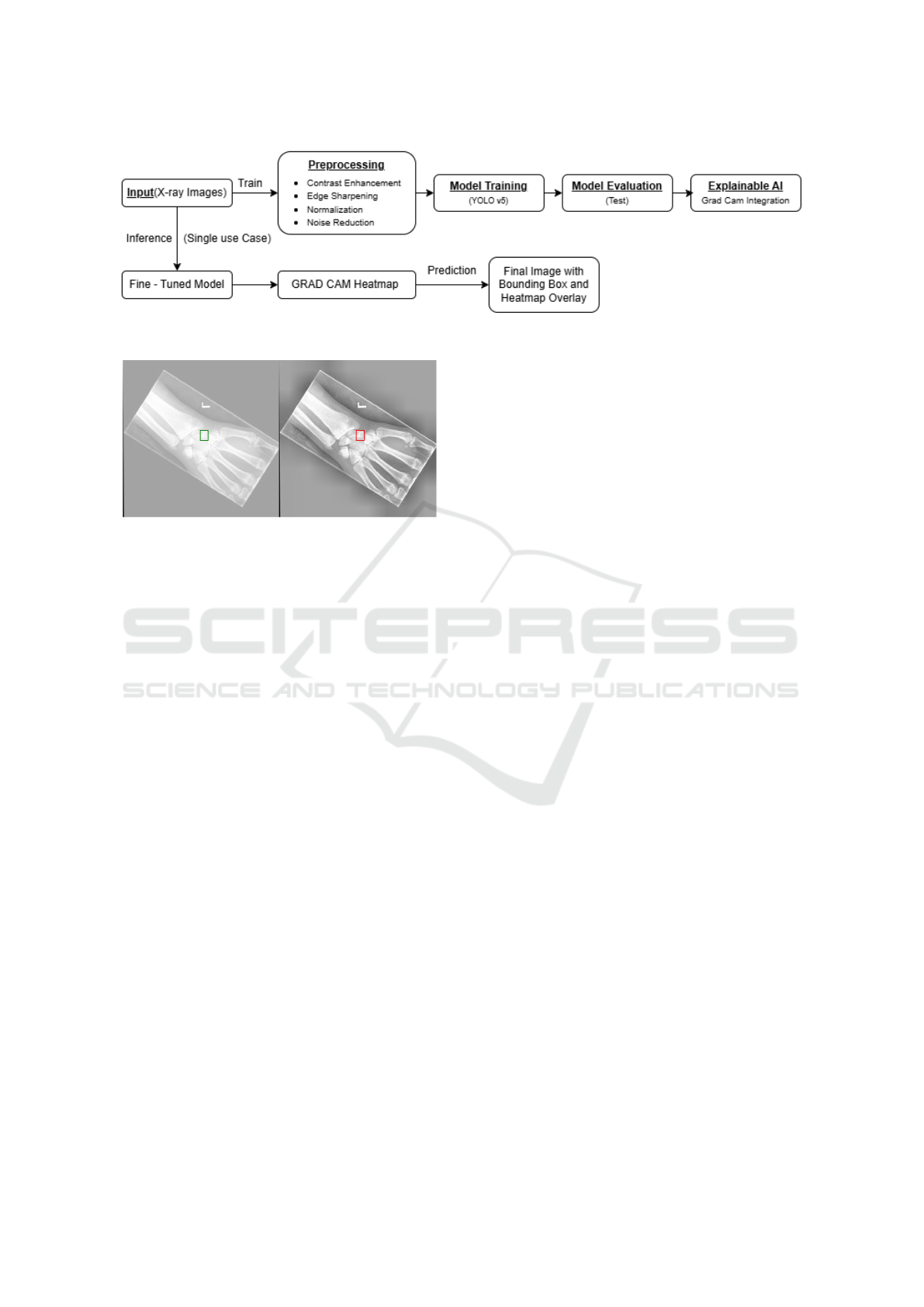
Figure 2: Flowchart Illustrating the Training Process and Single-Image Inference Outcomes
Figure 3: Comparison of Before (left) and After (right) pre-
processing.
tures, which often pose challenges for accurate detec-
tion.
The preprocessing steps included Contrast En-
hancement, Noise Reduction, Edge Sharpening, and
Image Normalization. Contrast enhancement was
achieved using histogram equalization, which im-
proved the visibility of bone structures by increasing
the dynamic range of pixel intensity values. This tech-
nique enhanced the contrast-to-noise ratio (CNR), en-
abling clearer differentiation between fracture lines
and surrounding bone. Noise reduction was per-
formed using a Gaussian filter to suppress high-
frequency noise introduced by imaging equipment or
environmental factors. This step preserved essential
spatial details critical for identifying fine bone struc-
tures, such as trabecular patterns, while improving
overall image clarity. To address inherent blurriness
caused by the finite size of X-ray focal spots, edge
sharpening was implemented using a Laplacian fil-
ter. This step enhanced bone boundaries and fracture
lines, enabling the detection model to focus on criti-
cal features for accurate identification. Finally, image
normalization was applied to standardize pixel inten-
sity values to a range of [0, 1], ensuring consistent
input to the detection models and reducing variability
across images.
As observed in Figure 3, focusing on preprocess-
ing techniques tailored to the requirements of bone
fracture detection ensured that the input data was op-
timized for object detection, providing a solid foun-
dation for training and evaluating advanced models.
4.2 Object Detection Model and Its
Applicability
To effectively detect fractures in hand X-ray images,
this study employs YOLOv5m. Building upon a ro-
bust preprocessing pipeline that enhances contrast, re-
duces noise, and sharpens critical edges, YOLOv5m
[28] was selected due to its proven ability to perform
well in tasks involving small and irregularly shaped
objects: characteristics common in bone fractures.
Interestingly, while a range of models including
other YOLO variants and two-stage detectors such
as Faster R-CNN were briefly explored, YOLOv5m
consistently outperformed them in both mean Aver-
age Precision (mAP) and inference speed. This un-
expected lead in performance is likely attributed to
its efficient use of anchor-based detection, optimized
feature aggregation through the Spatial Pyramid Pool-
ing (SPP) block, and its strong inductive bias toward
learning small object patterns, which aligns well with
the fracture detection task.
4.2.1 YOLOv5 Medium: Architecture and
Suitability
YOLOv5m utilizes an anchor-based detection mech-
anism and a Spatial Pyramid Pooling (SPP) module,
which allows the model to aggregate spatial informa-
tion across multiple scales. This design is particularly
effective in detecting fractures that vary in size, shape,
and intensity. The model’s backbone is optimized for
extracting deep spatial features, while the head simul-
taneously predicts bounding boxes and class proba-
bilities, enabling fast and reliable inference.
The anchor boxes were fine-tuned during training
to adapt to the dimensions specific to fractures in hand
radiographs. YOLOv5m’s modular design and effi-
cient convolutional layers allowed it to retain struc-
tural nuances in the X-ray images, improving its abil-
ity to generalize across varying fracture presentations.
KDIR 2025 - 17th International Conference on Knowledge Discovery and Information Retrieval
42
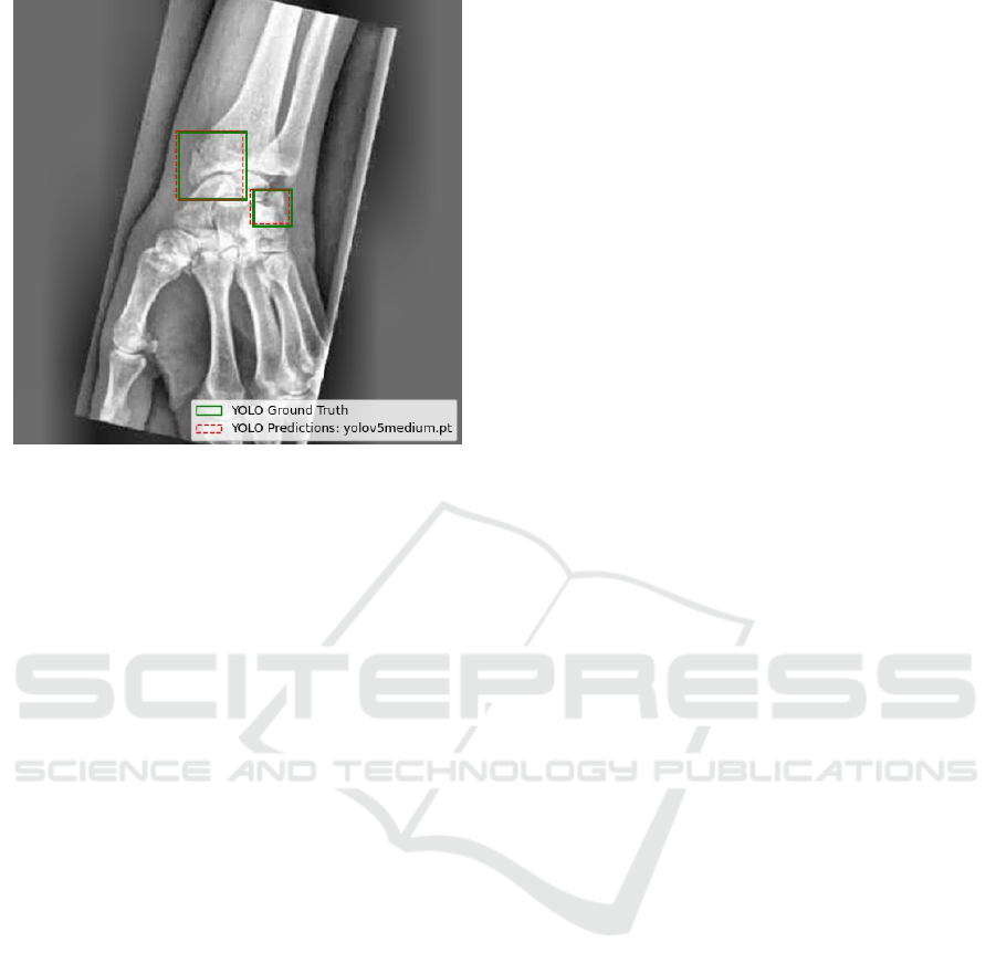
Figure 4: Yolov5m prediction vs Ground Truth.
4.3 Training and Validation
YOLOv5m was trained for 100 epochs using pre-
trained COCO weights as initialization. This trans-
fer learning approach allowed the model to converge
faster while benefiting from prior knowledge of gen-
eral object features. Training employed a batch size of
16 and a starting learning rate of 0.01 with momentum
set at 0.937, adhering to YOLOv5’s recommended de-
faults to ensure training stability and reproducibility.
To improve the model’s robustness and gener-
alization capability, several data augmentation tech-
niques were employed. Horizontal flipping simulated
anatomical variations in hand orientation, while Ran-
dAugment introduced random variations in brightness
and contrast to mimic real-world X-ray acquisition
inconsistencies. Mosaic augmentation, a technique
where four images are combined into one was used
during the initial 10 epochs to diversify object context
and improve detection under cluttered scenarios. This
augmentation was phased out in later epochs to allow
for more focused learning on fracture-specific fea-
tures.Validation was conducted after each epoch using
a holdout validation set, evaluating mAP, classifica-
tion accuracy, and localization precision. YOLOv5m
demonstrated consistent performance gains with each
augmentation step, converging steadily toward op-
timal detection capability. Its final performance
reached a mAP@50 of 95.87% with an inference
time of 690 ms per image, proving its suitability for
real-time clinical applications. By tailoring the train-
ing process specifically for YOLOv5m and integrat-
ing domain-specific preprocessing and augmentation
techniques, the model was optimized to detect even
the most subtle and overlapping fractures with high
confidence and speed.
4.4 Model Testing
The testing phase aimed to evaluate the generalizabil-
ity and performance of the trained model on unseen
data. During testing, the model generated predic-
tions in the form of bounding boxes and confidence
scores. These outputs were evaluated against their
corresponding ground truth annotations using stan-
dard object detection and localization metrics, which
are described in detail in Section 5.
Sample outputs were also manually visualized
to verify the correctness of the predicted bounding
boxes against the ground truth annotations. Figure 4
presents example outputs, demonstrating the overlap
between predicted and actual fracture regions, provid-
ing a qualitative check of the model’s performance.
4.5 Visualizing Model Decisions Using
Grad-CAM
To enhance the interpretability of the YOLOv5-based
fracture detection pipeline and foster clinician trust,
Grad-CAM was integrated into the inference pro-
cess of the model. Grad-CAM generates class-
discriminative heatmaps that visually highlight the re-
gions in an image most influential to a model’s deci-
sion, offering intuitive insights into its reasoning.
Unlike explainability methods such as LIME,
SHAP, or Integrated Gradients which are primarily
designed for structured data or classification tasks,
Grad-CAM is well-suited for spatial vision tasks like
object detection. Its ability to localize important fea-
tures makes it especially valuable in medical imaging,
where understanding the spatial rationale behind pre-
dictions is crucial.
To apply Grad-CAM within the YOLOv5 archi-
tecture, the inference pipeline was modified to ex-
tract gradient information from the final convolutional
block before the detection head. Gradients were com-
puted with respect to the confidence scores of high-
confidence bounding boxes post non-max suppres-
sion. The resulting heatmaps were normalized and
superimposed on the original X-ray images, providing
visual explanations for the predictions of the model.
This approach allowed to confirm that the model
consistently focused on clinically relevant features
such as cortical disruptions, fracture lines, or trabec-
ular misalignments while avoiding irrelevant regions
like overlapping soft tissues or background noise.
These visualizations not only validated true positives
Towards Transparent AI in Medical Imaging: Fracture Detection in Hand Radiographs with Grad-CAM Insights
43
but also provided insight into false negatives, particu-
larly in subtle or ambiguous cases.
By bridging the gap between AI predictions and
clinical reasoning, Grad-CAM significantly enhanced
the transparency of the system. It empowered clin-
icians to audit and cross-validate the model’s deci-
sions, promoting trust and supporting the safe inte-
gration of AI into real-world diagnostic workflows.
Overall, this integration reaffirmed YOLOv5’s robust-
ness for high-stakes medical imaging and demon-
strated Grad-CAM’s potential as a valuable diagnostic
companion tool.
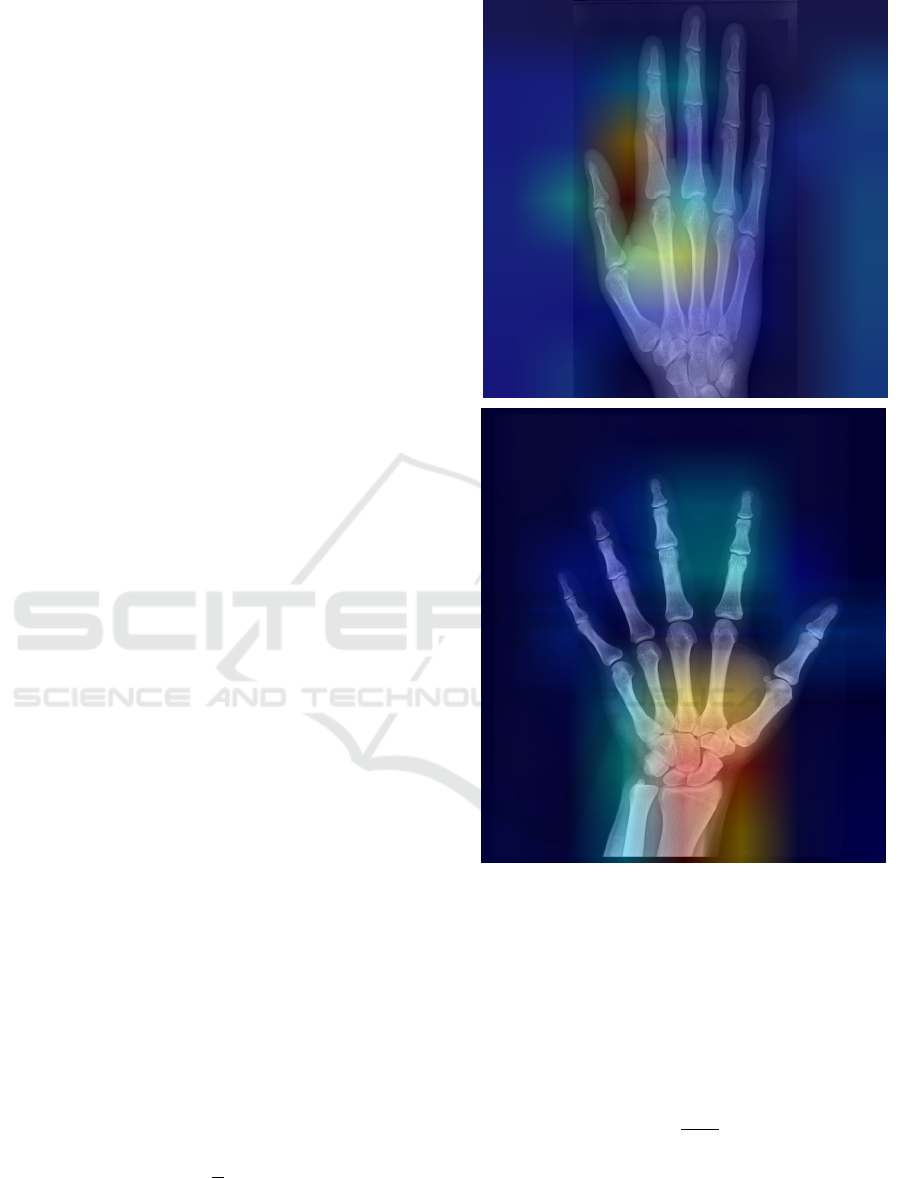
Figure 5 presents the XAI results, showcasing the
key image regions that influenced the model’s diag-
nostic decisions.
5 IMPLEMENTATION AND
EVALUATION METRICS
The training of all models was performed over 100
epochs using an NVIDIA RTX A4500 GPU. The soft-
ware environment comprised of a Windows operating
system, with code development and execution carried
out in Visual Studio Code. Key libraries, including
PyTorch and Torchvision, were utilized for model im-
plementation and dataset processing. CUDA was em-
ployed to accelerate computations on the GPU, while
Matplotlib was used for visualizing results and gen-
erating performance graphs, facilitating clear and in-
sightful analysis.
For inference evaluation, the trained models were
tested on an Intel Core i5 processor. Inference time
was used as an additional evaluation metric to assess
the real-time applicability of the models. This setup
provides a reliable and efficient framework for con-
ducting experiments, by ensuring a stable training and
evaluation environment.
The performance of all models was evaluated us-
ing standard object detection metrics, including mean
Average Precision (mAP) and Intersection over Union
(IoU). In bone fracture detection, mAP@50 measures
how well the model identifies fractures when there is
at least 50% IoU between the predicted and ground
truth bounding boxes. This metric focuses on whether
the model can reliably highlight fracture regions, even
with some localization error. A high mAP@50 means
the model is good at finding most fractures. It is given
by (1)
mAP@50 =
1
C
C
∑
c=1
AP
c
(50) (1)
whHere, C is the number of object classes, c de-
note a specific object class, and AP
c
(50) represent the
Figure 5: Grad-CAM highlights regions influencing pre-
dictions, with red areas showing key focus around the
metacarpal (top) and fracture site near the thumb (bottom).
Average Precision for the class c at an IoU threshold
of 50%.
mAP@50:95 provides a stricter and more compre-
hensive evaluation, as it considers detection perfor-
mance across multiple IoU thresholds (from 50% to
95% overlap). This is calculated by (2)
mAP@50 : 95 =
1
T · C
T
∑
t=1
C
∑
c=1
AP
c
(t) (2)
where T denote the number of IoU thresholds (e.g.,
[0.50, 0.55, ..., 0.95] in steps of 0.05), and AP
c
(t) rep-
resents the Average Precision for the class c at the IoU
threshold t. A higher mAP@50:95 indicates that the
KDIR 2025 - 17th International Conference on Knowledge Discovery and Information Retrieval
44
model not only identifies the fractures but also accu-
rately localizes their boundaries with minimal error.
IoU measures the accuracy of the bounding box
localization. It evaluates how much of the predicted
bounding box overlaps with the actual fracture area
and is given by (3). A higher IoU score means the
model is capturing the fracture’s shape and size more
accurately. This is vital for ensuring that subtle or
small fractures are not missed or mislocalized. The
IoU accuracy in this work has been computed at 50%
intersection minimum threshold to maintain object
detection quality while increasing tolerance for minor
localization errors.
IoU =
Area o f Overlap
Area o f U nion
(3)
The number of parameters across the models were
analyzed to assess the computational complexity and
efficiency. This evaluation provides insights into the
trade-offs between model size and performance. By
comparing parameter counts, it is easy to determine
which models offer a balance between accuracy and
resource requirements.
6 RESULTS AND DISCUSSION
The model testing results as seen in Table 1 pro-
vide important insights into the performance of vari-
ous object detection models for bone fracture detec-
tion. Among all the models, YOLOv5m emerged
as the best performer, achieving the highest mAP50
(95.87%), mAP50:95 (61.70%), and IOU50 (78.12%)
while maintaining a reasonable inference speed of
690 ms. This demonstrates that YOLOv5m effec-
tively balances accuracy, computational efficiency,
and generalization, making it the most suitable model
for this application. Its performance suggests that
its architecture with spatial pyramid pooling strategy
is well-aligned with the dataset’s characteristics. As
a result of multi-scale feature representation, it en-
ables the model to capture both fine-grained details
and broader contextual information, resulting in pre-
cise detection and localization of fractures without ex-
cessive computational overhead.
A clear trend also observed across the medium-
sized models (YOLOv5m, YOLOv8 Medium
(YOLOv8m), and YOLOv11 Medium (YOLOv11m))
is their consistent superiority in accuracy compared
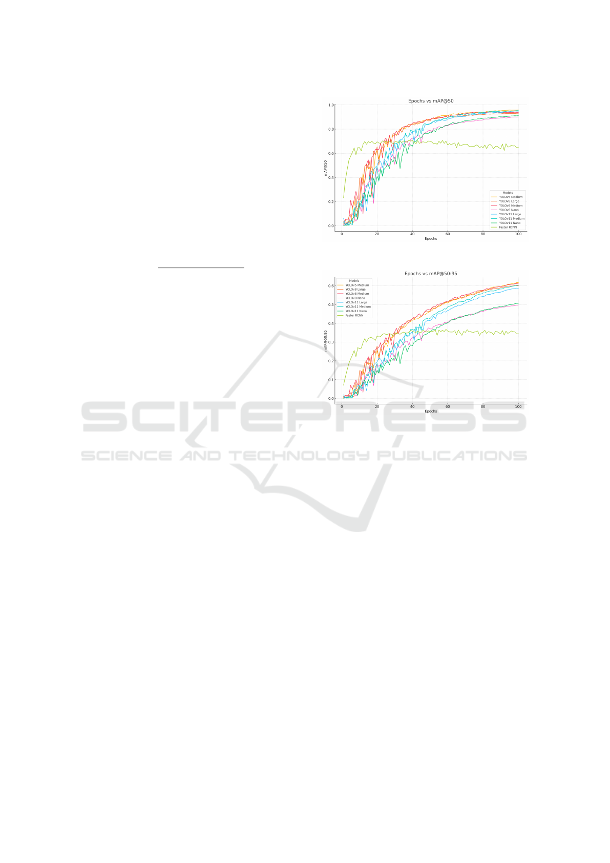
to both their smaller and larger counterparts. The
performance trend across epochs is visualized in
Figure 6, which shows how mAP@50 evolves during
training. Figure 7 further illustrates the trend by
comparing model performance (mAP@50:95) at dif-
ferent epochs. The results reinforces that YOLOv5m
Figure 6: Epochs vs mAP@50 for all models.
Figure 7: Epochs vs mAP@50:95 for all models.
not only converges faster, but also achieves the best
trade-off between accuracy and speed. Medium-sized
models have enough parameters to capture complex
features in the dataset without the risk of overfitting,
which is often observed in larger models. On the
other hand, smaller models, such as YOLOv8 Nano
(YOLOv8n) and YOLOv11 Nano (YOLOv11n),
excel in inference speed (166 ms and 180 ms, respec-
tively) but compromise significantly on accuracy,
particularly at higher IoU thresholds. This makes
nano models well-suited for applications reliant on
central server processing, where speed is prioritized,
with a modest tradeoff in precision and accuracy.
Interestingly, the larger models, including
YOLOv8 Large and YOLOv11 Large, underperform
compared to the medium models. Their lower
mAP@50:95 values indicate that these models may
suffer from overfitting due to their higher parameter
counts (43.7M and 25.34M, respectively). Larger
models typically require more extensive training data
and longer training times to generalize effectively,
which might not have been sufficiently addressed
in this study. Moreover, their higher inference
times (1,451 ms and 998 ms) further reduce their
practicality for time-sensitive applications.
Towards Transparent AI in Medical Imaging: Fracture Detection in Hand Radiographs with Grad-CAM Insights
45
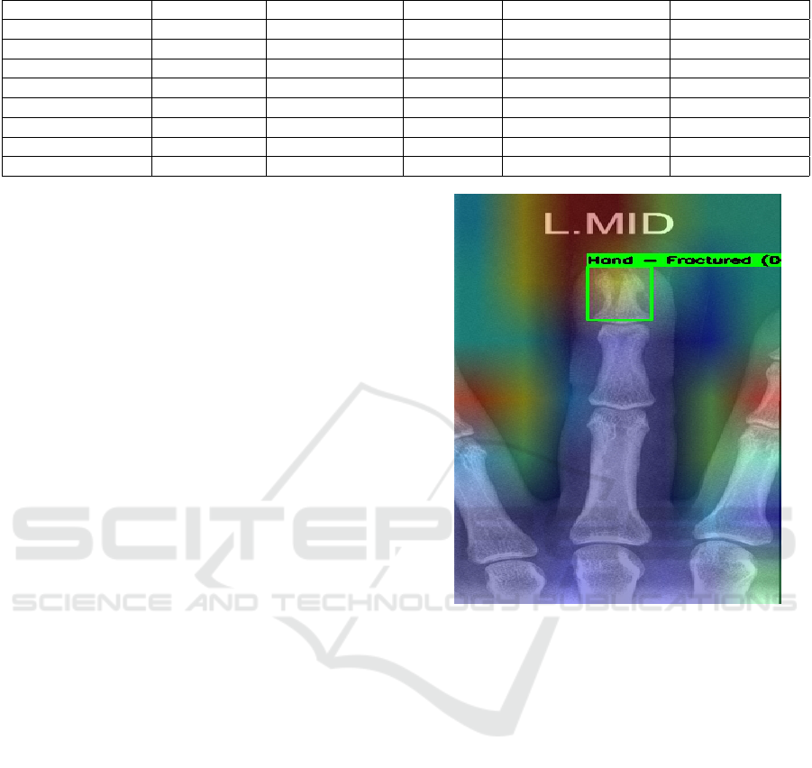
Table 1: Model Performance Comparison.
Model mAP@50 (%) mAP@50:95 (%) IOU50 (%) Inference Speed (ms) Num Parameters
YOLOv5 Medium 95.87 61.70 78.12 690 ms 21.2M
YOLOv8 Nano 90.07 49.75 67.87 166 ms 3.2M
YOLOv8 Medium 93.37 61.35 77.06 712 ms 25.9M
YOLOv8 Large 92.76 60.16 76.51 1,451 ms 43.7M
YOLOv11 Nano 91.18 50.66 68.59 180 ms 2.62M
YOLOv11 Medium 95.40 60.39 76.51 768 ms 20.09M
YOLOv11 Large 94.91 58.73 75.54 998 ms 25.34M
Faster R-CNN 70.32 36.17 17.51 3,151 ms 41.2M
Faster R-CNN, despite being a well-known two-
stage object detection model, performs poorly across
all metrics, with an mAP@50 of 70.32% and a par-
ticularly low IOU50 of 17.51%. Its inference time
of 3,151 ms is significantly slower than all YOLO
models, highlighting its computational inefficiency
for this task. The architecture of Faster R-CNN likely
struggles to adapt to the dataset’s requirements, as it
relies on generating region proposals in the first stage,
which can be less effective for subtle or small fea-
tures like bone fractures. Additionally, its large pa-
rameter count (41.2M) increases the risk of overfit-
ting, especially if the training data is not diverse or
large enough. The relationship between mAP values
and the number of epochs is illustrated in Figures 6
and 7, offering further insights into the learning pat-
terns and supporting similar conclusions.
In summary, the results emphasize the importance
of selecting a model that aligns with the specific
requirements of the application. While YOLOv5m
proves to be the most effective for bone fracture detec-
tion due to its balance of accuracy and speed, smaller
models like YOLOv8n offer exceptional speed at the
cost of precision, and larger models require more ex-
tensive optimization to perform well. Faster R-CNN,
meanwhile, demonstrates significant limitations for
this specific task, underlining the need for efficient,
single-stage architectures like YOLO when dealing
with datasets of this nature.
Furthermore, the incorporation of XAI through
Grad-CAM significantly enhanced the transparency
of our fracture detection pipeline by visually high-
lighting regions that influenced model predictions.
Figure 8 presents the combined output of YOLOv5m
predictions and Grad-CAM visualizations, clearly
demonstrating that the highlighted regions align well
with the annotated fracture areas, thereby validating
the model’s interpretability and attention to clinically
relevant features. Grad-CAM was applied to the fi-
nal convolutional layers of the YOLO-based mod-
els, enabling the identification of class-discriminative
regions that overlapped meaningfully with predicted
bounding boxes. This helped not only in de-
tecting fractures but also in localizing them accu-
Figure 8: Combined output of YOLOv5m and Grad-CAM
visualizations.
rately by highlighting potential cracks or fractures.
To quantitatively assess the alignment between the
YOLO-generated bounding boxes and the Grad-CAM
heatmaps, we employed the Pointing Game Accuracy
metric. This metric evaluates whether the maximum
activation point from the Grad-CAM heatmap falls
within the ground truth bounding box; a successful
hit indicates agreement between the model’s attention
and the annotated region. Our results showed a high
pointing game accuracy above 85 percent, demon-
strating that the regions the model focused on for
decision-making were clinically relevant. As illus-
trated in Figure 9, the overlap between the Grad-CAM
heatmaps and the predicted bounding boxes is clearly
visible and aligns well with the actual fracture sites.
We further verified this behavior across multiple im-
ages, consistently observing similar localization qual-
ity, which confirms the robustness and reliability of
our XAI integration. This synergy between detection
and interpretability not only validates the model’s per-
formance but also reinforces its suitability for real-
KDIR 2025 - 17th International Conference on Knowledge Discovery and Information Retrieval
46
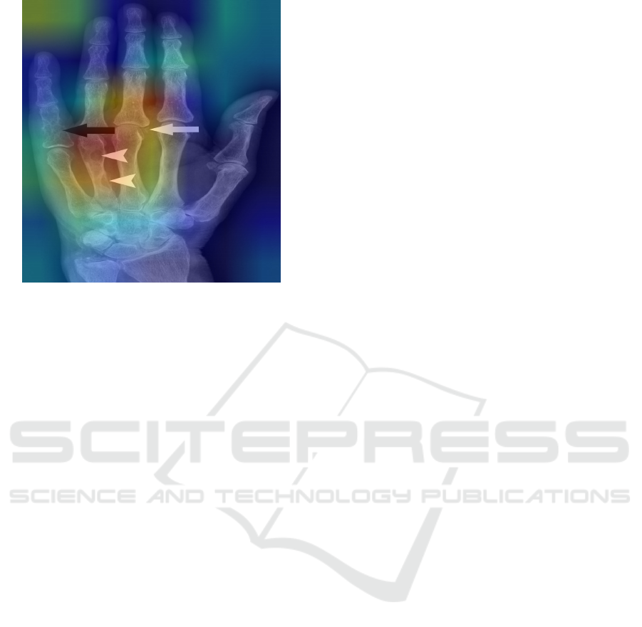
Figure 9: Grad-CAM visualization doesn’t align with the
micro fractures.
world clinical deployment.
In some instances, the Grad-CAM heatmaps did
not align with the YOLO bounding boxes. Upon in-
vestigation, two primary factors were identified. First,
many of the fractures were microfractures, which the
model was unable to detect. Second, shadows in the
X-rays or overlapping bone structures obscured the
fracture sites, preventing the model from perform-
ing effectively. This finding calls for future work to
develop enhanced imaging preprocessing techniques
and more robust model architectures that can reliably
detect microfractures and compensate for shadows or
overlapping anatomical features.
7 CONCLUSION
This study evaluates object detection models for de-
tecting finger fractures in X-ray images. By pre-
processing images to enhance anatomical details and
training several state-of-the-art models, we assessed
their performance in fracture detection and localiza-
tion.
Single-stage detectors, especially those using spa-
tial pyramid pooling for feature aggregation, con-
sistently outperformed two-stage models. Notably,
YOLOv5 surpassed newer models like YOLOv8 and
YOLOv11, indicating that its architecture may be bet-
ter suited for the specific features present in finger
fractures. Lightweight models like YOLO-Nano also
performed well despite having fewer parameters, sug-
gesting that smaller models can be effective when ap-
plied to narrowly defined tasks.
In contrast, Faster R-CNN, typically strong in
general object detection, underperformed in this task.
Its lower generalizability in this context reinforces
the need for architecture-specific tuning when dealing
with medical imagery. Our YOLOv5 based approach
achieved a detection accuracy of 95.8%, slightly
higher than the 95.1% reported by RoboFlow’s frac-
ture model on comparable datasets. These results are
significantly higher than the sub 70% accuracy com-
monly observed in broader fracture datasets, under-
scoring the benefits of task-specific optimization.
To enhance interpretability, we integrated Grad-
CAM with the YOLOv5 model, achieving a pointing
game accuracy of over 85%. The resulting heatmaps
reliably highlighted fracture regions, providing vi-
sual insight into model decisions and improving trans-
parency, an essential factor in medical AI applica-
tions.
This work demonstrates that carefully tuned ob-
ject detection models, particularly single-stage detec-
tors with spatial pooling mechanisms, can effectively
handle specialized medical tasks. It also highlights
the role of lightweight models and explainability tools
in building clinically relevant AI systems.
For future work, we propose transitioning from
detection to semantic segmentation of fractures. This
would allow pixel-level mapping of fracture morphol-
ogy, offering more detailed characterization, which is
critical for surgical planning and outcome prediction.
Integration of these models into clinical decision sup-
port systems could further streamline workflows and
enhance diagnostic precision.
REFERENCES
Abbas, W. et al. (2020). Lower leg bone fracture detection
and classification using faster rcnn for x-rays images.
IEEE Xplore.
Ahmed and Hawezi (2023). Detection of bone fracture
based on machine learning techniques. Measurement
Sensors, 27:100723.
Beyraghi, S. et al. (2023). Microwave bone fracture diag-
nosis using deep neural network. Scientific Reports,
13(1).
Borys, K. et al. (2023). Explainable ai in medical imag-
ing: An overview for clinical practitioners – saliency-
based xai approaches. European Journal of Radiol-
ogy, 162:110787.
Ghosh, S. et al. (2024). Automated bone fracture detection
in x-ray imaging to improve orthopaedic diagnostics
in healthcare. Procedia Computer Science, 233:832–
840.
Ju, R.-Y. and Cai, W. (2023). Fracture detection in pe-
diatric wrist trauma x-ray images using yolov8 algo-
rithm. Scientific Reports, 13(1):20077.
Towards Transparent AI in Medical Imaging: Fracture Detection in Hand Radiographs with Grad-CAM Insights
47

Karanam, S. R. et al. (2021). A systematic review on ap-
proach and analysis of bone fracture classification.
Materials Today: Proceedings.
Khatik, N. I. and Kadam, N. S. (2022). A systematic review
of bone fracture detection models using convolutional
neural network approach. Journal of Pharmaceutical
Negative Results, pages 153–158.
Lee, C. et al. (2020). Classification of femur fracture in
pelvic x-ray images using meta-learned deep neural
network. Scientific Reports, 10(1).
Ma, Y. (2021). Bone fracture detection through the two-
stage system of crack-sensitive convolutional neu-
ral network. Informatics in Medicine Unlocked,
22:100452.
Meena, T. and Roy, S. (2022). Bone fracture detection using
deep supervised learning from radiological images: A
paradigm shift. Diagnostics, 12(10):2420.
Moon, G. et al. (2022). Computer aided facial bone fracture
diagnosis (ca-fbfd) system based on object detection
model. IEEE Access, 10:79061–79070.
Morita, D. et al. (2024). Automatic detection of midfacial
fractures in facial bone ct images using deep learning-
based object detection models. Journal of Stomatol-
ogy, Oral and Maxillofacial Surgery/Journal of Stom-
atology Oral & Maxillofacial Surgery, pages 101914–
101914.
Nguyen, H. H. et al. (2024). Affnet - a deep convolutional
neural network for the detection of atypical femur
fractures from anteriorposterior radiographs. Bone,
187:117215.
Phanan (2024). bonefracturedetection v1 dataset. Open
Source Dataset, Roboflow Universe. Retrieved from
https://universe.roboflow.com/phanan/bonefracture
detection v1.
Su, Z. et al. (2023). Skeletal fracture detection with
deep learning: A comprehensive review. Diagnostics,
13(20):3245.
Valali et al. (2023). Bone fracture detection and classifi-
cation using deep learning models on x-ray images.
Diagnostics, 15(3):271.
Volkov, E. N. and Averkin, A. N. (2023). Explainable arti-
ficial intelligence in medical image analysis: State of
the art and prospects. In Proc. 2023 XXVI Int. Conf. on
Soft Computing and Measurements (SCM), Dubna,
Russia, pages 133–137.
Warin, K., Limprasert, W., Suebnukarn, S., Paipongna,
T., Jantana, P., and Vicharueang, S. (2023). Max-
illofacial fracture detection and classification in com-
puted tomography images using convolutional neural
network-based models. Scientific Reports, 13(1).
Wu, A. et al. (2021). Global, regional, and national bur-
den of bone fractures in 204 countries and territo-
ries, 1990–2019: a systematic analysis from the global
burden of disease study 2019. The Lancet Healthy
Longevity, 2(9):e580–e592.
Yadav, D. P., Sharma, A., Athithan, S., Bhola, A., Sharma,
B., and Dhaou, I. B. (2022). Hybrid sfnet model for
bone fracture detection and classification using ml/dl.
Sensors, 22(15):5823.
Zhang, X. et al. (2021). Window loss for bone fracture de-
tection and localization in x-ray images with point-
based annotation. In Proceedings of the AAAI Confer-
ence on Artificial Intelligence, volume 35, pages 724–
732.
Zou, J. and Arshad, M. R. (2024). Detection of whole body
bone fractures based on improved yolov7. Biomedical
Signal Processing and Control, 91:105995–105995.
KDIR 2025 - 17th International Conference on Knowledge Discovery and Information Retrieval
48
