
A Premature Recognition of Diabetic Retinopathy Using Deep
Learning and Grad Cam Technology
Abirami T, Adhishvar M, Dharani K and Darshana P R
Department of Electronics and Communication Engineering, Kongu Engineering College (Autonomous) Erode, India
Keywords: Diabetic Retinopathy, EfficientNetB2, Retinopathy, Transfer Learning, Retinal Images.
Abstract: This paper introduces a Flask-based web application for detecting and classifying diabetic retinopathy (DR),
a severe complication of diabetes mellitus, using retinal fundus images. The application integrates the
EfficientNetB2 neural network to categorize DR into five stages: No Diabetic Retinopathy (No DR), Mild,
Moderate, Severe, and Proliferative Diabetic Retinopathy, with an additional class to identify low-quality
images, preventing misclassification and ensuring reliable results. It employs preprocessing techniques such
as image scaling, data cleaning, and augmentation, along with class balancing strategies and hyperparameter
tuning to optimize model performance. The user-friendly Flask interface enables healthcare providers to
upload retinal images of both eyes and receive real-time diagnostic predictions, facilitating early diagnosis
and intervention. By improving diagnostic accuracy and accessibility, this system represents a crucial step
toward integrating artificial intelligence into practical medical applications to address a significant global
health challenge.
1 INTRODUCTION
Diabetic retinopathy is a frequent and significant
consequence of diabetes mellitus that affects the
retina and may lead to visual impairment or blindness
if not addressed. It is generally diagnosed by the
analysis of retinal fundus pictures, which allows
clinicians to track the disease's development.
However, the quality of these photographs is often
impaired by variables
such as poor lighting, low contrast, and noise,
making precise diagnosis difficult. In recent years,
deep learning methods, notably convolutional neural
networks (CNNs), have shown great promise in
automating the identification and categorization of
diabetic retinopathy, allowing healthcare providers to
make prompt treatments (Sharma, et al. , 2022). The
goal of this project is to create a web-based
application that uses the EfficientNetB2 neural
network architecture inside a Flask framework to
categorize diabetic retinopathy into five categories, as
well as to detect low-quality photos that may impair
the analysis's accuracy (Rajput, et al. , 2021). The
technology is intended to help physicians and
researchers diagnose and treat diabetic retinopathy
earlier by providing an accessible platform for picture
categorization, resulting in improved patient
outcomes.
1.1 Diabetic Retinopathy
Diabetic Retinopathy is a public health problem
worldwide. Diabetic retinopathy is the world's fifth
leading cause of visual impairment, and hence the
fourth leading cause of blindness(Agarwal, and,
Kumar, 2022). Cooperation between individuals in
charge of diabetes management and persons afflicted
by diabetic retinopathy is the most crucial function of
health systems in treating diabetes and preventing
irreversible blindness from it. Because each stage has
unique qualities and properties, clinicians may ignore
some of them, resulting in an incorrect diagnosis. As
a consequence, the concept of creating an automated
solution for DR detection emerges. More than half of
the most recent occurrences of this illness may have
been prevented with adequate and prompt treatment
and eye care.
1.2 EfficientNetB2 Model
EfficientNet-B2 is a profound learning show planned
for picture acknowledgment, adjusting exactness and
effectiveness. It employments compound scaling to
T, A., M, A., K, D. and P R, D.
A Premature Recognition of Diabetic Retinopathy Using Deep Learning and Grad Cam Technology.
DOI: 10.5220/0013633200004664
Paper published under CC license (CC BY-NC-ND 4.0)
In Proceedings of the 3rd International Conference on Futuristic Technology (INCOFT 2025) - Volume 3, pages 603-610
ISBN: 978-989-758-763-4
Proceedings Copyright © 2025 by SCITEPRESS – Science and Technology Publications, Lda.
603
optimize profundity, width, and determination, with
an input measure of 260x260 pixels (Khan, et al. ,
2022). Compared to littler EfficientNet models, it
offers higher precision (~79.8% Top-1 on ImageNet)
whereas remaining computationally effective (~9.2M
parameters). It is perfect for errands requiring both
accuracy and speed, such as therapeutic imaging and
real-time applications.
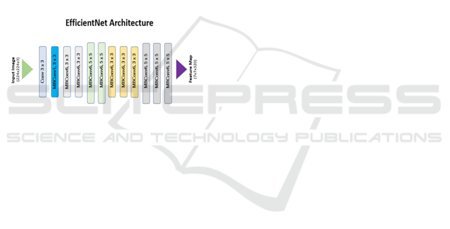
1.3 Architecture
The architecture of EfficientNet-B2 is built on the
principles of Mobile Inverted Bottleneck
Convolutions (MBConv) and Squeeze-and-
Excitation (SE) blocks, optimized for efficiency and
high performance. MBConv layers use a depthwise
separable convolution combined with an expansion
phase, reducing computational cost while capturing
rich feature representations.
Figure 1: EfficientNet Architecture
The SE blocks enhance the model by recalibrating
channel-wise feature responses, emphasizing critical
features and suppressing less important ones.
EfficientNet-B2 employs compound scaling, a unique
method to balance depth (number of layers), width
(number of channels per layer), and input resolution.
For B2, the input image size is 260x260 pixels,
providing higher resolution for detailed feature
extraction compared to smaller models like
EfficientNet-B0 and B1. The scaling ensures a
systematic increase in model capacity without
redundant computations, achieving efficiency. The
model contains stacked MB Conv layers grouped into
stages, each designed for different spatial resolutions,
followed by a global average pooling layer and a
fully connected classifier. With approximately 9.2
million parameters and 1.0 billion FLOPs,
EfficientNet-B2 is significantly lighter than
traditional models like ResNet but delivers
competitive accuracy (~79.8% Top-1 on ImageNet).
This architecture makes it highly suitable for image
classification tasks in resource-constrained
environments or real-time applications requiring both
speed and accuracy.have varying depths in various
designs. The top two levels have '4096' channels
apiece, while the third has '1000' channels. The
configuration of completely linked layers remains
consistent across all networks.
1.4 Retinopathy
Retinopathy, a word that refers to a variety of retinal
illnesses, is a serious medical problem with far-
reaching consequences for visual health. Retinopathy
is mostly connected with systemic disorders such as
diabetes, hypertension, and other vascular diseases
(Kumaresan, and, Palanisamy, 2022. It presents as
damage to the fragile blood vessels of the retina,
which may lead to visual impairment or even
blindness if not addressed. The retina, a critical
sensory tissue in the back of the eye, is responsible
for translating light impulses into visual information
for the brain. Therefore, any disturbance to its
complicated vascular network might result in
significant repercussions. As the frequency of
retinopathy-related disorders grows worldwide, there
is an increasing need for better diagnostic techniques
and strategies to diagnose and manage this ocular
problem early on, avoiding irreparable damage and
maintaining visual acuity. This introduction sets the
context for delving into the problems and
developments in retinopathy identification,
highlighting the importance of early detection in
reducing its effect on eye health.
1.5
Transfer Learning
Transfer learning, a strong paradigm in machine
learning, has emerged as a game-changing way to
improving the efficiency and performance of many
jobs. At its essence, transfer learning takes
information obtained from addressing one issue and
applies it to a new but related activity (Yadav, et al. ,
2022). Transfer learning, unlike standard machine
learning models that start from scratch, enables pre-
trained models on huge and varied datasets to be
adapted to new tasks, even when labeled data is
limited. This technology is especially useful in
sectors where getting large labeled datasets is
difficult or costly. In recent years, transfer learning
has achieved great success in a wide range of
applications, including image and audio recognition
and natural language processing. Its adaptability and
efficacy derive from its capacity to transfer learnt
features, representations, or information from one
domain to another, which speeds up the learning
process and considerably improves model
INCOFT 2025 - International Conference on Futuristic Technology
604
performance on certain tasks. This introduction lays
the groundwork for understanding the importance and
relevance of transfer learning, particularly in the
context of its use in retinopathy detection using
retinal imaging.
1.6 Retinal Images
Retinal pictures, which are complex photos of the
eye's innermost layer, the retina, provide crucial
insight into ocular health. The retina, which consists
of a complex network of blood arteries, neurons, and
light-sensitive cells, is critical to the visual process.
Retinal pictures capture the precise features and
structural properties of this essential tissue, providing
doctors with a non-invasive way to test and diagnose
a wide range of eye disorders. These scans give a lot
of information crucial for prompt intervention and
treatment, including identifying early symptoms of
disorders such as diabetic retinopathy and tracking
age-related changes. Advances in medical imaging
technology have not only improved the quality and
accuracy of retinal pictures, but they have also created
new opportunities for analyzing and interpreting
these images using artificial intelligence and machine
learning approaches. As we explore the convergence
of technology and ophthalmology, the importance of
retinal imaging grows, serving as a foundation in the
search of more efficient and accurate diagnostic
procedures for a wide range of ocular disorders.
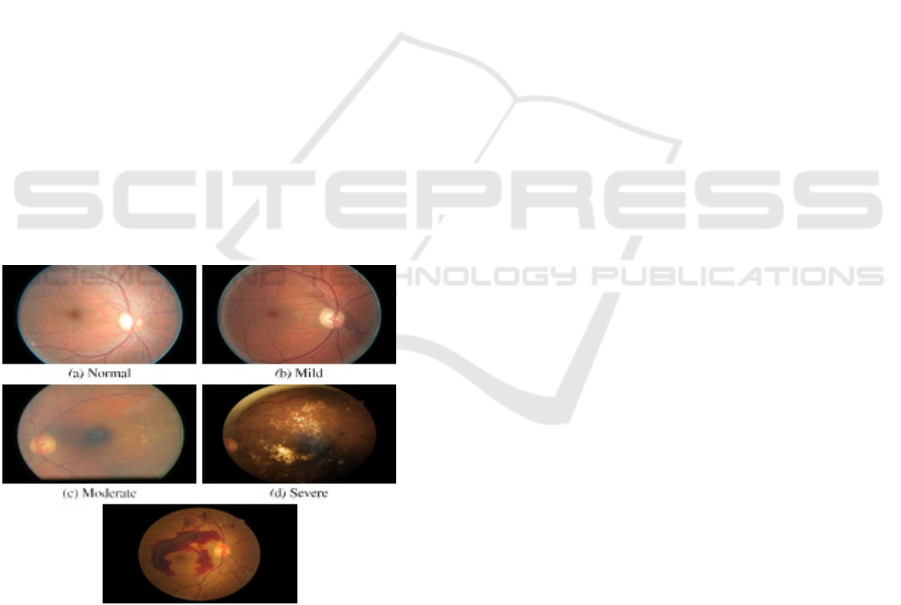
Figure 1: The different stages of DR
2 EXISTING SYSTEM
Diabetic retinopathy (DR) refers to the retinal
damage caused by diabetes. Diabetes has become a
major health problem across the globe, with an
unimaginable number of persons suffering from it.
Periodic eye examinations help physicians to
discover DR in patients at an early stage and
implement appropriate therapies. Advances in
artificial intelligence and camera technologies have
enabled us to automate DR diagnosis, potentially
benefiting millions of patients. This research
describes a unique approach for diagnosing DR based
on gray-level intensity and texture data taken from
fundus pictures using a decision tree-based ensemble
learning strategy. This research largely uses the Asia
Pacific Tele-Ophthalmology Society's 2019
Blindness Detection (APTOS 2019 BD) dataset. We
took numerous measures to select its contents so that
they were more suited for machine learning
applications. Our method uses several image
processing approaches, two feature extraction
strategies, and one feature selection strategy, yielding
a classification accuracy of 94.20% (margin of error:
±0.32%) and an F-measure of 93.51% (margin of
error: ±0.5%). Several more characteristics about the
suggested method's performance have been offered to
demonstrate its resilience and dependability. Details
on each approach used have been presented to make
the findings replicable. This approach may be a
beneficial tool for mass retinal screening to identify
DR, significantly lowering the rate of vision loss
associated with it.
3 PROPOSED SOLUTION
The suggested system is a web-based tool for
detecting and classifying diabetic retinopathy using
retinal fundus pictures. The system is designed using
the Flask framework, which provides a simple and
easy interface for users to submit photos of both their
right and left eyes. The system's core is built on the
EfficientNetB2, which has been pre-trained and fine-
tuned to categorize diabetic retinopathy into five
categories: no diabetic retinopathy (no DR), mild,
moderate, severe, and proliferative diabetic
retinopathy. In addition, the system includes a sixth
category for detecting low-quality photos that may
result in misclassification. The system preprocesses
the photos many times, including scaling, data
cleaning, and augmentation, to guarantee high-
quality inputs for reliable analysis. During the
training phase, class balance approaches and hyper
parameter adjustment are used to enhance the model's
performance (Patel, Shah, et al. , 2023). After the
photos are examined, the system returns a
categorization result for each eye. By providing a
user-friendly and accessible platform, this suggested
system seeks to help healthcare providers diagnose
A Premature Recognition of Diabetic Retinopathy Using Deep Learning and Grad Cam Technology
605
diabetic retinopathy and identify situations that need
further medical attention.
3.1 Load Data Module
This module is responsible for importing retinal
fundus pictures from multiple sources or datasets into
the system (Wang, et al. , 2022). It ensures that the
photos are ready for further processing and analysis
by reading them in the correct format and arranging
them for categorization.
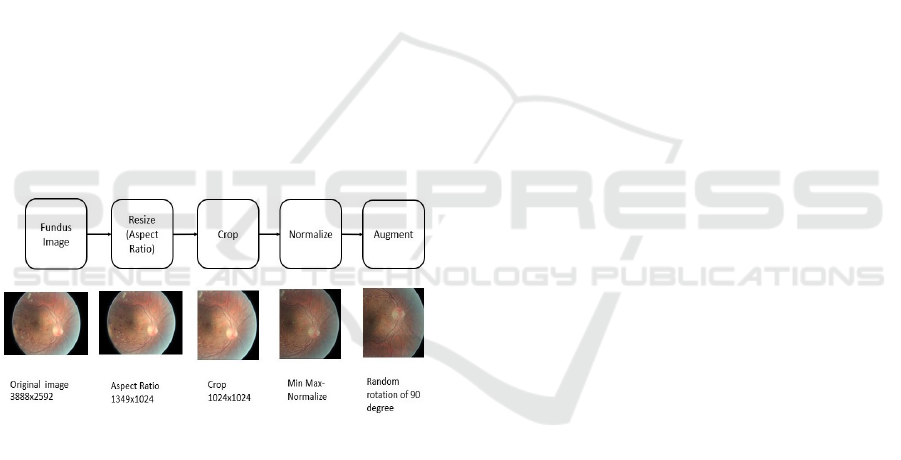
3.2 Data Preprocessing Module
This module does necessary preprocessing processes
to the supplied photos to improve their quality. It
entails activities such as scaling photographs to
standard dimensions, cleaning the dataset by
eliminating low-quality images, and using data
augmentation methods to enhance dataset variability
and improve model resilience. This module discovers
and picks the most relevant characteristics from the
preprocessed pictures that are important to the
classification job. By concentrating on key
characteristics, this module improves model
performance and reduces overfitting, resulting in
improved classification results.
Figure 3: Preprocessing steps
3.3 Training and Testing Module
This module splits the dataset into training and testing
subgroups. It trains the EfficientNetB2 neural
network on training data and assesses its performance
on testing data. The training method comprises hyper
parameter adjustment to improve the model's
accuracy in detecting diabetic retinopathy.
3.4 Model Evaluation Module
This module examines the trained model's
performance using a variety of measures, including
accuracy, precision, sensitivity, specificity, and F1-
score. It gives information on the model's
performance in categorizing the various phases of
diabetic retinopathy and identifies opportunities for
development.
3.5 UI Interface Module
The User Interface (UI) module provides a simple
web interface developed with Flask that enables
healthcare providers to simply submit retinal fundus
photos and examine categorization results. This
module enables smooth interaction and gives clear
visual feedback on analysis results, making it
accessible to users of diverse technical skill.
4 RESULT ANALYS IS
The system's results show that it effectively classifies
diabetic retinopathy into five distinct categories (No
DR, Mild, Moderate, Severe, and Proliferative), with
an additional category for detecting low-quality
images, which is critical for maintaining high
diagnostic accuracy (Kim, et al. , 2022). The use of
preprocessing procedures such as scaling, data
cleaning, and augmentation has shown to be critical,
increasing the model's capacity to handle a wide
variety of picture inputs and boosting the overall
system resilience. Hyperparameter adjustment
throughout the training phase increased the model's
performance, resulting in better generalization over
previously unknown data. The model's performance
was fully assessed using evaluation criteria such as
accuracy, precision, sensitivity, specificity, and F1-
score. High values across these measures imply that
the system can accurately distinguish between
different phases of diabetic retinopathy, lowering the
chance of misclassification and improving early
detection capabilities. The UI module allows
healthcare professionals of various technical
backgrounds to submit photos and obtain clear,
interpretable findings. This technique not only helps
with rapid diagnosis, but it also promotes educated
decision-making, resulting in improved patient care
and diabetic retinopathy treatment. Through the use
of the sophisticated features of the Efficient Net deep
learning architecture, the suggested method seeks to
dramatically improve the accuracy of diabetic
retinopathy identification. The accuracy in the current
approach is 75% and is based on a laborious manual
process carried out by skilled doctors. Understanding
how important it is to diagnose patients accurately
and quickly the suggested model uses Efficient Net
and a carefully selected dataset of retinal pictures to
obtain an astounding 81% accuracy rate as shown in
INCOFT 2025 - International Conference on Futuristic Technology
606
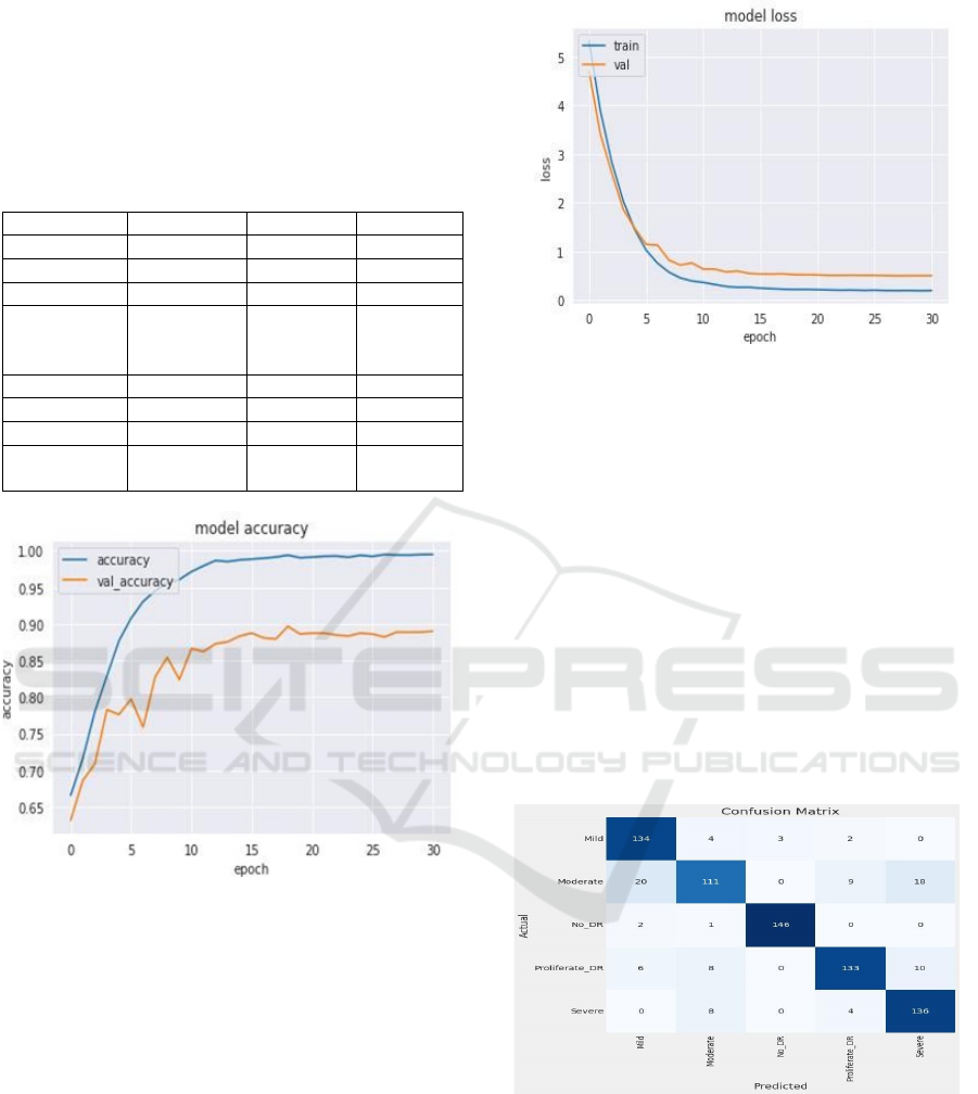
table 1. This enhancement tackles the inefficiencies
in the present manual approach, which frequently
results in delayed results and associated issues in
follow-up and prompt treatment. It also signals the
potential for more reliable detection of diabetic
retinopathy
Table 1: Performance Metrices
Cate
g
or
y
Precision Recall Fscore
Mil
d
0.83 0.94 0.88
Moderate 0.84 0.82 0.77
No DR 0.98 0.90 0.98
Proliferate
DR
0.90 0.85 0.87
Severe 0.83 0.92 0.87
accurac
y
0.87
macro av
g
0.88 0.88 0.87
weighted
av
g
0.88 0.87 0.87
Figure 4: Plot for Model Accuracy between Accuracy and
Epoch
The model represents how well the classification
system performs in distinguishing between different
stages of DR based on retinal images as shown in
figure 3. In our study, we utilized EfficientNet for
feature extraction and trained the model using a
carefully curated dataset of retinal images. The
overall accuracy of the model reached 81%, which
represents a significant improvement over traditional
manual diagnosis methods that typically achieve
around 75% accuracy. This improvement highlights
the model’s potential to automate the early detection
of diabetic retinopathy, thus providing faster and
more consistent diagnostic results. Additionally, this
higher accuracy is expected to facilitate timely
intervention, reducing the likelihood of vision loss
among diabetic patients.
Figure 5: Plot for Model Loss between Accuracy and Epoch
Model loss is a key metric in training neural
networks, indicating how well or poorly the model is
performing during the learning phase as shown in
figure 4. Our system utilized categorical cross-
entropy as the loss function, given the multiclass
nature of the diabetic retinopathy stages. Over
multiple training epochs, the model's loss consistently
decreased, showing that the model was effectively
learning the intricate patterns in the retinal images.
This reduction in loss is crucial, as it directly
correlates with the model's ability to make accurate
predictions. The final loss value suggests a well-
trained model, capable of generalizing well on new,
unseen data. The careful balancing of loss through
optimization techniques was instrumental in
achieving the desired classification performance.
Figure 6: Confusion Matrix
The classification report is shows the confusion
matrix in the figure 5 for our diabetic retinopathy
detection model provides a detailed performance
analysis across all severity levels: No DR, Mild,
Moderate, Severe, and Proliferate DR. Key metrics
such as precision, recall, and F1-score were
evaluated, highlighting the model’s effectiveness.
The model achieved an average precision of 0.88,
A Premature Recognition of Diabetic Retinopathy Using Deep Learning and Grad Cam Technology
607
demonstrating its ability to minimize false positives,
while the recall score of 0.87 reflects its capability to
capture most true positive cases. With an F1-score of
0.87, the model strikes a balance between precision
and recall, confirming its robust performance across
all stages. Specifically, for the Mild class, the model
achieved an F1-score of 0.88, and for Severe cases, it
reached 0.87, indicating that the system is effective at
identifying both early and advanced stages of the
disease(Singh, et al. , 2023) These metrics underscore
the model’s reliability and its potential for clinical
application in detecting diabetic retinopathy with
high accuracy.
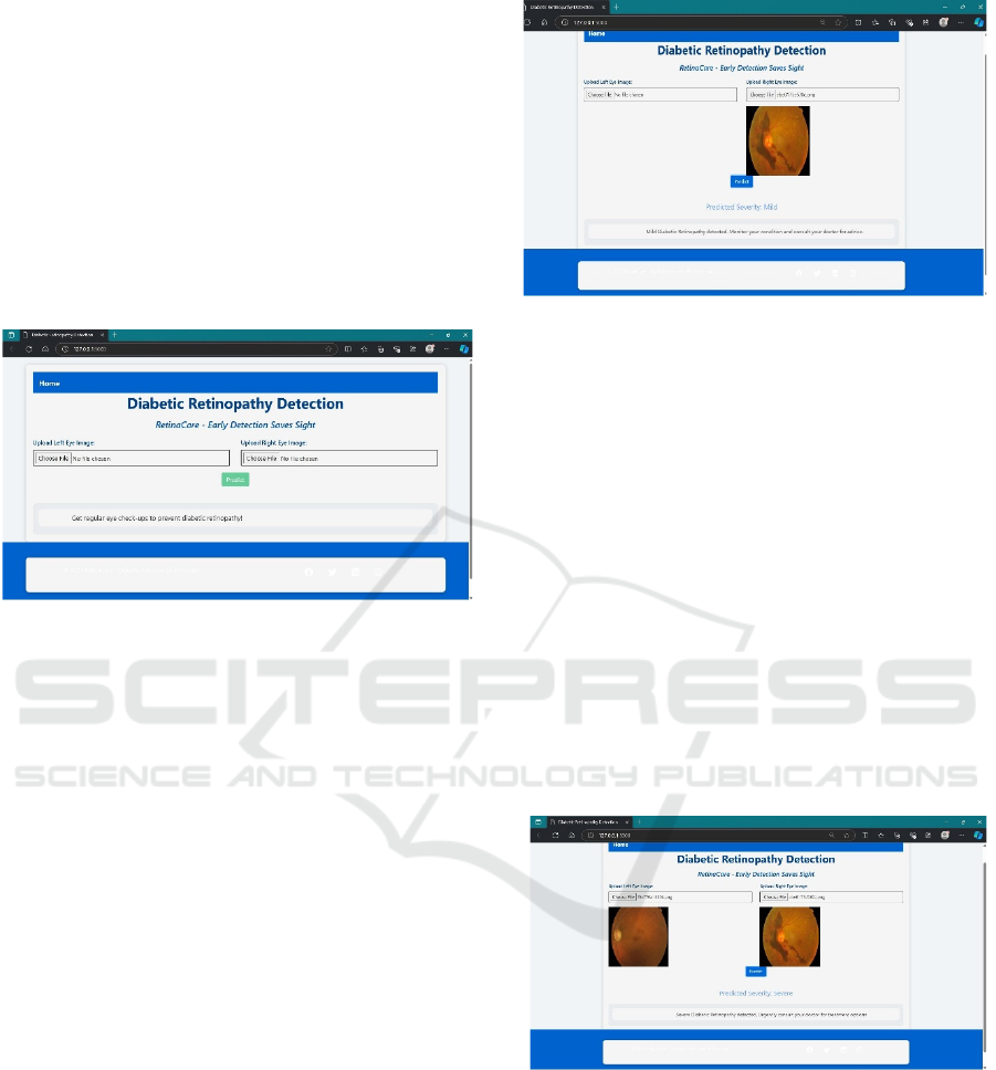
Figure 7: Frontend webpage to detect diabetic retinopathy
To implement the figure 6 shows the Diabetic
Retinopathy Detection webpage, use Flask for the
back-end and HTML with Bootstrap for the front-
end. The webpage allows users to upload retinal
images of both the left and right eye, which are
processed by a machine learning model to predict
Diabetic Retinopathy. In the back-end (app.py), Flask
handles routing, file uploads, and calls to the
predictive model. The images are securely stored and
processed to generate a result, which is displayed
back on the webpage. The front-end (index.html)
provides a clean, responsive interface using
Bootstrap, with simple input forms for image uploads
and a button to trigger the prediction. After
submission, the prediction result is dynamically
shown on the page. This project can be extended by
integrating the model for prediction, improving the
user interface, and deploying it to a cloud platform for
online use.
Figure 8: Prediction of Mild infection in the right eye
The webpage for diabetic retinopathy detection
allows users to upload images of their retina,
specifically from the left or right eye. Once an right
eye image is uploaded, the webpage of right eye in
the system processes it through a pre-trained machine
learning model designed to detect the presence and
severity of diabetic retinopathy as shown in figure 7.
The model then predicts the severity, which can range
from "Mild" to more severe stages. The result, along
with the uploaded image, is displayed directly on the
webpage. In the example shown, the right eye image
was analyzed, and the model predicted "Mild"
diabetic retinopathy, offering users valuable
information and advice to monitor their condition.
The system's simple interface, powered by Flask for
back-end operations and Bootstrap for front-end
design, ensures ease of use and efficiency in early
detection efforts.
Figure 9: Prediction of Severe infection by comparing the
both eye
The webpage of a image is processing both the left
and right eye retinal images to detect diabetic
retinopathy as shown in figure 8. The system analyzes
each image independently and provides a severity
prediction for the condition in both eyes. In this case,
the system predicted a "Severe" stage of diabetic
retinopathy based on the analysis of the uploaded
images. After uploading the retinal images, the user
can click the "Predict" button to trigger the model,
INCOFT 2025 - International Conference on Futuristic Technology
608
which will analyze the images and display the
severity level below. The result, "Severe Diabetic
Retinopathy detected", also includes a suggestion for
urgent medical consultation, emphasizing the
importance of early treatment in severe cases. The
interface is user-friendly, displaying both uploaded
images and the prediction results on the same page for
better clarity and guidance.
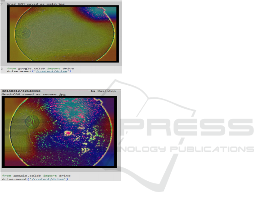
Figure 10: Grad-Cam output for mild
Figure 11: Grad-Cam output for severe
GRAD-CAM (Gradient-weighted Class
Activation Mapping) is a visualization technique used
to interpret deep learning models by highlighting
regions in input images that significantly influence
the model’s predictions. In diabetic retinopathy
detection, Grad-CAM is employed to identify and
visualize the retinal areas affected by mild and severe
DR, aiding clinicians in understanding AI-driven
diagnostic outputs
5 CONCLUSION
In conclusion, the suggested Flask web application
for diabetic retinopathy detection marks a big step
forward in harnessing deep learning technology to
assist healthcare professionals in the prompt
identification and categorization of this crucial eye
ailment. The system uses the EfficientNetB2 neural
network to successfully evaluate retinal fundus
pictures, dividing them into different phases of
diabetic retinopathy and detecting low-quality photos
that may influence diagnosis accuracy. The use of
robust preprocessing, feature selection, and model
assessment approaches improves the system's
dependability and efficacy. Furthermore, the user-
friendly interface allows physicians to input photos
and get actionable insights without having to navigate
difficult technological procedures. Finally, the goal of
this application is to enhance patient outcomes by
early diagnosis and intervention, therefore helping
continuing efforts to battle diabetic complications and
contributing to advances in medical imaging and
analysis.
6 FUTURE WORK
Future work on the proposed system will concentrate
on three major upgrades to increase its utility and
efficacy in detecting diabetic retinopathy. One
significant area will be to include more deep learning
architectures beyond EfficientNetb2, such as VGG-
16 or ResNet, to see whether these models can
improve classification performance. Furthermore,
increasing the dataset to include a broader variety of
retinal pictures from various demographics and
geographical areas would improve the model's
resilience and generalizability. Implementing real-
time analytic capabilities will also be investigated,
allowing healthcare practitioners to get instant
feedback during clinical tests. Furthermore, using
modern image processing methods, such as contrast
enhancement and noise reduction, may increase
picture quality and classification accuracy. Another
emphasis will be on developing a mobile application
version of the system to make it more available to a
wider range of users, including distant healthcare
practitioners. Finally, future development will
involve continuous user input and incremental
enhancements to the user interface to ensure that the
system stays intuitive and meets the requirements of
healthcare professionals. These developments are
intended to produce a complete and effective tool for
the early identification and control of diabetic
retinopathy, eventually leading to improved patient
outcomes.
A Premature Recognition of Diabetic Retinopathy Using Deep Learning and Grad Cam Technology
609

REFERENCES
M. Sharma et al., 2022, "Explainable AI for Diabetic
Retinopathy Classification Using Vision
Transformers," Computer Vision and Image
Understanding, vol. 224, pp. 102320.
S. Rajput, A. Arora2021 "Multi-Scale Attention Network
for Diabetic Retinopathy Classification," International
Journal of Computer Applications, vol. 75, no. 10, pp,
6-12.
K. Agarwal and T. Kumar, 2022, "Automatic recognition
of severity level for diabetic retinopathy diagnosis
using deep visual features," in 2nd International
Conference on Intelligent Computing and Control
Systems (ICICCS). IEEE.
A. Khan et al., 2022, "EfficientNetV2 for Diabetic
Retinopathy Detection: Improving Accuracy with
Compound Scaling," Medical Image Analysis, vol. 81,
pp. 102343.
T. Kumaresan and C. Palanisamy, 2022, "EyePACS: An
Adaptable Telemedicine System for Diabetic
Retinopathy Screening," International Journal of Bio-
Inspired Computation, vol. 10, no. 1, pp. 142-156.
P. Kumar and R. Yadav, 2022, "Grad-CAM Based
Visualization for Enhancing Explainability in Diabetic
Retinopathy Detection," Artificial Intelligence in
Medicine, vol. 126, pp. 102190.
V. Patel and K. Shah, 2023,"Diabetic Retinopathy Grading
Using Hybrid CNN-LSTM Model," Expert Systems
with Applications, vol. 202, pp. 117319.
L. Wang et al.,2022, "Multimodal Deep Learning for
Diabetic Retinopathy Classification Using Fundus and
Optical Coherence Tomography Images," IEEE
Transactions on Medical Imaging, vol. 41, no. 3, pp.
789-799.
J.Kim et al.,2022, "Federated Learning for Diabetic
Retinopathy Detection: Addressing Privacy Concerns
in AI Healthcare," Nature Machine Intelligence, vol. 4,
no. 7, pp. 570-578.
R. Singh et al.,2023 "Improved Robustness in Diabetic
Retinopathy Detection Using Adversarial
Training," Proceedings of the IEEE/CVF Conference
on Computer Vision and Pattern Recognition (CVPR),
pp. 2781-2789.
INCOFT 2025 - International Conference on Futuristic Technology
610
