
Epilepsy Diagnosis Using EEG Image Analysis
Seeba Doddmani, Sana Mulla, Dilipsingh Rajpurohit, Rajashri Khanai and Prema T. Akkasaligar
Department of Computer Science and Engineering, KLE Technological University, Belagavi, India
Keywords:
Epilepsy Detection, EEG Image Analysis, Machine Learning (ML), Convolutional Neural Network (CNN),
Scalogram Images, VGG-16 Model, Deep Learning, Feature Extraction, Transfer Learning, Bern-Barcelona
EEG Dataset, Seizure Classification, Signal Processing, Time-Frequency Analysis, Neural Networks,
Supervised Learning, Data Preprocessing, Binary Classification, Medical Diagnosis, Graph Spectral Features,
Long Short-Term Memory (LSTM), Real-Time Detection.
Abstract:
Epilepsy is a neurological disorder that affects millions of people around the world and is characterized by
recurrent seizures caused by abnormal brain activity. Electroencephalograms (EEG) are the primary diagnostic
tool, but traditional manual analysis is time-intensive and prone to errors. This project leverages machine
learning techniques to automate epilepsy detection using scalogram images generated from EEG signals. A
custom Convolutional Neural Network (CNN) model was developed and trained on the Bern-Barcelona EEG
dataset, achieving a training precision of 74.07% and a testing precision of 73.22%. The model demonstrates
good training performance and testing accuracy. An implemented VGG-16 gave a training accuracy of 81.13%
and a testing accuracy of 80.04%. This study aims to help clinicians improve diagnostic accuracy and provide
a scalable, real-time solution for epilepsy detection, particularly in underserved regions.
1 INTRODUCTION
Epilepsy, a chronic neurological disorder, affects
more than 50 million people worldwide, causing sig-
nificant health and social challenges. Diagnosis of
epilepsy traditionally relies on manual analysis of
EEG recordings, a process that is not only time-
consuming but also susceptible to errors due to the
complexity of EEG waveforms. With advances in ma-
chine learning, there is growing interest in automating
this process to improve diagnostic accuracy and effi-
ciency. This project focuses on converting EEG sig-
nals into scalogram images, which capture both tem-
poral and frequency domain features, and applying a
custom CNN model for seizure detection. The pro-
posed system aims to address the limitations of man-
ual analysis, offering a reliable and scalable solution
for clinicians, particularly in resource-limited areas
where access to specialized neurologists is scarce.
The high prevalence of epilepsy, coupled with
the challenges of timely and accurate diagnosis, mo-
tivates the need for automation in the detection of
epilepsy. Automated systems can significantly reduce
the burden on neurologists, improve diagnostic ac-
curacy, and ensure better access to healthcare in un-
derserved areas. Using machine learning, these sys-
tems can offer reliable and real-time diagnoses, facil-
itating earlier intervention and better management of
epilepsy.
Several approaches have been explored for
epilepsy detection, combining traditional machine
learning and deep learning techniques: Krishnasamy
et al. proposed supervised learning algorithms, in-
cluding Support Vector Machines (SVM) and CNNs,
achieving accuracy rates up to 99.7% but requiring
large datasets and computational resources. Pattnaik
et al. utilized scalogram images and transfer learn-
ing with ResNet50, achieving a classification accu-
racy of 95.23% with high sensitivity and specificity.
Sesha Sai et al. combined CNNs with SVMs for auto-
matic feature extraction, achieving 94.48% accuracy
but heavily dependent on data quality. Other stud-
ies, such as by Wang et al., explored hybrid CNN-
LSTM (Long Short Term Memory) models for cap-
turing both spatial and temporal features in EEG data.
Despite their promising results, these approaches
face several limitations: Heavy reliance on large,
high-quality datasets, making them less effective in
diverse clinical settings. Computational complex-
ity, particularly for deep architectures like Residual
776
Doddmani, S., Mulla, S., Rajpurohit, D., Khanai, R. and T. Akkasaligar, P.
Epilepsy Diagnosis Using EEG Image Analysis.
DOI: 10.5220/0013602300004664
Paper published under CC license (CC BY-NC-ND 4.0)
In Proceedings of the 3rd International Conference on Futuristic Technology (INCOFT 2025) - Volume 2, pages 776-783
ISBN: 978-989-758-763-4
Proceedings Copyright © 2025 by SCITEPRESS – Science and Technology Publications, Lda.
Network (ResNet) and hybrid models, which hin-
ders real-time application. Limited generalization to
unseen data, with significant performance degrada-
tion when applied to different patient demographics
or new datasets. Challenges in adapting models to
resource-limited environments due to high hardware
requirements.
Epilepsy detection faces several challenges: High
variability in seizure patterns across patients compli-
cates the creation of a universally robust model. EEG
data often contains noise and artifacts, requiring so-
phisticated pre-processing. Balancing model com-
plexity with computational efficiency is critical for
real-time applications. Addressing class imbalance in
datasets is essential to improve model generalization.
To address these challenges, this project proposes
a machine learning pipeline that converts EEG signals
into scalogram images to capture time-frequency do-
main features. A custom CNN is designed to classify
these images into seizure and non-seizure categories.
The pipeline incorporates preprocessing techniques
like normalization and leverages training strategies to
improve model generalization.
Developed a novel pipeline to transform EEG sig-
nals into scalogram images for time-frequency anal-
ysis. Designed and implemented a custom CNN ar-
chitecture optimized for binary classification. Evalu-
ated model performance on the Bern-Barcelona EEG
dataset, achieving a training accuracy of 97.77%.
Identified challenges in generalization through testing
accuracy analysis, providing insights for future im-
provement. Highlighted the potential for real-time de-
ployment of the proposed system in resource-limited
settings.
The rest of paper is organized as follows. Sec-
tion II provides a brief review of the recent works.
Section III details the problem statement, background
and the proposed methodology. Section IV describes
the experiment details along with the results and dis-
cussions. Finally, the paper concludes in Section V.
2 LITERATURE SURVEY
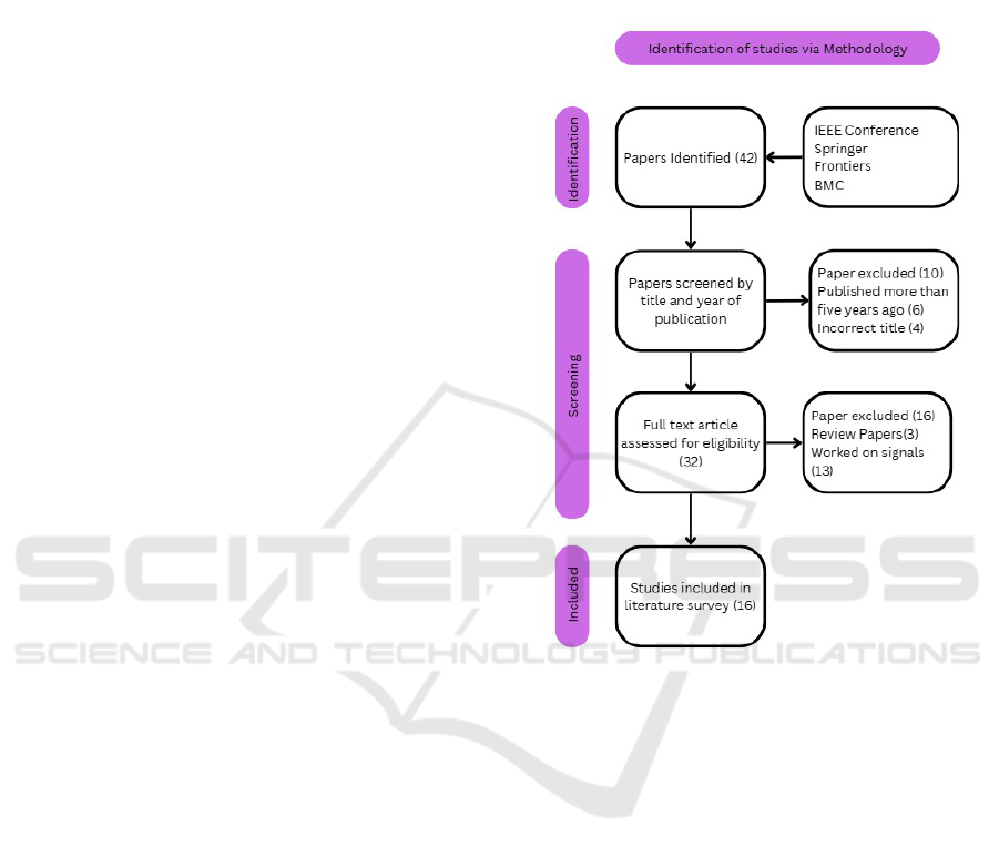
The methodology for identifying and selecting stud-
ies for the literature survey involved several stages
to ensure relevance and quality. Initially, 42 papers
were identified from reputable sources such as IEEE
Conference, Springer, Frontiers, and BioMed Cen-
tral(BMC). During the screening phase, 10 papers
were excluded—6 for being older than five years and
4 for having incorrect titles. Figure. 1 shows the
flowchart illustrating this process. After screening, 32
papers were assessed for eligibility, and 16 were fur-
ther excluded—3 were review papers, and 13 focused
on signals rather than images. Ultimately, 16 stud-
ies were included in the literature survey, providing a
robust foundation for the research.
Figure 1: Flowchart of literature survey paper selection
(Krishnasamy et al., 2024) explored supervised
learning algorithms for epileptic seizure detection,
achieving high accuracy rates (98.5% with SVM and
99.7% with fuzzy classifiers). Their system auto-
mates seizure detection effectively, reducing com-
plexity for experts, but struggles with real-time de-
ployment due to computational demands and limited
generalizability across datasets. Similarly, (Hafeez
and Shakil, 2024) utilized EEG-based brainwave im-
ages to classify stress levels using LSTM (70.67%
accuracy) and CNN (90.46% accuracy), demonstrat-
ing CNN’s capability for spatial feature extraction but
facing challenges with noise in EEG data and small
participant pools. (Pattnaik et al., 2024) achieved
95.23% classification accuracy using scalogram im-
ages analyzed with a pre-trained ResNet50 model,
showcasing the utility of transfer learning for EEG
signal analysis.
(Sadam and Nalini, 2024) combined CNN and
SVM to achieve 94.48% accuracy, highlighting the
advantages of hybrid models for automated feature
Epilepsy Diagnosis Using EEG Image Analysis
777

extraction but showing sensitivity to noisy datasets.
(Georgis-Yap et al., 2024) compared supervised and
unsupervised approaches like CNN, CNN-LSTM,
and TCN, finding comparable results for patient-
specific seizure prediction. While their methods re-
duce preprocessing needs, they require substantial
preictal data for optimal performance. (Krishnan
et al., 2024) applied GASF to convert EEG signals
into image representations, achieving up to 96% ac-
curacy. However, this approach is computationally
intensive, especially during image transformation and
feature extraction.
(Shankar et al., 2023) utilized CNNs with phase
synchronization matrices to classify seizures with
83.3% accuracy, demonstrating effective spatial fea-
ture extraction. However, intensive preprocessing and
the need for large datasets remain barriers to real-
time applications. (Khasawneh et al., 2022) achieved
99.8% precision using Faster R-CNN and transfer
learning for K-complex detection, with adaptability
across datasets. Nonetheless, overlapping image gen-
eration methods may introduce data leakage, compro-
mising model reliability. (Hu et al., 2020) proposed
a hierarchical neural network (HNN) using transfer
learning, attaining 98.97% accuracy on the CHB-
MIT dataset, though the reliance on large pre-trained
DNNs limits real-time applicability.
(Sharma and Meena, 2024) introduced a model
integrating GFT and DWT for feature extraction,
achieving over 98% accuracy across datasets. While
graph spectral features enhance detection accuracy,
the method’s complexity increases computational de-
mand, complicating real-time deployment. (Kunekar
et al., 2024) used LSTM networks to achieve 97% ac-
curacy, effectively handling temporal dependencies in
EEG data. However, dataset class imbalance could
hinder generalizability. (Jridi et al., 2024) employed
deep ResNet for multi-disorder detection, reaching
100% accuracy for epilepsy detection on the UBonn
dataset, but high computational requirements pose
challenges for implementation in resource-limited en-
vironments.
(Saleem et al., 2023) combined CNN with tra-
ditional classifiers, achieving 98.49% accuracy in
seizure detection. The hybrid model effectively iden-
tifies subtle EEG patterns but requires larger, more
diverse datasets to ensure generalizability. (Majzoub
et al., 2023) used AlexNet for multi-channel EEG sig-
nal classification, achieving 98.25% accuracy in bi-
nary classification. However, its accuracy drops to
92.98% with new patient data, highlighting the need
for diverse training samples. (Wang et al., 2023) de-
veloped a CNN-LSTM hybrid model, achieving 98%
accuracy in ternary classification and 100% in binary
classification. This model excels in capturing both
spatial and temporal features but demands high com-
putational resources.
Finally, (Supriya et al., 2021) adopted graph-
theory-based methods, such as VG and HVG, for fea-
ture extraction, achieving accuracies above 95%.
These methods are advantageous for their process-
ing speed and classification efficacy but are limited by
their sensitivity to dataset size and threshold depen-
dencies. Collectively, while these studies demonstrate
significant advancements in EEG-based seizure de-
tection and classification, the major limitations across
models include dependency on large datasets, compu-
tational demands, and challenges with generalization
across diverse patient populations.
This literature survey reviews advancements in
EEG-based epileptic seizure detection using super-
vised learning, deep learning, and hybrid models.
Techniques like CNN-SVM hybrids (94.48% accu-
racy), transfer learning with scalograms (95.23%),
and GASF image transformations (96%) demonstrate
high accuracy but face challenges with computational
demands and noisy data. Deep learning models like
CNNs and hybrid CNN-LSTM architectures achieve
exceptional spatial-temporal feature extraction but re-
quire large datasets. Despite progress, issues such
as dataset dependency, generalizability, and real-time
applicability remain significant hurdles.
3 PROBLEM STATEMENT AND
SYSTEM MODEL
Epilepsy is a chronic neurological disorder marked by
recurrent seizures resulting from abnormal electrical
activity in the brain. Diagnosing epilepsy relies heav-
ily on EEGs, which capture electrical patterns and
help identify abnormalities. Traditional approaches
involve manual inspection, a labor-intensive task
requiring specialized expertise.
EEG, or electroencephalography, plays a signifi-
cant role in medical diagnosis as a non-invasive and
cost-effective method for recording brain activity. It
is particularly useful in identifying abnormal brain
wave patterns, which can indicate potential seizures
or other neurological disorders. By capturing electri-
cal activity in the brain, EEG provides valuable in-
sights into the functional state of the brain, aiding
clinicians in the diagnosis and management of vari-
ous conditions.
Traditional methods of diagnosing epilepsy
present several challenges. The manual analysis of
long EEG recordings is a time-consuming process, re-
INCOFT 2025 - International Conference on Futuristic Technology
778
quiring significant effort from trained specialists. Fur-
thermore, the variability in seizure patterns across in-
dividuals adds complexity to achieving a consistent
and accurate diagnosis. These issues are compounded
by the limited availability of trained neurologists, par-
ticularly in rural or underserved areas, making timely
and effective diagnosis even more difficult.
Epilepsy is a neurological disorder that impacts
over 50 million people worldwide, characterized by
abnormal brain activity leading to seizures. Tradi-
tional epilepsy diagnosis using EEGs involves man-
ually analyzing extensive recordings to detect epilep-
tic events, a process prone to errors and inefficien-
cies. This project aims to automate the detection of
epilepsy by employing Machine Learning (ML) tech-
niques to classify epileptic and non-epileptic events
using 2D scalogram images derived from EEG sig-
nals. The goal is to create a robust, accurate system
that can enhance diagnostic efficiency, particularly in
resource-limited areas.
The primary objective of this work is to develop
a machine learning pipeline capable of automati-
cally classifying EEG-derived scalogram images as
”seizure” or ”non-seizure.” This automated approach
aims to improve diagnostic accuracy by increasing
sensitivity and specificity, thereby assisting clinicians
in making reliable diagnoses. Additionally, the solu-
tion is designed to be deployable in real-time systems,
enabling rapid analysis and decision-making in clini-
cal settings. To ensure versatility, the proposed solu-
tion is scalable and adaptable to diverse datasets and
hardware setups, including mobile and low-power de-
vices.
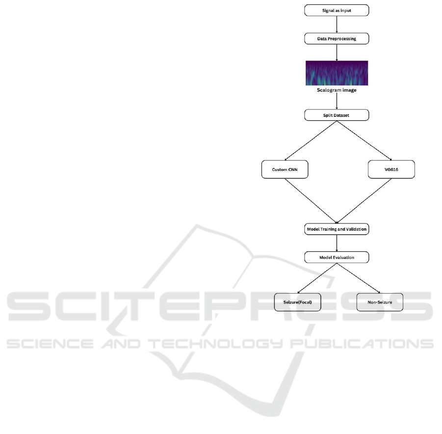
Figure.2. shows the system model that outlines
a process for classifying EEG signals, into two cate-
gories: seizure (focal) and non-seizure. The process
begins with signal input, followed by data preprocess-
ing to transform the signal into a scalogram image,
which visually represents the frequency content over
time. The dataset is then split into training and test-
ing subsets. Two models are employed for classifica-
tion: a custom CNN and a pre-trained VGG16 model.
Both models undergo training and validation to learn
patterns from the data. Finally, model evaluation is
conducted to assess performance, leading to the clas-
sification of the input signal as either seizure (focal)
or non-seizure.
4 PROPOSED METHODOLOGY
The proposed work is executed on device is powered
by an AMD Ryzen 7 4800H with Radeon Graphics
processor, operating at 2.90 GHz, and is equipped
Figure 2: System model for EEG detection
with 16 GB of RAM. It runs a 64-bit version of
Windows 11 Home and the NVIDIA GTX 3050
GPU. For implementation, we have used several
Python libraries are used for efficient data analysis
and visualization. NumPy supported numerical
computations, Pandas handled data manipulation.
Matplotlib are employed for creating visualizations,
tensorflow used for building the CNN Architecture.
The dataset originally consisted of EEG signals
represented by both X and Y components. These sig-
nals were processed and converted into scalogram im-
ages, with separate scalograms generated for the X
and Y components. Following this transformation,
normalization was applied to the images to standard-
ize pixel intensity values, ensuring they fell within a
consistent range to enhance model training.
4.1 Custom CNN
CNNs are a class of deep learning models specifically
designed for processing structured data like images.
A CNN consists of multiple layers, including convo-
lutional layers that extract spatial and hierarchical fea-
Epilepsy Diagnosis Using EEG Image Analysis
779
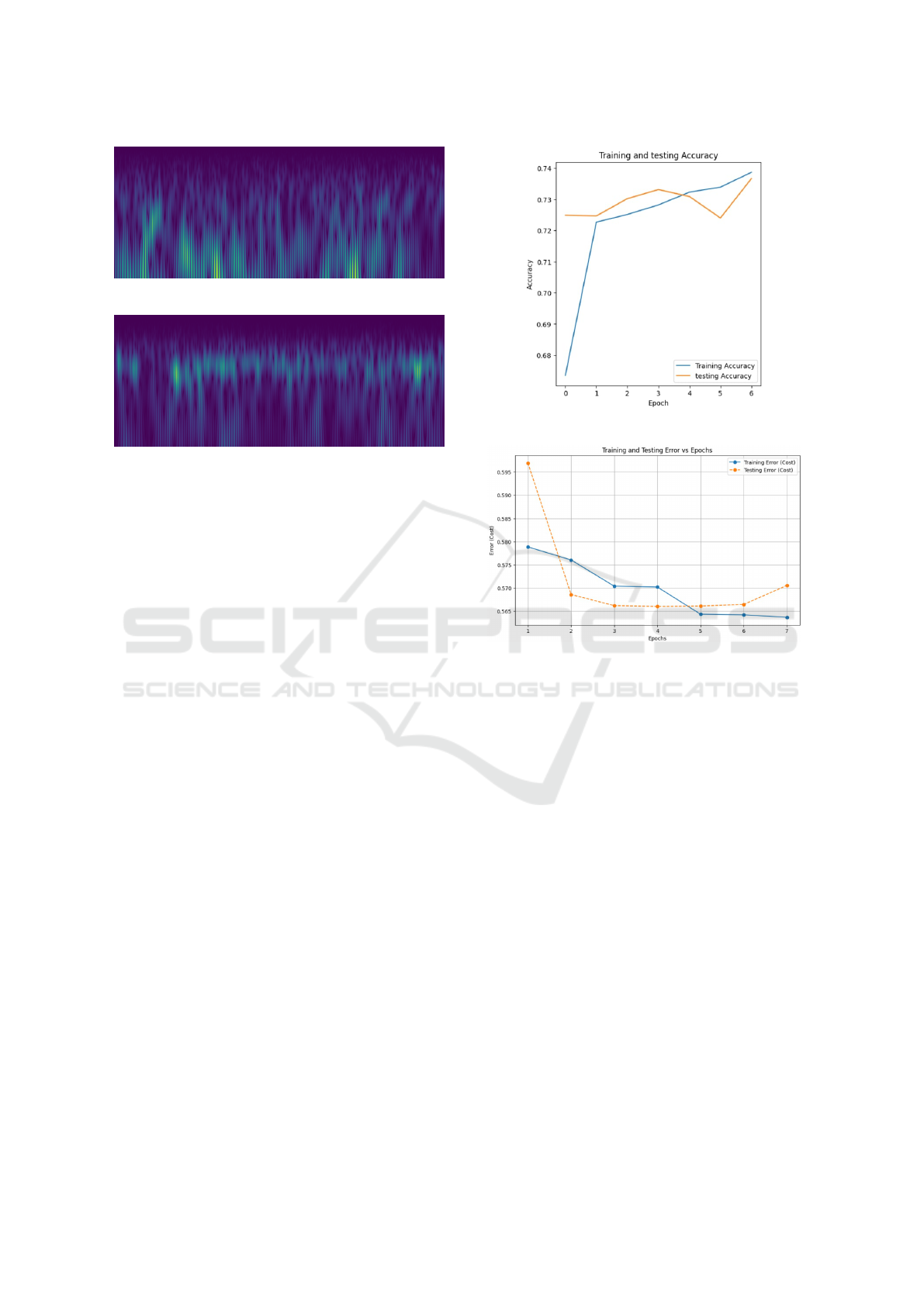
Figure 3: Focal Scalagram image (Seizure)
Figure 4: Non-Focal Scalagram image (Non-Seizure)
tures, pooling layers that reduce dimensionality, and
fully connected layers for final classification. The
training process involves optimizing a loss function,
typically using stochastic gradient descent and back-
propagation, to adjust weights and minimize classifi-
cation errors. Regularization techniques like dropout
are often applied to prevent overfitting. CNNs are
highly effective in capturing spatial patterns, making
them well-suited for tasks such as image recognition,
object detection, and, in this case, epilepsy detection
from EEG-based scalogram images.
The custom CNN for a multi-class classification
problem consists of sequential convolutional blocks
with increasing filters (32, 64, 128, and optionally
256), each containing a Conv2D layer with ReLU ac-
tivation, batch normalization for stability, and Max-
Pooling2D for down-sampling. To prevent overfit-
ting, L2 regularization is applied to both the convo-
lutional and dense layers, and dropout is used in the
dense layers for further regularization. The fully con-
nected layers include a flatten layer to convert fea-
ture maps into a 1D vector, followed by dense lay-
ers with ReLU activation to learn complex represen-
tations. The model uses the Adam optimizer, an adap-
tive method suitable for deep learning tasks.
After training the CNN model for 50 epochs with
early stopping at 11, it achieved a training accuracy of
74.07%, indicating that the model effectively learned
patterns from the training data and the testing accu-
racy is 73.22%.
Figure.5. This graph depicts the training and test-
ing accuracy of a model over several epochs (labeled
on the x-axis). The training accuracy (blue line)
starts higher and maintains relatively stable values
with slight fluctuations, while the testing accuracy
(orange line) begins lower but rises sharply, eventu-
Figure 5: Training vs testing accuracy for custom CNN
Figure 6: Training and testing error vs Epoch for Custom
CNN
ally fluctuating and surpassing the training accuracy
in later epochs. This pattern may suggest overfitting
at the final epochs, as the testing accuracy’s instabil-
ity could indicate sensitivity to the validation set. The
testing accuracy surpassing training at times suggests
randomness in the dataset split. However, the con-
vergence of both accuracies around epoch 4 indicates
balanced model performance during this phase.
Figure.6. The graph shows the training and test-
ing loss of a machine learning model over six epochs.
The training loss (blue line) starts at a moderate level,
gradually decreasing as the model learns from the
training data. The testing loss (orange line), which
measures the model’s performance on unseen data,
begins at a higher value but initially drops sharply, in-
dicating improvement in generalization. After a few
epochs, the testing loss increases slightly, where the
model begins to perform better on training data.
4.2 VGG 16
VGG16 is a pre-trained model and is well-suited for
study due to its ability to effectively extract spatial
features from images. EEG signals converted into vi-
INCOFT 2025 - International Conference on Futuristic Technology
780
sual representations, such as scalogram images, con-
tain patterns indicative of epileptic activity. VGG16’s
architecture, with its small 3x3 convolutional filters,
is adept at capturing these fine-grained spatial de-
tails. Additionally, using a pre-trained VGG16 model
through transfer learning allows leveraging its general
feature extraction capabilities while fine-tuning it for
the specific task of epilepsy detection. This approach
is particularly advantageous when working with lim-
ited data, as it reduces the need for extensive training
from scratch and ensures robust performance in clas-
sifying complex patterns in EEG images.
Learning Rate: 0.001, adjusted dynamically using
schedulers. Batch Size: 32 for balanced efficiency
and performance. Epochs: 25 with early stopping
to prevent overfitting. Optimizer: Adam for faster
convergence and adaptive learning. Image Resolu-
tion: 128x128 for consistent input size. Dropout layer
to prevent overfitting and also applied the L2 regu-
larization Loss Function : Binary cross entropy for
binary classification Activation Function : We used
both Relu and Sigmoid
Figure 7: Training vs testing accuracy for VGG-16
Figure.7. illustrates the training and validation ac-
curacy over epochs. Initially, both training and val-
idation accuracy increase, indicating that the model
is learning effectively. Around epoch 10, the vali-
dation accuracy starts to fluctuate, showing signs of
overfitting as the training accuracy continues to im-
prove steadily while the validation accuracy varies.
Despite these fluctuations, the validation accuracy re-
mains relatively close to the training accuracy, sug-
gesting that the model is performing reasonably well.
Figure.8. shows the training and validation loss
over epochs. Both losses decrease steadily, indicating
that the model is learning and improving its predic-
tions. However, the validation loss consistently re-
mains lower than the training loss after a few epochs,
which might suggest differences in how the training
and validation datasets are handled. The smooth de-
cline in loss for both suggests that the training process
is stable, and there are no significant issues like over-
Figure 8: Training vs testing loss for VGG-16
Figure 9: The architecture of VGG16
fitting or divergence in this range of epochs.
Figure.9. illustrates the convolutional neural net-
work (CNN) architecture used for binary classifica-
tion of scalogram images into focal and non-focal
categories. The model processes input scalograms
through a series of convolutional layers, each extract-
ing increasingly complex features, with max-pooling
layers reducing the spatial dimensions. The extracted
features are then flattened and passed through fully
connected layers, culminating in a sigmoid-activated
output layer for binary classification. This archi-
tecture is adapted from the sea ice classification of
SAR imagery presented by Khaleghian et al. (2021)
(Khaleghian et al., 2021).
Figure 10: ROC curve of VGG-16
Epilepsy Diagnosis Using EEG Image Analysis
781
Figure.10. shows the ROC curve demonstrates the
performance of the model in distinguishing between
classes, with an Area Under the Curve (AUC) of
0.87. This indicates that the model has a high ability
to discriminate between focal and non-focal classes,
performing significantly better than random guessing
(represented by the diagonal line). The curve’s prox-
imity to the top-left corner suggests a good balance
between the true positive rate (sensitivity) and false
positive rate, making the model reliable for classifica-
tion tasks.
Table 1: Comparison results of custom CNN and VGG16
Parameter VGG16 Custom CNN
Training Accuracy 81.13% 74.07%
Testing Accuracy 80.04% 73.22%
5 CONCLUSIONS
This study highlights the potential of machine learn-
ing in automating epilepsy detection using EEG-
based scalogram images. The proposed custom CNN
model achieved a training accuracy of 74.07% and a
testing accuracy of 73.22%, demonstrating its abil-
ity to learn meaningful patterns from the data. Addi-
tionally, the VGG-16 model outperformed the custom
CNN, achieving a training accuracy of 81.13% and a
testing accuracy of 80.04%.
The CNN model has 9 layers but yet works good
in comparison with the 16 layers of VGG-16. These
results underscore the utility of advanced image-
based analytics in healthcare while also emphasizing
the importance of optimizing models for enhanced
performance and generalization. This project lays
a foundation for developing scalable, real-time sys-
tems for epilepsy diagnosis, with future work focus-
ing on improving model robustness, leveraging larger
and more diverse datasets, and exploring deployment
strategies for real-world applications.
REFERENCES
Georgis-Yap, Z., Popovic, M. R., and Khan, S. S.
(2024). Supervised and unsupervised deep learning
approaches for eeg seizure prediction. Journal of
Healthcare Informatics Research, 8(2):286–312.
Hafeez, M. A. and Shakil, S. (2024). Eeg-based stress iden-
tification and classification using deep learning. Mul-
timedia Tools and Applications, 83(14):42703–42719.
Hu, D., Cao, J., Lai, X., Wang, Y., Wang, S., and Ding, Y.
(2020). Epileptic state classification by fusing hand-
crafted and deep learning eeg features. IEEE Trans-
actions on Circuits and Systems II: Express Briefs,
68(4):1542–1546.
Jridi, A., Djemal, R., and Belwafi, K. (2024). Neurologi-
cal disorder diagnosis through deep residual network-
based eeg signal analysis. In 2024 IEEE 7th Interna-
tional Conference on Advanced Technologies, Signal
and Image Processing (ATSIP), volume 1, pages 144–
149. IEEE.
Khaleghian, S., Ullah, H., Kræmer, T., Hughes, N., Eltoft,
T., and Marinoni, A. (2021). Sea ice classification
of sar imagery based on convolution neural networks.
Remote Sensing, 13(9):1734.
Khasawneh, N., Fraiwan, M., and Fraiwan, L. (2022). De-
tection of k-complexes in eeg waveform images using
faster r-cnn and deep transfer learning. BMC Medical
Informatics and Decision Making, 22(1):297.
Krishnan, P. T., Erramchetty, S. K., and Balusa, B. C.
(2024). Advanced framework for epilepsy detection
through image-based eeg signal analysis. Frontiers in
Human Neuroscience, 18:1336157.
Krishnasamy, L., Sriwastav, Y. K., Bharat, S. P., and
Ganachar, S. R. (2024). Epilepsy detection using su-
pervised learning algorithms. In 2024 IEEE Interna-
tional Conference on Contemporary Computing and
Communications (InC4), volume 1, pages 1–6. IEEE.
Kunekar, P., Gupta, M. K., and Gaur, P. (2024). Detection of
epileptic seizure in eeg signals using machine learning
and deep learning techniques. Journal of Engineering
and Applied Science, 71(1):21.
Majzoub, S., Fahmy, A., Sibai, F., Diab, M., and Mahmoud,
S. (2023). Epilepsy detection with multi-channel eeg
signals utilizing alexnet. Circuits, Systems, and Signal
Processing, 42(11):6780–6797.
Pattnaik, S., Rao, B. N., Rout, N. K., and Sabut, S. K.
(2024). Transfer learning based epileptic seizure clas-
sification using scalogram images of eeg signals. Mul-
timedia Tools and Applications, pages 1–15.
Sadam, S. S. P. and Nalini, N. (2024). Epileptic seizure de-
tection using scalogram-based hybrid cnn model on
eeg signals. Signal, Image and Video Processing,
18(2):1577–1588.
Saleem, A., Khan, M. A., and Yousaf, H. M. (2023). Ad-
vancing epilepsy disease classification through ma-
chine learning and deep learning models utilizing eeg
data. In 2023 17th International Conference on Open
Source Systems and Technologies (ICOSST), pages 1–
8. IEEE.
Shankar, A., Chakraborty, D., Saikia, M. J., Dandapat, S.,
and Barma, S. (2023). Seizure type detection using
eeg signals based on phase synchronization and deep
learning. In 2023 IEEE 19th International Conference
on Body Sensor Networks (BSN), pages 1–5. IEEE.
Sharma, R. and Meena, H. K. (2024). Enhanced epilep-
tic seizure detection through graph spectral analysis of
eeg signals. Circuits, Systems, and Signal Processing,
pages 1–21.
Supriya, S., Siuly, S., Wang, H., and Zhang, Y. (2021).
Epilepsy detection from eeg using complex network
techniques: A review. IEEE Reviews in Biomedical
Engineering, 16:292–306.
INCOFT 2025 - International Conference on Futuristic Technology
782

Wang, X., Wang, Y., Liu, D., Wang, Y., and Wang, Z.
(2023). Automated recognition of epilepsy from eeg
signals using a combining space–time algorithm of
cnn-lstm. Scientific Reports, 13(1):14876.
Epilepsy Diagnosis Using EEG Image Analysis
783
