
Automatic Detection of Cardiovascular Abnormalities in ECG Images:
CNN and MobileNet
Aditi Jambotkar
a
, Regina Fernandes
b
, Shreya Arun Miskin
c
, Prema T. Akkasaligar
d
and Rajashri Khanai
e
Department of Computer Science and Engineering, KLE Tech. University’s Dr. MSSCET, Belagavi, India
Keywords:
ECG Images, CNN, MobileNet, Cardiovascular Diseases.
Abstract:
Cardiovascular diseases are becoming a leading cause of death worldwide. Detection of irregular heart activi-
ties like arrhythmia and heart attacks are critical for timely treatment. Automation in detecting cardiovascular
abnormalities is essential for providing timely diagnosis, especially in resource-limited settings where trained
medical professionals may be scarce. The paper aims to detect cardiovascular abnormality in ECG images
automatically using deep learning techniques. It uses a Convolutional Neural Network(CNN) and MobileNet
for efficient and lightweight processing. The MobileNet model outperforms the CNN model, demonstrating
superior accuracy, precision and recall. The results show the potential of deep learning models in enhancing
the accuracy and automation of cardiovascular abnormality detection through ECG analysis. By automating
ECG interpretation, it enables early detection of abnormalities, reduces diagnostic delays, and improves pa-
tient care, particularly in resource-constrained settings.
1 INTRODUCTION
Electrocardiogram (ECG) imaging is an essential di-
agnostic tool used in cardiology to measure heart ac-
tivity. It monitors the electrical signals of the heart
and is basically used for detecting various abnormal-
ities in heart activities like arrhythmia and myocar-
dial infractions. Abnormalities in ECG images often
indicate severe cardiovascular conditions that require
immediate medical attention. Delayed diagnosis can
lead to critical consequences for patients. Automated
abnormality detection plays a crucial role in address-
ing this issue by enabling quick and accurate analysis
of ECG data, facilitating early and effective treatment.
Cardiovascular diseases are the leading cause of
death globally, taking an estimated 17.9 million lives
each year. According to (World Health Organiza-
tion, 2015), in 2000, around 14 million people died
from cardiovascular diseases globally, while in 2019,
it reached close to 18 million. The emerged need for
improved healthcare systems is more important than
a
https://orcid.org/0009-0002-9983-615X
b
https://orcid.org/0009-0002-4838-3824
c
https://orcid.org/0009-0001-6206-2309
d
https://orcid.org/0000-0002-2214-9389
e
https://orcid.org/0000-0002-5080-722X
ever, particularly as cardiovascular diseases remain to
be the leading cause of death worldwide. ECG imag-
ing is at forefront of heart care monitoring due to its
ability to capture the critical data of the heart.
(Agarwal et al., 2024) presents a novel approach
for detecting abnormalities in ECG images by uti-
lizing a MobileNet based CNN autoencoder. The
lightweight architecture is designed for efficient pro-
cessing, making it ideal for real-time applications and
devices with limited computational resources. The
autoencoder learns compact representations of ECG
images during the encoding phase and reconstructs
them during decoding, allowing the system to iden-
tify abnormalities based on discrepancies between the
original and reconstructed images. By leveraging Mo-
bileNet’s efficiency and the autoencoder’s capability
to highlight subtle deviations, the method achieves
high diagnostic accuracy. The study demonstrates the
potential of this approach for improving anomaly de-
tection in ECG images, ensuring reliability and scala-
bility across different datasets and clinical scenarios.
The MobileNet50 CNN autoencoder method for ECG
anomaly detection has several potential drawbacks. It
faces challenge with overfitting on limited or biased
datasets, reducing generalizability across diverse pop-
ulations. Handling noisy ECG signals, common in
real-world scenarios, also degrade performance. The
754
Jambotkar, A., Fernandes, R., Miskin, S. A., Akkasaligar, P. T. and Khanai, R.
Automatic Detection of Cardiovascular Abnormalities in ECG Images: CNN and MobileNet.
DOI: 10.5220/0013601600004664
Paper published under CC license (CC BY-NC-ND 4.0)
In Proceedings of the 3rd International Conference on Futuristic Technology (INCOFT 2025) - Volume 2, pages 754-761
ISBN: 978-989-758-763-4
Proceedings Copyright © 2025 by SCITEPRESS – Science and Technology Publications, Lda.

method’s reliance on a complex deep learning archi-
tecture introduces latency and impacting real-time ap-
plications. Additionally, without thorough compar-
ison to simpler techniques or clinical validation, its
real-world efficacy remains uncertain. Addressing
these issues with robust datasets enhance its practical
applicability. The deep-learning models for MI
detection and QRS complex detection face many chal-
lenges. Deep learning techniques used like 1D CNNs
and hybrids of CNN-LSTM are limited by high com-
putational resources and data dependency. Image-
based methods and low-quality signal processing ma-
jorly have difficulties with noise reduction, feature ex-
traction etc. The models perform better in some en-
vironments but struggle with over-fitting which im-
pacts the real-world applications. Enhanced machine
learning and deep learning algorithms need thorough
validation in clinical settings.
The challenges observed in the problem space in-
clude data availability and format, where the open-
access datasets for ECG images are limited. ECG
data is usually recorded on paper in clinical settings
which makes it difficult to digitize, store and ana-
lyze the data. ECG images usually consists of text-
annotations, grid lines and other background elements
that often interfere with the image extraction. The
other challenges include model performance and val-
idation, where achieving high sensitivity is crucial as
false negatives in MI detection can have serious health
consequences. These challenges highlight the impor-
tance of using reliable image processing and machine
learning methods to accurately identify heart diseases
using ECG images.
The present study introduces an advanced abnor-
mality detection model that utilizes CNNs for accu-
rate and reliable classification. The model is specif-
ically designed to handle ECG image data, ensuring
that it undergoes thorough preprocessing to improve
input quality. By leveraging the preprocessing steps,
the model enhances the consistency and quality of the
ECG image data. Furthermore, the CNN-based archi-
tecture excels in extracting meaningful and relevant
features from the processed ECG images, enabling ef-
ficient classification of abnormalities. This approach
aims to address challenges in medical diagnostics by
providing a robust and precise solution for ECG anal-
ysis.
The paper is organized as follows. Section 2 pro-
vides a brief review of the literature survey on the
recent works. Section 3 contains the problem state-
ment, background and provides proposed methodol-
ogy. Section 4 discusses the implementation details
along with results and discussions. Finally, the paper
concludes in Section 5.
2 RELATED WORKS
The detection of ECG peaks has seen exceptional
progress with the application of advanced image
processing techniques and deep learning algorithms.
This survey explores a wide range of methods pro-
posed by researchers for accurate and efficient detec-
tion of ECG features which enables the detection of
arrhythmia and other cardiac conditions.
(Sane et al., 2021) developed a computerized
method for detecting Myocardial Infarction(MI) by
using a dataset containing 12-lead ECG images. They
included a two-step approach which involved im-
age processing to extract ECG signals from the im-
ages, followed by a one-dimensional CNN to classify
MI. The model is validated on the PTB diagnostic
database. This method avoids the need for manual
computation and handcrafted features. The advantage
of this model is that it is adaptable to various datasets
and provides real-time assessment, which makes it
well-suited for critical health conditions. However,the
limitation lies in the challenge of extracting signals
from ECG images and the lack of open access to ECG
image datasets.
(Zhou et al., 2020) introduced a novel deep-
learning algorithm for the real-time prediction of R-
peaks in ECG signals using a combined model of
CNN and LSTM network. The designed strategy is to
predict the next R-peak by computing the variability
of previous ECG intervals which indicate the poten-
tial future health problems like depression, anxiety,
asthma and sudden infant death. It also proves to be
a strong indicator for the onset of myocardial infrac-
tions. The model is validated on MIT-BIH arrhyth-
mia dataset and the combined model outperforms the
standalone models like CNN or LSTM by combin-
ing their strengths. CNN has been used for filtering
noise and extracting visual pattern, LSTM for han-
dling temporal dynamics in ECG signals. The advan-
tage of the model includes its ability to perform real-
time monitoring. However, the accessibility of high-
quality ECG data and computational resources are the
key-issues.
(Yuen et al., 2019) developed a innovative CNN-
LSTM model for identifying QRS complexes in noisy
ECG signals. In this approach, CNN is used to extract
features, LSTM identifies the QRS complex timings,
and a multi-layer perceptron makes the final predic-
tions. The model performs well in scenarios where
the training and testing datasets have data from dif-
ferent patients. It is tested using the MIT-BIH dataset
and evaluated based on metrics like precision, recall,
and F1 score. Advantages of this approach include
adaptability to noisy signals which makes it suitable
Automatic Detection of Cardiovascular Abnormalities in ECG Images: CNN and MobileNet
755

to generalize on unseen patient data.
(Cai and Hu, 2020) introduced two deep learn-
ing models for QRS complex detection in ECG sig-
nals: a CNN and a hybrid Convolutional Recurrent
Neural Network(CRNN). The CNN is fundamentally
conposed of convolutional blocks and Sqeeze-and-
Exitation networks, while the CRN combines both
convolutional and recurrent layers to improve feature
extraction and temporal dependency learning. The
model is evaluated on four open access ECG datasets.
Advantages of this model involves its noise resistance
and high accuracy, however it faces issues such as
over-fitting, computational complexity which limits
their effectiveness in real-time applications.
(Zahid et al., 2022) created a reliable system to
detect R-peaks in low-quality Holter ECG signals. It
uses a 1D CNN combined with a verification model
to reduce false alarms. The approach includes a
encoder-decoder structure that generates a 1D seg-
mentation map for precise localization of R-peaks
from ECG inputs. The model is tested on China Phys-
iological Signal Challenge(CPSC) and MIT-BIH Ar-
rhythmia database, achieving a excellent performance
on F1 score on CPSC database while showing better
results on MIT-BIH database. The advantages of this
approach lies in the adaptability to low-quality signals
and its generalization across different datasets, which
makes it suitable for real-time applications. How-
ever,the issue of over-fitting remains as a challenge,
particularly in variable ECG environments.
(Das, 2024) proposed a comprehensive system
for blood pressure prediction using three machine
learning algorithms: Support Vector Classifier (SVC),
Random Forest Classifier, and Naive Bayes Classi-
fier. The approach is tested on a dataset consisting
of 3000 image samples, of which 2005 represented
cases of high blood pressure and 495 represented nor-
mal blood pressure. The strength of the model lies in
the synergistic use of multiple algorithms, which sig-
nificantly enhances the accuracy and reliability of pre-
dictions by leveraging the strengths of each method.
SVC effectively handles the separation of complex
data, Random Forest contributes robustness through
ensemble learning, and Naive Bayes adds simplicity
and speed to the classification process. One major
drawback is the increased computational complexity,
which arises from training multiple algorithms and
integrating their outputs. Efficiently handling large
datasets remains challenging due to high memory us-
age and processing time.
(Yang et al., 2021) proposed a hybrid deep learn-
ing model for non-invasive, cuff-less blood pressure
estimation. The study focuses on using raw ECG im-
ages directly as input for deep learning models. Two
types of experiments are caried out. In the first, phys-
ical characteristics and features from the ECG signals
are extracted and used with traditional ML techniques
like SVR, LASSO, Ridge regression, KNN, Multi-
ple Linear Regression, and AdaBoost. In the second,
DL models such as CNNs, LSTMs, and fully con-
nected networks are applied for testing and analysis
of model. The advantage of the model is its ability
to automatically extract features from raw PPG and
ECG signals which reduces the complexity of use for
users. However, a disadvantage is the model’s re-
liance on insufficient data for optimal performance.
Table 1 illustrates recent 2024 literature surveys for
cardiovascular diseases detection in ECG Images.
In conclusion, the literature survey provides a
comprehensive overview of research on abnormalities
in ECG images, highlighting a wide range of stud-
ies that identify key trends and systematic methodolo-
gies. By addressing the existing gaps in this domain,
the study aims to contribute significantly to the ad-
vancement of accurate and efficient detection of car-
diovascular abnormalities.
3 PROPOSED METHODOLOGY
The study aims on detecting abnormalities in ECG
images by utilizing advanced and enhanced deep
learning techniques to identify irregular heart activ-
ities such as arrhythmia and MI. Consider a patient
experiencing chest pain. An ECG is conducted, and
the image is analyzed for any signs of MI or other
irregular heart conditions. Currently, the analysis is
performed manually by trained professionals. How-
ever this can lead to delays and many inconsisten-
cies. An automated model quickly detects whether
the ECG contains any abnormalities which takes less
usage of time and treatment decisions can be taken on
time.
The objectives are as follows :
• To preprocess the data by cleaning it to improve
the image quality of ECG images for better per-
formance of the model.
• To design and implement robust deep learning
models for accurate detection and classification of
abnormalities in ECG signals.
• To assess the developed model’s performance by
using the evaluation metrics.
Quality of ECG images is assumed that the ECG
images provided are of acceptable resolution and
sufficient quality for processing is present. The ECG
images are assumed to have consistent dimensions
INCOFT 2025 - International Conference on Futuristic Technology
756
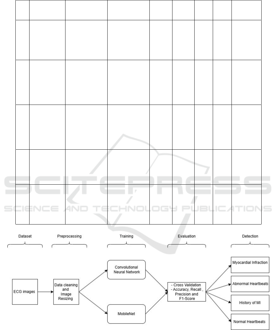
Table 1: Literature Surveys for Cardiovascular Diseases Detection.
Year Paper Title Dataset Approach Accuracy Precision Recall F-score Gaps identi-
fied
2024 Detection of
Cardiovascular
Disease Using
ECG images
(Jessy et al.,
2024)
ECG dataset col-
lected from hospitals
SVM, K-NN, DT,
RF, Naive Bayes
98.23% 98.31% 97.50% 97.90% Limited gen-
eralizability;
focus on
classification
only
2024 Interpreting
Deviant Heart
Patterns: Mo-
bileNet CNN
Autoencoder
(Agarwal et al.,
2024)
ECG images dataset
with anomalies
MobileNet (CNN) 76.93% 55.23% 70.00% 61.63% Low accuracy
and not effec-
tive on larger
ECG datasets
2022 Pan-
Tompkins++:
A Robust Ap-
proach to Detect
R peaks in ECG
Signals (Imtiaz
and Khan, 2022)
MIT-BIH Arrhyth-
mia, European ST-T,
PhysioNET PTB,
Atrial Fibrillation
dataset
CNN, CRNN 97.49% 95.89% 96.00% 95.94% High process-
ing demands
2022 Energy Efficient
Compression Al-
gorithm of ECG
Signals (Fathi
et al., 2022)
Real-time ECG im-
ages dataset
Krawtchouk and
AALO algorithm
92.00% 91.00% 90.00% 90.50% Lack of
anomaly
detection
capabilities
2022 Intelligent Sys-
tem for ECG
Classification
Using CNN
(Hammad and
Abdulbaqi, 2022)
Publicly available
ECG dataset
1D CNN 90.00% 88.00% 87.00% 87.50% Limited in
handling
diverse ECG
signal varia-
tions
Figure 1: Proposed Methodology for Cardiovascular Abnormality Detection.
after pre-processing, making them compatible with a
CNN model. It is assumed that a sufficient amount of
labeled ECG image data is provided for testing and
training.
The proposed solution utilizes deep learning
models, specifically CNN and MobileNet, to au-
tomate the detection of ECG signal abnormalities,
offering a fast, accurate, and reliable diagnostic tool
Automatic Detection of Cardiovascular Abnormalities in ECG Images: CNN and MobileNet
757

for cardiovascular diseases. The methodology begins
by preprocessing ECG signals to ensure consistency
across ECG samples. CNN extracts critical temporal
and spectral features, such as P and T waves and QRS
complexes, which are vital for identifying cardiac
conditions. Its convolutional layers capture complex
patterns, pooling layers reduce dimensionality while
preserving essential information. The fully connected
layers classify signals into normal or abnormal cate-
gories. Complementing this, MobileNet a lightweight
architecture, is fine-tuned on the ECG dataset for
efficient real-time analysis, using depthwise separa-
ble convolutions to optimize feature extraction with
minimal computational overhead. A global average
pooling layer and customized dense layers adapt
MobileNet for ECG classification. Both models are
trained on the same dataset with early stopping to
avoid overfitting and ensure optimal performance.
Comprehensive evaluation metrics, including ac-
curacy, precision, recall, F1-score, and confusion
matrix, are used to assess their effectiveness. The
framework uses CNN’s robustness and MobileNet’s
efficiency to deliver a scalable and accessible solution
for ECG analysis.
The schematic diagram illustrating the proposed
methodology is depicted in Figure 1. Initially, ECG
images are drawn from the dataset consists of la-
beled samples of ECG signals. Preprocessing steps
such as resizing the images to a standard dimension
and normalizing pixel intensity values are performed
to ensure data consistency and quality. These steps
aim to enhance the clarity of the ECG images, which
is crucial for accurate feature extraction. The pre-
processed ECG images are then trained using CNN
and MobileNet models. The CNN architecture in-
corporates convolutional layers that use filters to ex-
tract local features, pooling layers to reduce spa-
tial dimensions, and fully connected layers to pre-
dict the class labels by analyzing the acquired fea-
tures. Activation function, such as ReLU, introduce
non-linearity, enabling the network to learn complex
patterns and relationships in ECG images. CNNs ex-
cel in automatically extracting hierarchical represen-
tations of features from ECG images, which facili-
tates effective classification of normal and abnormal
ECG. Similarly, the MobileNet model is employed to
efficiently analyze ECG images with reduced com-
putational complexity. It uses depth-wise separable
convolutions to optimize the feature extraction pro-
cess while maintaining high classification accuracy.
This makes it particularly suitable for real-time appli-
cations in clinical settings. Finally, the trained mod-
els are evaluated using performance metrics such as
accuracy, precision and recall to validate their effec-
tiveness. The evaluated models are then used to detect
abnormalities in new ECG images, classifying them
into normal or abnormal categories. The results are
visualized to provide clinicians with interpretable in-
sights, aiding in decision-making for cardiac health
monitoring and diagnosis.
4 RESULTS AND DISCUSSION
The experiments are conducted on a machine with
an Intel(R) core(TM) i3-7020U processor operating
at 2.30 Ghz equipped with 4 GB RAM and running
on Windows 10. The implementation is carried out
using Python, with Tensorflow and keras serving as
the primary libraries for developing the deep learn-
ing models. Additional libraries such as NumPy and
Pandas are used for efficient data manipulation and
analysis.
Figure 2: Sample Images: (a) Myocardial Infraction, (b)
Abnormal Heartbeats, (c) History of MI, (d) Normal Heart-
beats.
The dataset used in this study is sourced from
mendeley data and comprises a collection of 12-lead
ECG images, categorized into various classes such
as normal, myocardial infarction, abnormal heartbeat,
and history of myocardial infarction. The Figure 2
shows the sample images. The proposed method is fo-
cused on the targeted analysis of ECG images, specif-
ically concentrating on identifying abnormalities in-
dicative of cardiac conditions. The entire dataset
consists of 1,377 ECG images, divided into train-
ing and testing subsets. The training dataset com-
prises 929 samples, used to train the model by extract-
ing and learning features, essential for distinguish-
ing normal from abnormal ECG images. The testing
dataset includes 448 samples, reserved for evaluating
the model’s performance and generalization capabil-
ity. The dataset ensures a balanced representation of
normal and abnormal ECG images to support effec-
INCOFT 2025 - International Conference on Futuristic Technology
758

tive classification and robust model performance. In
the study, a comprehensive approach is applied for
the detection and classifications of ECG peaks, aim-
ing to distinguish between normal and abnormal pat-
terns. The approach focuses on automating the ECG
images, which is critical in diagnosing cardiac condi-
tions. Two deep learning models, a custom CNN and
MobileNet architectures are implemented to perform
the classification task.
The first step in this study involves designing and
training a custom CNN to classify the ECG images
into four predefined categories. The architecture con-
sisted of an input layer accepting images of size(100,
100, 3) followed by three CNN filter sizes of 32, 64
and 128. These layers are accompanied by ReLU ac-
tivation functions and max-pooling layers to reduce
the spatial dimensions of the feature maps. The fi-
nal layers include dense layer with 128 neurons us-
ing ReLU activation function. The output layer con-
sists of four neurons employing the softmax function
for multi-class classification, where the four neurons
indicate myocardial infarction, abnormal heartbeats,
history of myocardial infarction, and normal heart-
beats. The model is trained over 20 epochs. The per-
formance of the CNN model is remarkable achieving
a test accuracy of 90.98% and a minimal test loss of
0.029. It is observed that, as epochs increase, accu-
racy increases and loss decreases.
The next step employs MobileNet, a lightweight
and efficient CNN architecture, for image classifi-
cation. The approach is particularly effective for
medical imaging tasks due to its computational
efficiency and high accuracy. The base MobileNet
model is pre-trained on ImageNet data but initialized
with custom weights in this implementation. The
fully connected top layers are removed to allow
fine-tuning for type specific task. A custom classifier
is added, which includes a global average pooling
layer followed by two dense layers, one with 128
neurons using ReLU activation and the other with
softmax activation to output probabilities for multiple
classes. The MobileNet model is compiled using the
Adam optimizer with a learning rate of 0.0001. The
categorical cross entropy loss function is employed
as the dataset is multi-class in nature. Accuracy
is chosen as the evaluation metric. To prevent
overfitting and ensure early convergence, an early
stopping callback is applied monitoring validation
loss and restoring the best weights if no improvement
is observed after given consecutive epochs. Training
the model occurred over a minimum of 50 epochs,
although the early stopping criterion terminated
training early upon validation loss stabilization. The
training results showed a progressive improvement
in accuracy and reduction in loss over epochs. The
final evaluation gives test accuracy of approximately
91.73% and a test loss of 0.445. Figure 3, shows the
accuracy of the MobileNet model and it is observed
that, as epochs increase, accuracy also increases.
Figure 4, shows the loss of the MobileNet model and
it shows that, as epochs increase, loss decreases.
The confusion matrix in Figure 8 shows the
performance of the model on the ECG dataset. It
is observed that the model accurately classifies the
majority of normal and abnormal heartbeats, as well
as MI and patients with MI history. However, there
are a few misclassifications, with a small number of
abnormal heartbeats being misclassified as normal,
and vice versa. Overall, the loss is minimal, indi-
cating that the model performs well in detecting and
classifying ECG images. These highlight the capabil-
ity of the MobileNet model in effectively classifying
images. Compared to traditional CNN’s, MobileNet
offered a significant advantage in terms of reduced
computation and memory requirements while main-
taining high accuracy. The efficiency is achieved by
employing depth wise separable convolutions which
reduces the number of parameters and computational
cost. The inclusion of dropout layers and global
average pooling further reduced overfitting while
enabling robust feature extraction. In summary, the
MobileNet-based models successfully demonstrated
high accuracy and efficiency, underlining its potential
for real-world applications in detecting abnormalities
in ECG images.
The Table 2 clearly highlights the superiority
of MobileNet over CNN in detecting ECG abnor-
malities. MobileNet achieves a higher accuracy of
91.73% compared to CNN’s 90%, and excels with an
F1-Score of 96% versus CNN’s 95%. MobileNet also
outperforms CNN in precision and recall, achieving
96% for both, compared to CNN’s 95% precision and
94% recall, further emphasizing its effectiveness in
minimizing false positives and negatives. When com-
pared to other existing methods, such as the approach
proposed by (Agarwal et al., 2024), which focuses
on ECG anomaly detection using MobileNet50 CNN
autoencoder, and the method by Sane et al. (Cai and
Hu, 2020), which centers on detecting myocardial
infarction from 12-lead ECG images, MobileNet
demonstrates superior performance. The methods in
(Agarwal et al., 2024) and (Cai and Hu, 2020) report
lower accuracies and F1-Scores, with precision and
recall metrics that fall short of MobileNet’s consistent
96% values. Overall, MobileNet outperforms CNN
and existing methods in accuracy, precision, recall,
and F1-Score, solidifying its position as a highly
Automatic Detection of Cardiovascular Abnormalities in ECG Images: CNN and MobileNet
759
effective model for detecting abnormalities in ECG
images.
Figure 3: Accuracy of MobileNet model.
Figure 4: Loss of MobileNet model.
5 CONCLUSIONS
The study utilizes advanced DL models: CNN and
MobileNet, for classification of ECG images into four
classes to detect abnormalities. The CNN model, fea-
turing three convolutional layers to extract key pat-
Figure 5: Confusion Matrix of MobileNet.
Table 2: Quantitative Comparison of CNN and MobileNet.
Performance parameters (Sane
et al.,
2021)
(Agarwal
et al.,
2024)
CNN MobileNet
Accuracy 86.21% 76.93% 90% 91.73%
Precision 91.30% 55.23% 95% 96%
Recall 85% 70% 94% 96%
F1-Score 88.05% 61.63% 95% 96%
terns like P waves, QRS complexes, and T waves, is
followed by dense layers for classification. Trained
over 20 epochs, it achieved a test accuracy of 90.98%,
highlighting its efficacy in diagnosing cardiovascu-
lar conditions. MobileNet, a lightweight and pre-
trained DL architecture, is also employed for this
study. Known for its computational efficiency, Mo-
bileNet demonstrated robust performance by lever-
aging its depthwise separable convolutional layers to
extract features efficiently. Fine-tuned for the ECG
classification task, MobileNet achieved a test accu-
racy of 91.73%, proving its adaptability and effective-
ness in handling medical image data. MobileNet’s
lightweight design and faster inference time make
it highly suitable for real-time applications, such as
portable ECG monitoring devices and telemedicine
platforms. Among the two approaches, MobileNet
emerges as the more practical solution for real-world
deployment due to its efficient architecture and scal-
ability. However, the MobileNet model demonstrates
superior accuracy, showcasing its capability for de-
tailed and precise classification, which is particularly
beneficial in controlled or research settings. Future
advancements aim to improve ECG analysis by devel-
oping more accurate and efficient tools for detecting
cardiac anomalies. These innovations will leverage
AI and real-time monitoring to enable early diagno-
sis and better outcomes. However, their applicability
may be limited to ECG-specific data, requiring com-
plementary tools for broader diagnostic needs.
INCOFT 2025 - International Conference on Futuristic Technology
760

REFERENCES
Agarwal, M., Gill, K. S., Malhotra, S., and Devliyal, S.
(2024). Decoding Abnormal Heart Patterns: ECG
Anomaly Detection using MobileNet50 CNN Autoen-
coder Method. In 2024 IEEE International Confer-
ence on Information Technology, Electronics and In-
telligent Communication Systems (ICITEICS), pages
1–4.
Cai, W. and Hu, D. (2020). Qrs Complex Detection using
novel Deep Learning Neural Networks. IEEE Access,
pages 97082–97089.
Das, S. (2024). Prediction of Blood Pressure using Machine
Learning. International Journal of Intelligent Systems
and Applications in Engineering, pages 579–586.
Fathi, I. S., Makhlouf, M. A. A., Osman, E., and Ahmed,
M. A. (2022). An Energy-Efficient Compression Al-
gorithm of ECG Signals in remote Healthcare Moni-
toring Systems. IEEE Access, pages 39129–39144.
Hammad, A. H. and Abdulbaqi, A. S. (2022). Intelli-
gent System for Classification ECG based on Con-
volutional Neural Networks (CNN). In 2022 8th In-
ternational Conference on Contemporary Information
Technology and Mathematics (ICCITM), pages 101–
106.
Imtiaz, M. N. and Khan, N. (2022). Pan-tompkins++: A
robust approach to detect R-Peaks in ECG Signals. In
2022 IEEE International Conference on Bioinformat-
ics and Biomedicine (BIBM), pages 2905–2912.
Jessy, K. J., Manikandan, G., Hemalatha, S., and Veronica,
V. (2024). Detection of Cardiovascular Disease using
ECG Images in Machine Learning and Deep Learn-
ing. International Journal of Scientific Research in
Science and Technology, pages 131–139.
Sane, R. K. S., Choudhary, P. S., Sharma, L. N., and Danda-
pat, P. S. (2021). Detection of Myocardial Infarction
from 12 lead ECG Images. In 2021 National Confer-
ence on Communications (NCC), pages 1–6.
World Health Organization (2015). Target 3.4: Re-
duce by one third Premature Mortality from Non-
Communicable Diseases through Prevention and
Treatment and Promote Mental Health and Well-
Being. Accessed: 2024-12-11.
Yang, S., Zhang, Y., Cho, S.-Y., Correia, R., and Morgan,
S. P. (2021). Non-invasive Cuff-Less Blood Pressure
Estimation using a hybrid Deep Learning Model. Op-
tical and Quantum Electronics, page 93.
Yuen, B., Dong, X., and Lu, T. (2019). Inter-patient CNN-
LSTM for QRS Complex Detection in noisy ECG Sig-
nals. IEEE Access, pages 169359–169370.
Zahid, M. U., Kiranyaz, S., Ince, T., Devecioglu, O. C.,
Chowdhury, M. E. H., Khandakar, A., Tahir, A., and
Gabbouj, M. (2022). Robust R-Peak Detection in low-
quality Holter ECGs using 1d Convolutional Neural
Network. IEEE Transactions on Biomedical Engi-
neering, pages 119–128.
Zhou, P., Schwerin, B., Lauder, B., and So, S. (2020). Deep
Learning for real-time ECG R-Peak Prediction. In
2020 14th International Conference on Signal Pro-
cessing and Communication Systems (ICSPCS), pages
1–7.
Automatic Detection of Cardiovascular Abnormalities in ECG Images: CNN and MobileNet
761
