
Hybrid CNN-ResNet50 Model for Brain Tumor Classification Using
Transfer Learning
Sheenam Middha
1a
, Sonam Khattar
1b
and Tushar Verma
2c
Department of Computer Science and Engineering, Chandigarh University, Mohali, Punjab, India
Keywords: Hybrid Deep Learning CNN Model, MRI Images, Brain Tumor, Classification.
Abstract: A tumor is fatal cancers that can affect both adults and minors. A brain tumor's treatment depends on an early
and precise diagnosis. Finding the brain tumor with computer-aided technologies is a crucial first step for
physicians. Experts can spot tumors more quickly and easily thanks to these devices. But conventional
procedures also prevent mistakes from happening. This article uses magnetic resonance imaging (MRI) to
diagnose brain tumors. A hybrid approach that uses CNN models—one of the deep learning networks—for
diagnosis has been put forth. One of the CNN models, Resnet50 architecture, serves as the foundation.97.67%
accuracy rate is achieved with this model. The model that performed the best out of all of them has been used
to classify the images of brain tumors. Consequently, further analyses in the literature indicate that the
suggested method is practical and useful for brain tumor detection in computer-aided systems.
1 INTRODUCTION
A brain tumor is an abnormal development of cells
inside the brain. While some brain tumors are benign,
some could be cancerous. Brain tumors that originate
from the actual tissue of the brain are known as
primary brain tumors. Metastasis is the term used to
describe a malignant tumor that has moved from
another area of the body to the brain. The type,
location, and range of the tumor can all affect the
available treatment options. Therapy or symptom
reduction is the main goal of treatment. The tumor
symptoms include migraines and recurrent
headaches. It may still lead to visual impairment. At
this point, science may not know enough about what
caused the tumor's extraordinary growth in the first
place. Tumors can be classified based on two factors,
such as where they originated from and whether or
not they are cancerous. A benign tumor is a non-
cancerous tumor that does not impact any other
portion of the human body (Chen, Liu, et al. 2018),(
Sultan, Upadhyay, et al. 2019)
(Hossain, Shishir, et al. 2019). They have a modest
pace of expansion and are easily recognizable.
a
https://orcid.org/0000-0002-0639-5539
b
https://orcid.org/0000-0002-5444-4358
c
https://orcid.org/0000-0002-6696-4537
Malignant brain tumors, which are founded on
cancer and can impact other brain regions, can be
extremely violent and terrifying since they can be
difficult to diagnose. When it comes to detecting a
tumor, the physicians will decide between an X-ray
and magnetic resonance imaging (MRI). If no
examination is able to provide sufficient information,
an MRI scan may be appropriate. The MRI scan uses
radio waves and magnetism features to create
flawless images.
MRI scan of the brain can provide a safe and
painless experiment that uses magnetic fields and
radio waves to provide detailed images of the human
brain. As an alternative to a Computed Tomography
(CT) scan, an MRI scan doesn't use radiation. MRI
scanners typically have a large magnet field in the
shape of a doughnut with a channel in the middle. The
patients will be positioned on a table that slides into
the channel for this testing procedure. Numerous
locations with better opening in MRI machines are
available, which can help individuals who are
claustrophobic (Anaraki, Ayati, et al. 2019),
(Özyurt, Sert, et al. 2019). A brain examination
called an MRI machine is offered in radiology centers
and hospitals. During the testing procedure, radio
444
Middha, S., Khattar, S. and Verma, T.
Hybrid CNN-ResNet50 Model for Brain Tumor Classification Using Transfer Learning.
DOI: 10.5220/0013594400004664
Paper published under CC license (CC BY-NC-ND 4.0)
In Proceedings of the 3rd International Conference on Futur istic Technology (INCOFT 2025) - Volume 2, pages 444-448
ISBN: 978-989-758-763-4
Proceedings Copyright © 2025 by SCITEPRESS – Science and Technology Publications, Lda.
waves are used to pinpoint the magnetic location of
the atoms in the human body. These signals can then
be chosen by a powerful antenna and transmitted to a
computer. The computer is capable of carrying out
millions of estimations, producing clear and white
photographs of the body.
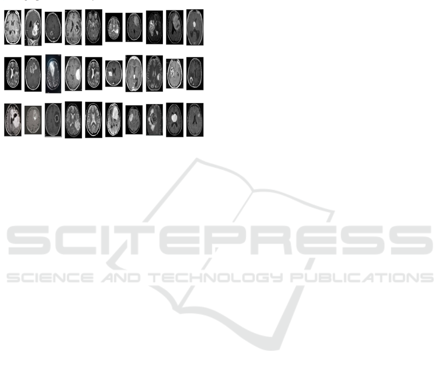
Figure 1: MRI images
Stages Of Brain Cancer
• Under a microscope, grade I tumors appear
nearly normal and are characterized by
sluggish growth. These tumors are classified
as benign. Since these tumors usually have
distinct borders, surgical removal of them is
less difficult.
• Low-grade malignant tumors, or grade II
tumors, develop more slowly than other
tumor types and have the ability to invade
adjacent brain tissue. Under a microscope,
the cells of Grade II tumors appear slightly
aberrant, suggesting a degree of malignancy.
• Anaplastic or malignant tumors, commonly
referred to as grade III tumors, grow quickly
and are more aggressive. Under a
microscope, the cells in these tumors appear
incredibly aberrant, and they are probably
going to migrate into adjacent brain tissue.
Grade III cancers include anaplastic
oligodendroglioma and anaplastic
astrocytoma. Aggressive treatment is needed
for these tumors, which includes radiation,
chemotherapy, and surgery. A thorough
treatment strategy and close monitoring are
crucial because the overall prognosis is less
favorable and there is a higher chance of
recurrence as compared to lower-grade
cancers.
• Grade IV tumors that are highly aggressive
include the widely recognized glioblastoma
multiforme. These tumors exhibit highly
rapid growth and, when observed under a
microscope, the cells appear highly
abnormal. Grade IV tumors often induce
angiogenesis to facilitate their rapid
proliferation. Despite intensive treatment, the
outlook for Grade IV tumors is quite
unfavorable and often involves a
combination of radiation, chemotherapy, and
surgery. The average survival duration for
individuals diagnosed with glioblastoma is
typically between 15 and 18 months.
The paper is organized in such a way that section 2
provides a full overview of the pertinent work, and
Section 3 provides a thorough overview of the
proposed system along with implementation details.
In Section 4, the comprehensive experimental
outcomes are displayed. The results are shown in
Section 5.
2 LITRATURE SURVEY
(Zotin et al. 2018) proposed FCM clustering-based
medical image processing system for MRI brain
tumor edge identification is presented. The input
image is enhanced by BCET after being denoised
with a median filter. After segmenting the picture
using the FCM clustering approach, the Canny edge
detector is used to create an edge map of the brain
tumor. The suggested approach works better since the
Canny method is used on perfect set of images that
are divided into homogeneous regions and have
superior quality because of the FCM and BCET.
Consequently, the suggested approach yields good
estimators, presenting great image quality for medical
specialists to analyze. An analysis of the edge maps
by a medical expert revealed that the segmentation
accuracy is 10-15% better in specific tumor pathology
cases compared to the comparable expert estimations.
The experimental study that was carried out proved
how stable the edge map produced by the suggested
technique was against the effects of noise.
An innovative CNN architecture that differs from
the ones typically utilized in computer vision is
introduced by (Havaei et al. 2017). Our CNN utilizes
both local and more global contextual aspects at the
same time. Moreover, our networks have a final layer
that is a convolutional version of a fully connected
layer, which allows a 40-fold speedup over most
typical CNN implementations. To address tumor
label imbalance issues, we also provide a two-phase
training protocol. Finally, we study a cascade design
in which a second-class CNN uses the output of a
first-class CNN as an additional information source.
Hybrid CNN-ResNet50 Model for Brain Tumor Classification Using Transfer Learning
445

(Hollon et al. 2020) provided a parallel approach
that uses deep convolutional neural networks (CNNs)
in conjunction with label-free optical imaging
technique stimulated Raman histology to detect
disease at predict almost real-time. In particular, our
CNNs—which were trained on more than 2.5 million
SRH images—can diagnose brain tumors in the
operating room in less than 150 seconds, which is
orders of magnitude quicker than traditional methods
(which take, say, 20–30 minutes).
(Arif F et al. 2022) In order to enhance
performance and streamline the medical picture
segmentation process, a deep learning classifier and
Berkeley's wavelet transformation (BWT) have been
the foundation of the suggested system's research.
Utilizing the gray-level-co-existence matrix (GLCM)
approach, significant features are identified from each
segmented tissue and then optimized using a genetic
algorithm. Based on factors including accuracy,
sensitivity, specificity, spatial overlap, AVME, FoM,
Jaccard's coefficient, and coefficient of dice, the
creative outcome of the employed approach was
evaluated.
(Alsaif et al. 2022) The suggested approach
performs exceptionally well for the initial cluster
centers and size. Segmentation is carried out utilizing
BWT methods, which have lower computational
speed and accuracy. This paper suggests a method to
divide the brain tissue that involves very little human
intervention. The primary motive of this approach is
to expedite the process of patient identification for
neurosurgeons or other human experts. Comparing
the testing results to the most advanced technology,
the accuracy is 98.5%. There is still room for
improvement in terms of computational time, system
complexity, and memory usage when executing the
algorithms. The same methodology can also be
applied to the identification and examination of
various illnesses present in other bodily organs, such
as the kidney, liver, or lungs. It is possible to employ
several classifiers with optimization techniques.
Utilizing the Faster R-CNN deep learning
architecture, (R. Sa et al. 2017) propose a method to
identify intervertebral discs in X-ray pictures.
Scientists employ this CNN to enhance the accuracy
and efficiency of intervertebral disc recognition, a
vital stage in diagnosing spinal problems. Their
methodology demonstrates significant improvements
in detection accuracy compared to traditional
approaches, highlighting the potential of Faster R-
CNN for application in medical image processing.
The study demonstrates how sophisticated deep
learning methods may improve radiology's capacity
for diagnosis. This problem was resolved by (R. Sa et
al. 2017). Traditional machine learning methods
require a manually generated feature for
classification. However, without requiring human
feature extraction, deep learning systems can be
developed to yield accurate classification results.
Since there are a lot of MRI pictures in the first
dataset, we use a 23-layer CNN to build our models
at first.
(Alanazi, Muhannad Faleh et al. 2022) To
evaluate how well convolutional neural networks
(CNNs) perform on brain magnetic resonance
imaging (MRI), they are built from the ground up
using various layers. The 22-layer, binary-
classification (tumor or no tumor) isolated-CNN
model is then utilized once more to re-adjust the
weights of the neurons for the purpose of classifying
brain MRI pictures into tumor subclasses using the
transfer-learning concept. This results in a high
accuracy of 95.75% for the transfer-learned model
developed for the MRI images from the same MRI
machine. The created transfer-learned model has also
been validated using brain MRI images from another
machine to verify its general competence, flexibility,
and reliability for future real-time application. The
results show that the proposed model achieves a high
accuracy of 96.89% for a previously unobserved
brain MRI dataset. Thus, the recommended deep
learning.
3 METHODOLOGY
The Hybrid approach for Brain Tumor detection
using CNN with ResNet50 was proposed and detailed
description is given below:
This methodology follows three step process.
Firstly, trained the data. Secondly, various pooling
techniques are applied and finally classifiers are used
to find features. The Concatenating pooling layer
from the ResNet50 model yielded the final features.
Ultimately, a concatenated feature vector measuring
4096 × 1 is obtained. Because each pre-trained CNN
model's final pooling layers seek to gather the best
features for classifying the target class rather than
irrelevant features.
Figure 2 presents the suggested hybrid deep
learning model. It performs Radiography
classification method using two base models and a
heading model. Concatenating with CNN models
with ResNet50 results in a single feature vector. The
output metrics are examined using the deep neural
network classifier. Because of their easy training
times and straightforward structure, two pre-trained
models are recommended.
INCOFT 2025 - International Conference on Futuristic Technology
446
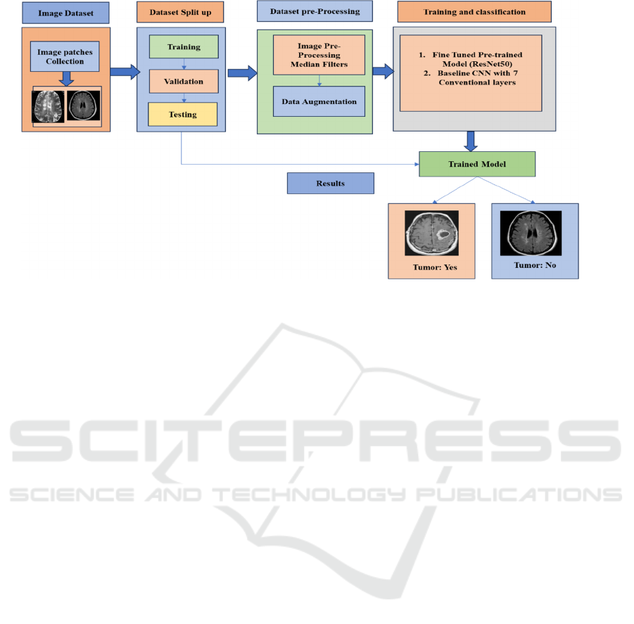
Figure 2: Proposed model for detection of Brain Tumor images
The first layer of this neural network architecture
is an input layer that can handle 224x224 images with
three colour channels. It makes use of a ResNet50
model that has already been trained and produces 7x7
feature maps with 2048 channels. These
characteristics are then further refined by a Conv2D
layer with 16 filters, which adds non-linearity while
lowering complexity. The spatial dimensions are then
reduced to 3x3 while maintaining the depth using a
MaxPooling2D layer. Following the application
of another MaxPooling2D layer that further decreases
the dimensions to 1x1, another Conv2D layer with 32
filters is applied. The spatial data is condensed by the
global average pooling layer into a 32-dimensional
vector. To avoid overfitting, this vector goes through
a dropout layer after passing through a dense layer
with 512 units.
4 RESULTS AND DISCUSSIONS
The trials were conducted in the Google Colab
environment. Computation was done with both CPU
and GPU. Utilized were a Tesla K80 accelerator,
Xeon CPU running at 3.35 GHz, and a 20 GB RAM.
The accuracy of our model during training and
validation is shown in Figure 3. The Keras callback’s
function computed it. Accuracy in training and
validation was observed when using varying numbers
of epochs. We discovered that the Hybrid model had
the maximum accuracy for both training and
validation after 6 epoches.
We can observe from the previously mentioned
graphs that validation accuracy increases in tandem
with training accuracy. As loss decreases, so does the
validation loss. To improve the results, we can adjust
the hyperparameters of the learning rate, train the
model on more photos, or simply train it for more
epochs. Our test accuracy is 97.5 percent thanks to the
evaluate () technique.
5 CONCLUSIONS
Due of various diversities of medical images, image
segmentation is important in medical image
processing. We employed MRI scans for brain tumor
segmentation. Brain tumor segmentation and
classification are the two main uses of MRI. This
paper uses CNN modes with seven layers to classify
photos of brain tumors. Using Resnet50 architecture
as a foundation, a hybrid model is introduced. The
developed hybrid model has a 97.67% accuracy rate
and Loss is 0.02%. Additionally, many models are
used to classify images of brain tumors. The hybrid
model that was created has the highest accuracy rate.
The accuracy of previous architectures, including the
classical Resnet architecture, has significantly
improved with the release of the upgraded Resnet50
architecture
REFERENCES
W. Chen, B. Liu, S. Peng, J. Sun, and X. Qiao, “Computer-
aided grading of gliomas combining automatic
segmentation and radiomics,” Int. J. Biomed. Imaging,
2018.
H. H. Sultan, N. M. Salem, and W. Al-Atabany, “Multi-
classification of brain tumor images using deep neural
Hybrid CNN-ResNet50 Model for Brain Tumor Classification Using Transfer Learning
447

network,” IEEE Access, vol. 7, pp. 69215–69225,
2019.
T. Hossain, F. Shishir, M. Ashraf, M. Al Nasim, and F.
Shah, “Brain tumor detection using convolutional
neural network,” in Proc. IEEE Int. Conf., 2019, pp. 1–
6.
A. K. Anaraki, M. Ayati, and F. Kazemi, “Magnetic
resonance imaging-based brain tumor grades
classification and grading via convolutional neural
networks and genetic algorithms,” Biocybern. Biomed.
Eng., vol. 39, no. 1, pp. 63–74, 2019.
F. Özyurt, E. Sert, E. Avci, and E. Dogantekin, “Brain
tumor detection based on convolutional neural network
with neutrosophic expert maximum fuzzy sure
entropy,” Measurement, vol. 147, 2019.
Khattar, S., & Bajaj, R. (2023, December). A Deep
Learning Approach of Skin Cancer Classification using
Hybrid CNN-Inception Model. In 2023 Global
Conference on Information Technologies and
Communications (GCITC) (pp. 1-7). IEEE.
A. Zotin, K. Simonov, M. Kurako, Y. Hamad, and S.
Kirillova, “Edge detection in MRI brain tumor images
based on fuzzy c-means clustering,” Procedia
Computer Science, vol. 126, pp. 1261-1270, 2018.
H. Greenspan, A. Ruf, and J. Goldberger, “Constrained
Gaussian Mixture Model Framework for Automatic
Segmentation of MR brain Images,” IEEE Transactions
on Medical Imaging, vol. 25, no. 9, pp. 1233-1245,
2018.
M. Havaei, A. Davy, D. Warde-Farley, A. Biard, A.
Courville, Y. Bengio, C. Pal, P. M. Jodoin, and H.
Larochelle, “Brain tumor segmentation with deep
neural networks,” Medical Image Analysis, vol. 35, no.
1, pp. 18-31, 2017.
T. C. Hollon, B. Pandian, A. R. Adapa, E. Urias, A. V. Save,
S. S. S. Khalsa, D. G. Eichberg, R. S. D’Amico, Z. U.
Farooq, S. Lewis, et al., “Near real-time intraoperative
brain tumor diagnosis using stimulated raman histology
and deep neural networks,” Nat Med., vol. 26, no. 1, pp.
52–58, 2020.
Goudarzi, H., Pournajaf, K., & Zarrin, M. (2022). "A F.
Muhammad Arif, A. S. Shamsudheen, O. Geman, D.
Izdrui, and D. Vicoveanu, “Brain tumor detection and
classification by MRI using biologically inspired
orthogonal wavelet transform and deep learning
techniques,” J. Healthcare Eng., 2022
F. Muhammad Arif, A. S. Shamsudheen, O. Geman, D.
Izdrui, and D. Vicoveanu, “Brain tumor detection and
classification by MRI using biologically inspired
orthogonal wavelet transform and deep learning
techniques,” J. Healthcare Eng., 2022
INCOFT 2025 - International Conference on Futuristic Technology
448
