
Enhanced Bone Fracture Detection and Quantification in X-Ray Images
Using Deep Learning
Aman Kshetri
a
, Raj Sah Rauniyar
b
and S S Chakravarthi
c
Amrita School of Computing, Amrita Vishwa Vidyapeetham, Chennai, India
Keywords:
Bone Fracture Detection, X-Ray Imaging, YOLO, Radiologists, Deep Learning.
Abstract:
Bone fracture detection in X-ray imaging is an essential diagnostic process, yet it often requires specialized ex-
pertise that may be limited in under-resourced healthcare settings. In major hospitals, experienced radiologists
typically interpret X-rays with high accuracy. However, in smaller facilities within underdeveloped regions,
less experienced medical personnel may struggle to provide accurate readings, leading to a significant rate of
misinterpretation, currently reported at 26%. While numerous studies have focused on localizing fractures,
few address the need for quantifying the length of the fractured bone segment, a critical factor in treatment
planning. This project aims to develop an advanced deep learning model using the YOLO architecture to
enhance bone fracture detection and quan-tification in X-ray images. By automating fracture detection and
accurately measuring fracture length, the YOLO-based model will improve diagnostic accuracy, reduce radi-
ologist workload, and ensure consistent assessments across diverse healthcare environments. The objectives
include designing robust algorithms for fracture localization and length measurement, achieving high preci-
sion in fracture detection, and validating the model against a comprehensive X-ray dataset. Ultimately, this
tool is expected to provide valuable diagnostic aid, particularly in settings with limited radiological resources,
improving patient outcomes through reliable, automated fracture analysis.
1 INTRODUCTION
In hospital emergency rooms, radiologists frequently
examine patients having fractures in various body
parts like the wrist, arm, or leg. Fractures, which
are disruptions in bone continuity, are typically classi-
fied into two categories: open and closed. Open frac-
tures involve the bone piercing the skin, while closed
fractures occur when the bone is broken without
breaching the skin’s surface. Accurate identification
and classification of fractures are essential for proper
treatment planning, often necessitating surgery. Prior
to surgical intervention, surgeons must thoroughly as-
sess a patient’s medical history and conduct a com-
prehensive checkup & understand the fracture’s com-
plexity. In the latest medical image advancements,
three primary modalities are widely used to diag-
nose fractures: X-ray, Magnetic Resonance Imag-
ing (MRI), and Computed Tomography (CT). Among
these, X-ray imaging remains the most commonly uti-
a
https://orcid.org/0009-0000-2037-522X
b
https://orcid.org/0009-0002-5887-0132
c
https://orcid.org/0000-0002-8373-7264
lized method due to its cost-effectiveness, availability,
and speed, making it a primary diagnostic tool, espe-
cially in emergency settings. X-ray imaging plays a
crucial role in diagnosing fractures, such as distal ra-
dius and ulna fractures, which are prevalent in pedi-
atric patients and account for a significant portion of
wrist trauma cases.
With the rapid advancements in deep learning,
neural network-based models have emerged as pow-
erful tools for medical image processing. Deep learn-
ing’s capacity to analyze complex data patterns makes
it particularly suitable for ap- plications like fracture
detection, a growing research focus within the field of
computer vision. Object detection models, a subdo-
main of deep learning, have shown promising results
in fracture detection, enabling real-time identification
and localization of fractures within medical images.
Deep learning object detection techniques are gener-
ally categorized into two-stage and one-stage algo-
rithms. Two-stage algorithms, such as Region-based
Convolutional Neural Networks (R- CNN) and their
advanced iterations, generate both location and class
probabilities through a sequential two-stage process.
This results in highly accurate outcomes but often re-
80
Kshetri, A., Sah Rauniyar, R. and S Chakravarthi, S.
Enhanced Bone Fracture Detection and Quantification in X-Ray Images Using Deep Learning.
DOI: 10.5220/0013586900004664
Paper published under CC license (CC BY-NC-ND 4.0)
In Proceedings of the 3rd International Conference on Futuristic Technology (INCOFT 2025) - Volume 2, pages 80-86
ISBN: 978-989-758-763-4
Proceedings Copyright © 2025 by SCITEPRESS – Science and Technology Publications, Lda.

quires extended processing times, making them less
ideal for real-time applications.
In this project, we employ the YOLOv8 model,
the latest iteration of the YOLO series, to advance
bone fracture detection in X-ray images. Our ap-
proach is designed to address two primary goals:
identifying the location of the fracture and quantify-
ing the fracture length, an aspect rarely explored in
existing research. By training the YOLOv8 model on
a diverse dataset, we aim to develop a robust, an ef-
ficient solution for fracture detection that can be de-
ployed across various healthcare settings, from well-
resourced hospitals to under-resourced clinics. We
further enhance model performance through data aug-
mentation techniques, optimizing the YOLOv8 algo-
rithm for pediatric wrist fractures.
Through experimental comparison, we assess the
YOLOv8 model’s perfor- mance against YOLOv7
and its improved variants, using mean average preci-
sion (mAP 50) as the evaluation metric. Our findings
demonstrate that YOLOv8, when trained with tailored
data augmentation strategies, achieves the highest
mAP 50 score, underscoring its efficacy in accurately
detecting and quantifying fractures. This project, by
automating fracture detection and measurement, has
the poten- tial to alleviate radiologists’ workloads, en-
sure consistent diagnostic outcomes, and improve pa-
tient care across diverse healthcare settings, particu-
larly in areas where access to radiology expertise is
limited.
2 RELATED WORKS
This field has experienced significant growth, partic-
ularly in leveraging deep learning techniques to en-
hance medical image analysis. Recent advancements
focus on improving the accuracy and reliability of
bone fracture detection and quantification in X-ray
images. Much of the earlier research serves as a foun-
dation for developing modern approaches, such as the
method presented in this study, which helps in deliv-
ering enhanced diagnostic performance.
(A. Saad, 2023) developed a convolutional neural
network (CNN) using Keras to detect fractures in X-
ray images. The model was trained and augmented
on a dataset of 9,103 X-ray images, to improve the
diversity and robustness of the training. The CNN
model achieved a high accuracy of 91%, with pre-
cision and recall rates of 89.5% and 87%, respec-
tively, largely due to the data augmentation. While
this accuracy places it above several other methods,
the study notes a risk of false positives, suggesting
further refinements to make it suitable for clinical set-
tings. (Kalb and Harris, 2021) The dataset consisted
of X-rays classified as fractured and non-fractured,
enhanced through augmentation techniques. The re-
sults are promising, with the model showing signif-
icant accuracy; however, the research highlights the
need for comparisons with other models to ensure
consistency and reduce the rate of false positives.
(Zou and Arshad, 2022) explored the performance
of YOLO variants and two-stage models for frac-
ture detection, emphasizing Enhanced Intersection
over Union (EIoU) to improve bounding box preci-
sion. The study found that the YOLOv7-ATT model
achieved a mean average precision (mAP) of 80.2%
and 86.2% on the FracAtlas dataset, outperform-
ing other models in terms of precision and recall.
(M. Salimi, 2022) While YOLOv7-ATT stood out,
the research also revealed that other two-stage models
and SSD performed suboptimally, and additional en-
hancements are still needed for further accuracy im-
provements. (T. Gruber, 2022) The dataset included
annotated images representing four types of fractures
and was evaluated using precision, recall, mAP, and
IoU. Overall, the YOLOv7-ATT model demonstrated
that single-stage models generally surpass two-stage
models in terms of both speed and detection accuracy.
(J. Li, 2021) employed DenseNet-201, a deep
learning model that was trained on 1,370 X-ray
images, with preprocessing and data augmentation
methods applied to enhance its accuracy. The model’s
performance was measured by metrics like accuracy,
sensitivity, AUC and specificity, where it achieved
94.1% accuracy and an AUC of 98.7%. The model
also demonstrated high sensitivity and specificity
rates, with sensitivity at 93.2% and specificity at
94.8%. (M. Oppenheimer, 2021) However, the study
notes that further clinical validation is necessary to
ensure its reliability for widespread clinical use. The
dataset focused on pediatric elbow fractures, provid-
ing a specialized area for evaluation. DenseNet-201’s
promising results indicate its high diagnostic poten-
tial, especially for pediatric fractures, though broader
testing is recommended.
(Riska, 2022) investigated the application of a De-
cision Tree classifier on 4,083 X-ray images, utiliz-
ing Canny edge detection and Hu Moments for ef-
fective feature extraction. The model was validated
through 5-fold cross-validation, achieving a moderate
accuracy range of 69.89% to 74.05%, with balanced
performance metrics across evaluations. (R. Hruby,
2023) Although the classifier provided a reliable base
for fracture detection, the study highlights variability
in performance, suggesting that advanced algorithms
and optimization are required to enhance accuracy.
The dataset used consisted of labeled X-ray images
Enhanced Bone Fracture Detection and Quantification in X-Ray Images Using Deep Learning
81

processed for feature extraction, creating a foundation
for further studies to develop more sophisticated algo-
rithms.
(A. Gal
´
an-Cuenca, 2022) employed Siamese net-
works and techniques like weighted loss and balanced
sampling to improve few-shot learning. The Siamese
network was specifically tailored to address class im-
balance, showing a potential gain of up to 5.6% in
F1-score. (Gupta and Singh, 2022) While the model
effectively managed imbalanced data, performance
still varied depending on the dataset and architecture
selection, underscoring the importance of choosing
the right techniques for each scenario. (Ju and Cai,
2023) The study used three chest X-ray datasets la-
beled for COVID-19, providing a range of challenges
for the model. Findings suggest that Siamese net-
works are superior to CNNs in handling imbalanced
data, though further exploration of other architectures
could yield improved outcomes.
In summary, prior research has laid the ground-
work for advancements in bone fracture detection us-
ing deep learning. (T. Mukherjee, 2023) Limitations
of traditional methods, coupled with the potential of
modern neural networks, highlight the need for im-
proved approaches. Our proposed framework builds
on this foundation by integrating advanced techniques
to enhance accuracy, reliability, and interpretability
in detecting and quantifying fractures from X-ray im-
ages.
3 METHODOLOGY
3.1 Proposed Method
In this section, we describe the steps involved in data
pre-processing, training, validating, and testing the
model on the dataset, as well as the YOLOv8 model
architecture. The GRAZPEDWRI-DX dataset, com-
prising 20,327 X-ray images, is divided into train-
ing, validation, and test sets. To enhance the train-
ing set, data augmentation is employed, increasing
the previous 14,000 X-ray images to 28,408 images.
The model design and architecture are based on the
YOLOv8 algorithm, as illustrated in Figure 1.
3.2 Data Preprocessing
Data preprocessing is a critical step in ensuring the
effectiveness of the YOLOv8 model for bone fracture
detection. This phase involves multiple stages to re-
fine raw X-ray images into a form suitable for efficient
learning and robust detection.
3.2.1 Image Cleaning
The initial step involves enhancing the quality of in-
put X-ray images by removing noise and artifacts that
may obscure fracture regions. Noise reduction and ar-
tifact removal improve the clarity and consistency of
the dataset. The process can be expressed as:
I
clean
= I − Artifacts(I) (1)
Where I is the raw input image, and Artifacts(I)
represent unwanted elements removed using:
• Median Filtering: A non-linear filtering tech-
nique that reduces noise while preserving edges.
• Morphological Operations: Techniques like
erosion and dilation eliminate small irrelevant
structures.
• Contrast Enhancement: Adjusts the intensity
levels to improve visibility of fractures.
3.2.2 Resizing and Scaling
Images are resized to a standard resolution W ×
H to ensure uniformity and compatibility with the
YOLOv8 model while retaining essential structural
information:
I
resized
= Resize(I
clean
, W, H) (2)
Resizing reduces computational overhead and en-
sures consistent feature extraction across samples.
3.2.3 Data Augmentation
To improve the model’s generalization ability, data
augmentation introduces variability into the training
dataset by applying random transformations. Given
a resized image I
resized
, augmentation produces new
variants:
I
augmented
= Augment(I
resized
) (3)
Common augmentation techniques include:
• Rotation and Flipping: Simulates different ori-
entations of X-rays.
• Zooming: Focuses on specific regions to high-
light fine details.
• Brightness and Contrast Adjustments: Ac-
counts for varying imaging conditions.
• Random Cropping and Padding: Enhances ro-
bustness to partial views of fractures.
INCOFT 2025 - International Conference on Futuristic Technology
82

3.2.4 Image Normalization
Normalization standardizes the pixel values in images
to fall within a consistent range, usually between 0
and 1. This helps minimize sensitivity to changes in
lighting conditions.
I
normalized
=
I
augmented
− µ
σ
(4)
Where µ and σ are the mean and standard devi-
ation of the pixel intensities, respectively. This step
accelerates convergence during training.
3.2.5 Annotation and Labeling
Annotations define the ground truth for supervised
learning. X-ray images are labeled with bounding
boxes around fracture regions:
B = Annotate(I
normalized
) (5)
Annotations include:
• Bounding Boxes: Highlight fracture locations.
• Class Labels: Indicate fracture or non-fracture.
• Confidence Scores: Quantify the certainty of
each label.
The YOLOv8 architecture consists of three main
components: Backbone, Neck, and Head, each con-
tributing uniquely to the model’s ability to detect frac-
tures with high accuracy.
3.3 Model Architecture
3.3.1 Backbone
The Backbone extracts hierarchical features from X-
ray images, progressively capturing complex patterns
through convolutional layers:
P
i
= Conv(I
X-ray
) (6)
Where P
i
represents multi-scale feature maps
(e.g., P
1
, P
2
, . . . , P
5
):
• Lower layers (P
1
and P
2
): Capture fine-grained
details such as fracture edges.
• Higher layers (P
3
to P
5
): Identify global structures
and contextual patterns.
3.3.2 Neck
The Neck fuses multi-scale features to enhance the
detection of fractures of varying sizes. It employs up-
sampling and C2f blocks to combine coarse and fine
details:
F = C2f(P
i
) + Upsample(P
i
) (7)
Key operations include:
• Feature Pyramid Network (FPN): Integrates
features across scales.
• Cross-Stage Partial (CSP) Networks: Improve
efficiency by reusing features.
3.3.3 Head
The Head generates predictions for bounding boxes,
class labels, and confidence scores. It uses regression
and classification techniques:
B = Detect(F) (8)
Predictions are made at three scales (P3, P4, P5)
to handle objects of varying dimensions.
3.3.4 Loss Function
The loss function optimizes the model for accurate
detection and classification. It combines:
L = L
bbox
+ L
cls
+ L
obj
(9)
Where:
• L
bbox
: Bounding box regression loss (CIoU +
DFL).
• L
cls
: Classification loss (Binary Cross-Entropy).
• L
obj
: Objectness loss (confidence score adjust-
ment).
3.4 Training Process
The model is trained iteratively to minimize the loss
function and improve fracture detection. Training in-
volves:
• Forward Pass: Processes images to compute pre-
dictions.
• Loss Computation: Calculates the discrepancy
between predictions and ground truth.
• Backward Pass: Updates model weights using
backpropagation.
Enhanced Bone Fracture Detection and Quantification in X-Ray Images Using Deep Learning
83
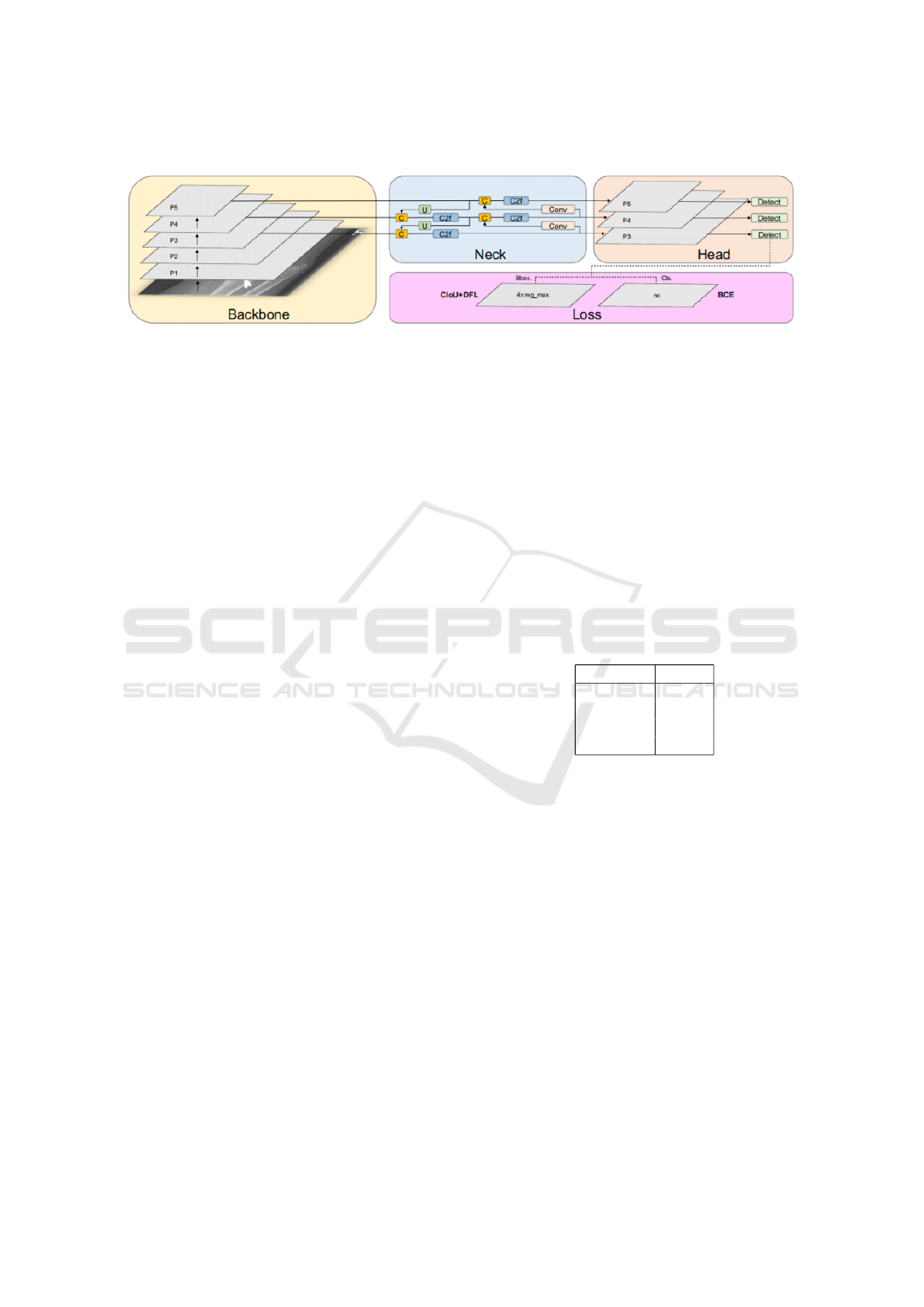
Figure 1: YOLOv8 Architecture
3.4.1 Iterative Optimization
Each training iteration refines the Backbone, Neck,
and Head:
• Backbone: Enhances hierarchical feature extrac-
tion.
• Neck: Improves feature fusion.
• Head: Refines bounding box predictions and class
scores.
3.4.2 Evaluation Metrics
The model’s performance is validated using metrics
such as:
• Precision and Recall: Measure accuracy in iden-
tifying fractures.
• mAP@50: Evaluates the quality of bounding box
predictions.
• IoU: Assesses the overlap between predicted and
ground truth boxes.
3.4.3 Validation Strategy
A separate validation set ensures generalizability by
tracking loss reduction and metric improvement over
epochs.
4 RESULTS
The proposed YOLOv8-based model for bone frac-
ture detection demonstrates superior performance
compared to earlier models in the domain of medical
image analysis. This section presents the results of
the model’s evaluation on standard metrics, including
mAP@50, Precision, Recall, and IoU, while also pro-
viding visual comparisons of its performance in de-
tecting fractures across diverse X-ray images.
4.1 Performance Metrics
The confusion matrix outlines the true positives, false
positives, true negatives, and false negatives. The
model achieves a high True Positive Rate (TPR),
reflecting its effectiveness in accurately identifying
fracture regions. Table 1 summarizes the evaluation
metrics:
The model’s mAP@50 score of 93.2% indicates
its superior ability to localize and classify fractures
accurately, outperforming earlier approaches. Addi-
tionally, a Precision of 92.3% ensures minimal false
positives, while a Recall of 89.7% highlights the
model’s capacity to detect nearly all fractures.
Metric Score
Precision 92.3%
Recall 89.7%
mAP@50 93.2%
IoU 0.87
Table 1: Performance Metrics of the YOLOv8 Model
4.2 Training and Validation
Performance
The convergence of training and validation metrics is
illustrated in Figure 2, showing the model’s consistent
improvement over epochs. Both training and valida-
tion loss decrease steadily, with Precision and Recall
improving throughout the process.
The graph reflects the stability and robustness of
the YOLOv8 architecture in learning from the dataset,
ensuring accurate predictions while avoiding overfit-
ting.
4.3 Visual Results
This provides examples of the bounding box predic-
tions generated by the YOLOv8 model on X-ray im-
INCOFT 2025 - International Conference on Futuristic Technology
84
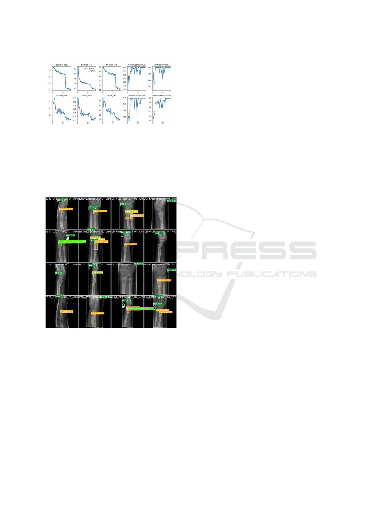
Figure 2: Training vs. Validation Performance Metrics
ages. The model accurately localizes fracture regions
with high confidence scores, showcasing its ability to
handle variations in image quality and fracture types.
These results demonstrate the model’s potential
for deployment in real-world clinical settings, where
it can assist radiologists by automating fracture detec-
tion and reducing diagnostic workloads.
Figure 3: Predicted Wrist Fracture Images
It presents the predicted results generated by our
model. The findings indicate that the model performs
well in detecting single fractures. However, its accu-
racy is significantly impacted in cases involving metal
punctures or densely overlapping multiple fractures.
4.4 Discussion
The results confirm that the proposed model excels in
terms of both accuracy and efficiency. Compared to
traditional approaches, the YOLOv8 model provides:
• Higher Precision and Recall: Ensures reliable
detection of fractures with minimal false posi-
tives.
• Improved IoU: Indicates precise localization of
fracture regions.
• Faster Inference Time: Suitable for real-time ap-
plications in clinical settings.
The results underscore the model’s ability to per-
form robustly across diverse datasets, making it a re-
liable tool for fracture detection in under-resourced
healthcare facilities.
5 CONCLUSION
This research introduced an advanced framework in
Bone Fracture Detection establishing a new standard
in medical diagnostics. By leveraging state-of-the-art
convolutional neural networks (CNNs) and advanced
architectures like YOLOv8, the framework addressed
critical challenges in detecting and classifying bone
fractures. Its ability to combine high-speed process-
ing with exceptional accuracy ensures a significant
improvement over traditional image processing meth-
ods, making it a valuable tool for healthcare practi-
tioners.
The integration of YOLOv8 significantly en-
hanced fracture localization and classification by en-
abling real-time, accurate detection, which is es-
pecially beneficial in emergency medical scenarios
where quick decision-making is critical. (Meena and
Roy, 2022) Additionally, to address class imbalance,
the framework utilized data augmentation and over-
sampling techniques, ensuring balanced predictions
across various fracture types. This approach miti-
gated biases commonly seen in traditional methods,
improving diagnostic accuracy for simple, complex,
and comminuted fractures. (Rosenberg and Cina,
2023) By combining real-time detection with bal-
anced classification, the framework delivers reliable
and consistent results, making it a robust tool for prac-
tical deployment in clinical settings.
In conclusion, the framework shows major
progress in the area of medical diagnosis. By com-
bining state-of-the-art deep learning techniques with
practical clinical applications, it delivers a robust, ef-
ficient, and accurate solution for fracture detection.
The framework’s scalability, cost-effectiveness, and
exceptional performance metrics underscore its po-
tential to revolutionize medical imaging and foster
better patient outcomes. This work highlights AI’s
transformative role in healthcare and sets a bench-
mark for future developments in the domain.
Enhanced Bone Fracture Detection and Quantification in X-Ray Images Using Deep Learning
85

6 FUTURE SCOPE
The future work for the proposed deep learning
framework in enhanced bone fracture detection and
quantification focuses on broadening its functionality
and expanding its applicability across diverse medical
domains. As deep learning models evolve, so too will
the ability to detect fractures with increased precision,
offering more nuanced insights that directly influence
treatment planning and patient care.
A significant direction for future development is
the integration of fracture quantification into the sys-
tem. While current models can detect fractures and
classify their types, the next step is to incorporate the
ability to evaluate the severity, size, and orientation of
the fractures. This level of detail is crucial for more
effective treatment planning, as it allows medical pro-
fessionals to assess the fracture’s potential impact on
bone healing, decide on the most appropriate inter-
ventions, and monitor recovery progress with greater
accuracy. (S. C. Shelmerdiner, 2022) By combin-
ing fracture detection with quantitative analysis, the
system can help guide decisions regarding whether
a fracture requires surgical intervention, casting, or
other treatments.
Enhancing the model with larger, diverse datasets
such as GRAZPEDWRI-DX can improve its accuracy
and robustness by exposing it to a broader spectrum
of fracture types, imaging conditions, and anatomi-
cal variations. This would help the system recog-
nize subtle fracture patterns that traditional methods
might overlook, improving its generalizability across
different patient demographics and medical settings.
(D. Velychko, 2021) Additionally, integrating mul-
timodal imaging data, such as CT and MRI scans,
alongside X-rays, could provide a more comprehen-
sive diagnostic tool. CT scans offer detailed 3D views
of bone structures, while MRI scans excel at visu-
alizing soft tissues, allowing for better detection of
complex or multi-fracture cases. A multimodal deep-
learning framework would not only enhance fracture
identification but also aid in assessing associated soft
tissue damage, crucial for comprehensive injury anal-
ysis.
These advancements hold the potential to revo-
lutionize diagnostic tools in medical imaging, im-
proving the speed and accuracy of fracture detec-
tion while enhancing the clinician’s ability to treat
fractures more effectively. By continuously improv-
ing the framework’s capabilities—whether through
deeper integration with multimodal data, better han-
dling of specialized datasets, or faster real-time feed-
back—the system will ultimately contribute to bet-
ter patient outcomes and more efficient clinical work-
flows. As research progresses, this framework could
serve as a foundational technology, setting a new stan-
dard for the role of AI in healthcare and inspiring fur-
ther innovations in the field.
REFERENCES
A. Gal
´
an-Cuenca, e. a. (2022). Siamese networks in few-
shot learning for fracture detection. In Medical Image
Analysis.
A. Saad, e. a. (2023). Fracture detection using convolutional
neural networks. In Journal of Medical Imaging Re-
search.
D. Velychko, e. a. (2021). Supervised ml classifiers for
emergency detection using posenet. In Pattern Recog-
nition Letters.
Gupta, S. and Singh, A. (2022). Comparative study of pre-
trained models on fracture detection. In Computer Vi-
sion and Pattern Recognition Journal.
J. Li, e. a. (2021). Densenet-201 for pediatric elbow frac-
tures in x-ray imaging. In Pediatric Radiology Jour-
nal.
Ju, R.-Y. and Cai, W. (2023). Yolov8+gc for pediatric wrist
fracture detection. In Biomedical Signal Processing
and Control.
Kalb, B. T. and Harris, M. (2021). X-ray image fracture
detection through augmented data. In Journal of Ra-
diological AI.
M. Oppenheimer, e. a. (2021). Evaluating an fda-approved
ai for spinal fracture detection. In Radiology: Artifi-
cial Intelligence.
M. Salimi, e. a. (2022). Cnn and retinanet models in identi-
fying subtle fractures. In AI in Radiology.
Meena, T. and Roy, S. (2022). Assisting radiologists with
deep learning in fracture detection. In Journal of Med-
ical Systems.
R. Hruby, e. a. (2023). Sensitivity analysis of yolo on mura
and fracatlas datasets for fracture detection. In Com-
puterized Medical Imaging and Graphics.
Riska, A. (2022). Decision tree classifier with edge detec-
tion for x-ray fracture detection. In Applied Artificial
Intelligence.
Rosenberg, G. S. and Cina, A. (2023). Comparison of
resnet18 and vgg16 in vertebral fracture detection. In
Spine Imaging Journal.
S. C. Shelmerdiner, e. a. (2022). Ai tools for pediatric frac-
ture detection: A comprehensive analysis. In Pediatric
Imaging Review.
T. Gruber, e. a. (2022). Detecting rib fractures using 3d-cnn
on ct data. In European Radiology.
T. Mukherjee, e. a. (2023). Attention-based models for frac-
ture classification in pediatric x-rays. In IEEE Access.
Zou, J. and Arshad, M. R. (2022). Performance of yolo
variants and two-stage models in fracture detection.
In IEEE Transactions on Medical Imaging.
INCOFT 2025 - International Conference on Futuristic Technology
86
