
Detection of BABESIA BIGEMINA in Cattle Blood: AI and Impedance
Methods
Jacob Varghese
1,2 a
, Allen George Thomas
1,2
, Khadeeja Nilofer Saleem Mukkunnoth
1,2
,
Sivan Murali
1,2
and Lakeshmy G B
1,2
1
TKM Institute of Technology, APJ Abdul Kalam Technological University, Thiruvananthapuram, Kerala, India
2
Department of Biomedical Engineering, TKM Institute of Technology, Kollam, Kerala, India
Keywords:
Anaplasma, Babesia Bigemina, Haemoprotozoans, IoU, Mean Average Precision, Theileria, YOLO V8.
Abstract:
Haemoprotozoans are a diverse group of blood-borne parasites that cause significant economic losses in the
veterinary field. In cattle, the three most common haemoprotozoans are Babesia, Theileria, and Anaplasma.
Detection and treatment of these parasites are currently time-consuming and require laboratory facilities,
which can delay treatment and lead to poorer outcomes, including increased anaemia and death. Infections
also lead to diminished productivity, compromised reproductive performance, and increased vulnerability to
secondary infections. To address this challenge, the project aims to develop novel technologies that will help
in early detection of the parasite. The paper presents the design of an embedded AI software to detect the
presence of Babesia bigemina protozoan within the cattle blood. The software used YOLO V8 model to train
the system, and the software was integrated into a 3D printed open flexure microscope. The model yields a
mean average precision of 66.2 percent for an IoU threshold of 0.5 and 34.7 percent for an IoU threshold of 0.5
to 0.9. The project also proposed research on the change in conductivity and impedance of the infected cattle
blood and concluded that the presence of foreign particles, such as protozoans, in the blood samples resulted
in a decrease in conductivity by values ranging between 2.2 to 3 milli siemens and an increase in impedance
by a value within a range of approximately 330 to 450 milli ohm compared to normal blood samples.
1 INTRODUCTION
Haemoprotozoans are a diverse group of single-celled
eukaryotic organisms transmitted by blood-feeding
invertebrates, causing diseases in various animals
and humans. They include genera like Babesia,
Hepatozoon, Theileria, and Trypanosoma, with dif-
ferent species identified in wildlife such as cattle,
dog and rats(A. S. Nair, 2011). Haemoprotozoan
species affecting cattle include Theileria annulata,
Trypanosoma evansi, and Babesia bovis. These par-
asites cause significant economic losses globally due
to their impact on livestock health and productivity.
Theileriosis, trypanosomosis, and babesiosis are ma-
jor haemoprotozoan diseases, with difficulties in di-
agnosis due to low parasitemia and concurrent in-
fections. Tick-borne diseases, such as babesiosis,
anaplasmosis, and theileriosis, are major constraints
in the dairy industry, leading to decreased produc-
tivity and increased control costs. The hot and hu-
a
https://orcid.org/0009-0001-2862-681X
mid climate in tropical regions like India provides a
favorable environment for haemoprotozoan parasites
transmitted by vectors like ticks, posing a constant
threat to susceptible animals(A. Tlili and Jaffrezic-
Renault, 2006). Haemoprotozoa species detected in
cattle in Northern Kerala were Theileria like piro-
plasms and Babesia bigemina, with PCR revealing
Trypanosoma evansi, Theileria sp., and B. bigem-
ina(A. S. Nair, 2011). Detection and diagnosis of
these diseases can be challenging as they require lab-
oratory facilities and time consuming that even in-
creases the rate of mortality. There is an alarming
need of an on-field device that will accurately and pre-
cisely detect the presence of haemoprotozan within
the animal blood. This concept is the main build-
ing block of our device(C. S. Bhatnagar and Meena,
2015). A Non-invasive portable device can do the vet-
erinarians and the farmers a good turn as they give out
rapid results in the field detection fundamentally. The
results would not only be rapid but also with tower-
ing accuracy. In particular this device facilitates to
characterize to redline the different types of proto-
Varghese, J., Thomas, A. G., Mukkunnoth, K. N. S., Murali, S. and G B, L.
Detection of BABESIA BIGEMINA in Cattle Blood: AI and Impedance Methods.
DOI: 10.5220/0013584700004664
Paper published under CC license (CC BY-NC-ND 4.0)
In Proceedings of the 3rd International Conference on Futuristic Technology (INCOFT 2025) - Volume 1, pages 729-734
ISBN: 978-989-758-763-4
Proceedings Copyright © 2025 by SCITEPRESS – Science and Technology Publications, Lda.
729

zoans and its variations. Finding tick-borne diseases
in cattle while out in the field poses significant chal-
lenges. Typically, diagnosing these diseases requires
access to laboratory facilities equipped with special-
ized tools like microscopes for examining blood sam-
ples and PCR (Polymerase Chain Reaction) tests for
identifying specific pathogens(B. R. Maharana and
Sudhakar, 2016). However, such facilities may not
always be readily available, especially in remote or
rural areas where these diseases are prevalent. Fur-
thermore, the cost associated with conducting these
tests can vary depending on factors such as the type
of test required and the location of the testing facility.
In some cases, farmers may need to travel long dis-
tances to access testing centres, incurring additional
expenses and time delays. Unfortunately, the delay
in initiating treatment procedures due to the time-
consuming nature of laboratory testing can have se-
vere consequences. Which may even lead to the death
of the animal, causing a devastative economic loss for
the farmer. The primary outreach of this project is
to concisely overcome the current challenges in de-
tecting the presence of haemoprotozoan within the
cattle blood, especially during the on-field examina-
tion. Thereby narrowing down the constraints which
leads to the lagging of treatment initiation(B. R. Ma-
harana and Sudhakar, 2016). Hence, the main aim of
this project is to develop an on field low-cost portable
device that could accurately and precisely detect the
presence of protozoans from the blood sample under
study. Additionally, the device may also be able to
differentiate the genera of protozoans, since the drug
administration for each protozoan is specific. The
main objectives of the project are Analysing the re-
lationship between protozoan infection and change
in the electrical conductivity and impedance property
of the blood due to the infection(I. Szyma
´
nska and
Kaliszan, 2007). Developing a novel methodology
based on the observation from the first objective to de-
sign a circuit to detect and measure the corresponding
change in blood properties(G. C. McConnell, 2009).
Designing and developing a software to accurately de-
tect each genus of protozoan and concluding whether
the blood sample under study is protozoan infected or
not(B. K. Yap, 2018). For the time being the study
only concentrates on one species. To successfully im-
plement the designed software to a low-cost portable
3D printed microscope with high resolution.
2 METHODOLOGY
The proposed solution for the particular problem that
this project addresses, can be done mainly in two
Figure 1: Circuit diagram of Microcontroller based Human
Blood Conductivity Measurement System
ways, that is, one complete hardware and the other
an AI software system embedded with 3D printed mi-
croscope. In this chapter the design of the hardware,
components used to develop the circuit, role of each
component in the circuit etc will be discussed first.
Following session of the same chapter will introduce
about the software design as well as the design and
structure of the 3D printed microscope.
2.1 System Hardware: Detection of
Haemoprotozoan in Cattle Blood by
Analyzing the Conductivity and
Impedance of the Blood Sample
This project aims to designs a circuit to measure
the conductivity and impedance of liquids(B. S.
R. Bharati and Bhaskar, 2013), particularly blood
samples, using important components like the LM741
op-amp, Arduino ATmega328, and a display as men-
tioned in figure 1. The system consists of three parts,
namely:
• Signal Conditioning Circuit
• Conductivity Cell (Electrodes)
• Microcontroller and Display
Working Principle:
An AC signal is imposed on electrodes immersed in
the sample. The ions in the liquid facilitate the flow of
current, and the voltage across the electrodes reflects
the liquid’s impedance. The resulting signal is am-
plified, rectified, and converted to a readable format,
from which conductivity is calculated(McAdams and
Jossinet, 1995).
Signal Conditioning:
The signal conditioning unit consists of
• Wein Bridge Oscillator: It provides a 1kHz sine
wave at an amplitude of 10Vpp by using the
LM741 op-amp and an RC feedback network.
The frequency is given by
f =
1
2πRC
(1)
INCOFT 2025 - International Conference on Futuristic Technology
730

• Impedance Bridge: An AC signal from the oscil-
lator excites the bridge, one arm of which con-
tains the sample. Sample conductivity changes
the impedance of the bridge, hence the voltage
distribution.
The last component includes the Amplification and
Processing which is followed by rectification and fil-
tering to get a DC voltage representative of conduc-
tivity. This DC voltage is digitized through an ADC
for processing.
2.2 System Software: Real Time
Monitoring and Detection of the
Haemoprotozoan from the Cattle
Blood Sample Using Low-Cost
Portable Microscope Embedded
with AI Software
2.2.1 Blood Cell Classification Software
The developed software to classify the blood cells in-
fected with haemoprotozoan will be architected to ef-
ficiently handle various tasks involved in processing
the data, training and evaluation of models, and mak-
ing inference(J. Knapper, 2022). The followings are
included:
1. Modular Components: Dedicated modules for
augmentation, model creation, training, evalua-
tion, and inference of data. Reusability in func-
tions and classes promotes maintainability and
reusability of code.
2. Flexibility: Allows customization of model ar-
chitectures, hyperparameters, pre-trained models,
loss functions, and optimization techniques.
3. Performance Optimization: Batch processing,
parallelization, and GPU acceleration are em-
ployed to ensure efficiency(C. Honrado, 2018).
4. Error Handling and Platform Independence: Ro-
bust error handling ensures smooth operation,
while platform independence allows compatibil-
ity across operating systems.
2.2.2 Camera Module
The camera module includes a 4-megapixel CMOS
sensor that can capture images at 1920×1920 and
record videos at 640×480 pixels as shown in fig-
ure 2. This high resolution enables detailed analysis
and live demonstrations. The CMOS sensors convert
light into a digital signal, providing high-quality im-
ages suitable for microscopy applications(Berney and
O’Riordan, 2008).
Figure 2: CMOS sensor obtained from a 1600x digital USB
microscope.
2.2.3 Parasite Detection Methodology
1. Data Preparation: Blood sample images contain-
ing Babesia parasites are formatted in the YOLO
structure, with annotation files marking bounding
boxes around parasites. The dataset is split into
training and validation sets.
2. Model Selection: YOLOv8, a state-of-the-art ob-
ject detection model, is chosen for its high accu-
racy and real-time detection capabilities.
3. Model Training: The model is trained for 100
epochs using the AdamW optimizer. Data aug-
mentation techniques involved include blur, me-
dian blur, grayscale conversion, and CLAHE to
enhance robustness.
4. Evaluation: Model performance was measured in
terms of precision, recall, and mean average pre-
cision.
5. Deployment: The developed model is deployed
after validation in the identification of haemopro-
tazoon infections from blood sample images.,
6. Improvement: To perform well continuously,
make iterations in fine-tunes, additional data, or
advance the object detection algorithm.
2.2.4 Affordable Portable Microscope Design
The microscope utilizes a 3D-printed Open Flexure
Microscope which resolves samples 2–5 µm in size.
This open-sourced created, by Dr. Richard Bowman
at the University of Bath, is a cost effective, portable
solution, well suited for education and research. Im-
ages are acquired live and analyzed by the software to
identify in real time infections caused by haemoproto-
zoa(J. Knapper, 2022), (C. Honrado, 2018), (Berney
and O’Riordan, 2008).
Detection of BABESIA BIGEMINA in Cattle Blood: AI and Impedance Methods
731
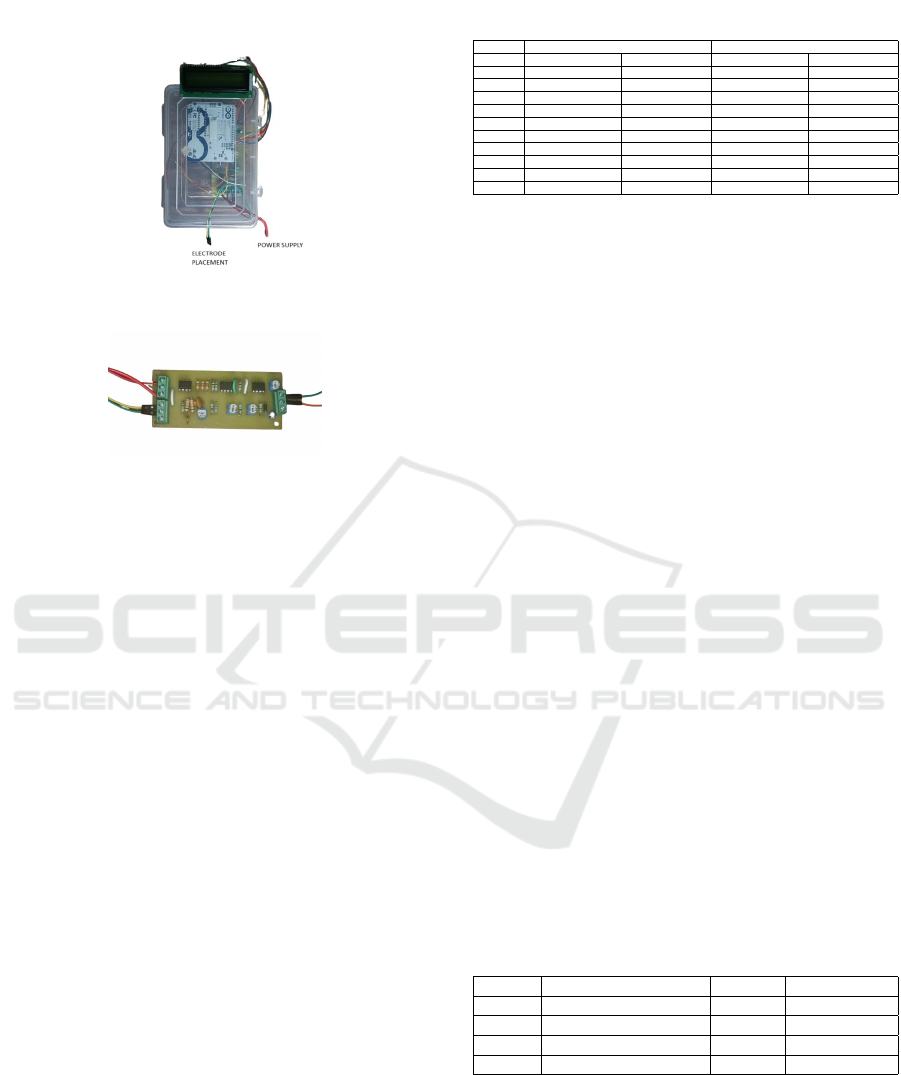
Figure 3: Prototype of the hardware
Figure 4: PCB and circuit components
3 RESULT
3.1 Blood Conductivity and Impedance
The objective of this project is to measure the conduc-
tivity and impedance of blood samples using a con-
ductivity measuring circuit with a Wein bridge oscil-
lator, LM741 IC, copper electrode arrangement, and
an Arduino microcontroller as shown in the figure 3.
A conductivity measuring circuit was constructed
comprising a Wein bridge oscillator with an LM741
IC as the signal source generator. A copper electrode
arrangement was used as the conductivity cell, im-
mersed in the blood samples. An Arduino microcon-
troller was employed to process the signals and cal-
culate conductivity and impedance values. Figure 4
shows the PCB fabrication.
3.1.1 Observations
The conductivity measurements ranged from 2.2 ms
to 5.8 ms. A total of 20 readings were obtained within
this range, with incremental changes in conductivity
values. Impedance values were calculated as the re-
ciprocal of conductivity.
The presence of foreign particles, such as proto-
zoans, in the blood samples resulted in a decrease in
conductivity to a range of 2.2 to 3ms and an increase
in impedance within a range of 330 to 450 mohms
compared to normal blood samples. This suggests
that the electrical properties of blood are influenced
by the presence of contaminants, highlighting the im-
Table 1: Observations of conductivity and impedance mea-
surements of blood samples.
Reading Normal Blood Sample Infected Blood Sample
Conductivity (mS) Impedance (mω) Conductivity (mS) Impedance (mω)
1 4.4 227.27 2.8 357.14
2 4.35 229.89 3.2 312.50
3 4.3 232.56 3.0 333.33
4 4.25 235.29 2.4 416.67
5 4.2 238.10 2.9 344.83
6 4.1 243.90 3.4 294.12
7 4.15 240.96 2.3 434.78
8 4.3 232.56 3.3 303.30
9 3.95 253.60 2.6 384.62
10 3.8 263.16 2.2 454.44
portance of monitoring conductivity and impedance
for detecting abnormalities.
3.2 Result of the Software System
The project integrated YOLOv8 into a 3D-printed in-
verted microscope to develop an automated system
for detecting Babesia Bigemina in blood samples.
Positive and negative samples were tested, and the
software effectively analyzed, classified, and detected
the parasite. Due to dataset availability, the study fo-
cused on Babesia Bigemina.
3.2.1 Dataset and Training
The dataset consisted of blood sample images with
bounding boxes for Babesia parasites. Training and
validation sets were made.
• Model Training: YOLOv8 was trained for 100
epochs using the AdamW optimizer with auto-
matic learning rate and momentum.
• Data Augmentation: Techniques include blur,
median blur, grayscale conversion, and contrast-
limited adaptive histogram equalization.
3.2.2 Model Evaluation and Results
The model’s performance was evaluated on the val-
idation set based on metrics such as precision, re-
call, mAP50, and mAP50-95 computed at various IoU
thresholds. Observations can be seen in Table 4.2.
Table 2: Results of the Model.
SL. No Performance Metrics Babesia Extracellular
1 Precision 0.685 0.462
2 Recall 0.736 1.000
3 mAP50 0.892 0.995
4 mAP50-95 0.621 0.895
3.2.3 Metric Definitions
• Precision: Ratio of true positives to total predicted
positives, measuring prediction accuracy.
INCOFT 2025 - International Conference on Futuristic Technology
732

Figure 5: Precision-Recall Curve
Figure 6: Normalized confusion matrix
• Recall: Ratio of true positives to total actual pos-
itives, assessing the model’s ability to detect all
instances.
• mAP50: Mean average precision at IoU 0.5, pro-
viding a balance between precision and recall.
• mAP50-95: Mean average precision across IoU
thresholds (0.5–0.95), offering a comprehensive
evaluation.
• Class-Specific Metrics: Precision, recall, and
mAP for individual classes, enabling detailed per-
formance analysis.
3.2.4 Observations
For YOLOv8, satisfactory metrics were obtained with
more room for improvement in detecting Babesia par-
asites. Future optimizations can be done for better re-
sults with increased accuracy and generalization.
4 DISCUSSION AND
CONCLUSION
4.1 Conductivity and Impedance of
Protozoan Affected Blood
The project developed hardware and software tech-
nologies for the detection of haemoprotozoan in cattle
Figure 7: F1- Confidence Curve
blood. The hardware consists of a conductivity mea-
surement circuit consisting of a Wein bridge oscil-
lator, LM741 IC, copper electrodes, and an Arduino
microcontroller. This setup facilitates portable blood
conductivity and impedance measurement, which is
of great value during the identification of changes in
blood properties.
While blood impedance measurement is preferred
for the detection of blood-borne parasites, results may
be affected by factors such as temperature and for-
eign particles that limit accuracy. These disadvan-
tages could be overcome by incorporating advanced
sensors and electrodes.
In conclusion, as much as the LM 741-based con-
ductivity circuit is not fully accurate for the detection
of haemoprotozoa, it effectively detects changes in
impedance and conductivity of blood.
4.2 Real Time Monitoring and
Detection of Haemoprotozoan Using
Embedded Ai System
The proposed study is to design a portable, non-
invasive device for the detection of haemoprotozoan
parasites in cattle blood, keeping in mind the limita-
tions of existing methods of diagnosis that are time-
consuming, expensive, and require laboratory facil-
ities. The focus is on the development of an on-
field, low-cost device for identifying protozoan gen-
era so that appropriate drugs can be administered.
The study focuses on the relationship between pro-
tozoan infection and changes in blood conductivity
and impedance properties. A software integrated
with a low-cost 3D-printed high-resolution micro-
scope is developed to analyze blood samples and
detect infections. The project emphasizes the po-
tential of biomedical engineering in enhancing vet-
erinary healthcare, supporting cattle farming-a key
contributor to Indian GDP and milk/meat produc-
tion(E. O. Adekanmbi and Srivastava, 2023).
Haemoprotozoan diseases have a major impact on
livestock, resulting in financial losses from infections
and mortalities(Garcia and Sabuncu, 2019). Facili-
ties for diagnosis are generally insufficient, relying on
conventional techniques of microscopic examination
and serological tests like ELISA, which are laborious
and time-consuming(Technologies, 2017). Modern
molecular techniques such as PCR detect the disease
during its latent phase more effectively(E. O. Adekan-
mbi and Srivastava, 2023). Electrochemical
Impedance Spectroscopy and immunosensor-based
methods show potential in the diagnosis of diseases
such as Babesia bovis through the analysis of electri-
cal properties(Cole, 1941)(Macdonald and Johnson,
Detection of BABESIA BIGEMINA in Cattle Blood: AI and Impedance Methods
733

2005)(Garcia and Sabuncu, 2019).
Although the novel diagnostic approach was in-
troduced, certain constraints occurred. The presence
of other pathogens, such as viruses, bacteria, or other
protozoan genera, was not considered; temperature
dependencies during experiments were not consid-
ered, either. Low-cost components reduced accuracy,
and software analysis was restricted to Babesia, ex-
cluding complex genera like Theileria and Anaplasma
due to the limitation of the dataset. These factors con-
strained the model’s total mean average precision. Fu-
ture research can address these limitations by replac-
ing low-cost components with precise ICs or sensors
like AD5933(H. Cho and Baek, 2021), designing spe-
cific electrodes for impedance-based detection, and
expanding datasets through more blood sample col-
lection and annotation.
Enhancing training models to differentiate haemo-
protozoan genera accurately and employing advanced
microscopic technologies like lensless microscopy or
muscope can broaden the device’s applications.
These methodologies could also be applied to the
diagnosis of human parasites such as Plasmodium and
Trypanosoma, laying the foundation for innovative
veterinary diagnostics with significant societal and
economic benefits.
REFERENCES
A. S. Nair, R. Ravindran, B. L. e. a. (2011). Haemopro-
tozoa of cattle in northern kerala, india. Tropical
Biomedicine.
A. Tlili, A. Abdelghani, S. A. and Jaffrezic-Renault, N.
(2006). Impedance spectroscopy and affinity measure-
ment of specific antibody–antigen interaction. Mate-
rials Science and Engineering: C, 26(2):546–550.
B. K. Yap, S. N. A. M. Soair, e. a. (2018). Potential point-of-
care microfluidic devices to diagnose iron deficiency
anemia. Sensors, 18(8):2625.
B. R. Maharana, A. K. Tewari, B. C. S. and Sudhakar, N. R.
(2016). Important hemoprotozoan diseases of live-
stock: challenges in current diagnostics and therapeu-
tics: an update. Veterinary World, 9(5):487–495.
B. S. R. Bharati, C. S. P. and Bhaskar, P. (2013). Atmel
microcontroller based human blood conductivity mea-
surement system. IJEE.
Berney, H. and O’Riordan, J. J. (2008). Impedance mea-
surement monitors blood coagulation. Analog De-
vices.
C. Honrado, L. Ciuffreda, D. S. e. a. (2018). Dielectric char-
acterization of plasmodium falciparum-infected red
blood cells. Journal of the Royal Society Interface,
15(147):20180416.
C. S. Bhatnagar, B. Bhardawaj, D. S. and Meena, S. K.
(2015). Incidence of haemoprotozoan diseases in cat-
tle in southern rajasthan, india. International Journal
of Current Microbiology and Applied Sciences.
Cole, K. S. (1941). Dispersions and absorption in di-
electrics. Journal of Chemical Physics, 9:341–351.
E. O. Adekanmbi, M. W. U. and Srivastava, S. K. (2023).
Dielectric characterization of babesia bovis using the
dielectrophoretic crossover frequency. Electrophore-
sis, 44(11-12):1001–988.
G. C. McConnell, R. J. Butera, e. a. (2009). Bioimpedance
modeling to monitor astrocytic response to chroni-
cally implanted electrodes. J. Neural Eng., 6.
Garcia, A. and Sabuncu, A. C. (2019). Electrical system for
bioelectric impedance using ad5933 impedance con-
verter.
H. Cho, S. R. L. and Baek, Y. (2021). Anemia diagnostic
system based on impedance measurement of red blood
cells. Sensors (Basel), 21(23):8043.
I. Szyma
´
nska, H. Radecka, J. R. and Kaliszan, R. (2007).
Electrochemical impedance spectroscopy for study of
amyloid β-peptide interactions. Biosensors and Bio-
electronics, 22(9):1955–1960.
J. Knapper, J. Stirling, D. G. R. e. a. (2022). Smart feedback
for reliable scanning: developing the openflexure mi-
croscope.
Macdonald, J. R. and Johnson, W. B. (2005). Fundamentals
of Impedance Spectroscopy, pages 1–26.
McAdams, E. T. and Jossinet, J. (1995). Tissue impedance:
a historical overview. Physiol. Meas., 16:A1–A13.
Technologies, K. (2017). Impedance Measurement Hand-
book, 6th Edition. Keysight Technologies, Santa Rosa,
CA.
INCOFT 2025 - International Conference on Futuristic Technology
734
