
Morphological and Anatomical Features of Leaf and Stem of
Dioscorea Nipponica Makino
Dilfuza
Berdibaeva
a
, Gulnoza
Atabaeva
b
, Gulom
Jumaboyev
c
, Mavluda Amanova
d
and Gulbahor Sultanova
e
Tashkent State Agrarian University, 100140, University str. 2, Tashkent, Uzbekistan
Keywords: Dioscorea Anatomy, Stem Structure, Leaf Morphology.
Abstract: In this article Dioscorea nipponica Makino. information on the anatomical structure of the leaf and stem of
the species, the location, structure and functions of each tissue. The results of this study support the taxonomy
of D. nipponica species based on anatomical structure. This study showed that the leaves and stems of
Dioscorea nipponica have characteristics similar to those of Eudicot. Our observations showed that the leaf
surface of Dioscorea nipponica is covered with solitary trichomes. The structure of the vascular bundle is
represented by a definitive V-shaped arrangement of metaxylem vessels, tracheids and phloem units. Raphid
crystals are present in the leaf mesophyll tissue. It was also found that the stem shape is undulating,
parenchyma cells are saturated with granular substance, conducting tissues are arranged in 2 rows, conducting
vessels are paired
.
1 INTRODUCTION
Dioscorea nipponica Makino. - is a perennial
herbaceous liana of the Dioscoreae family
(Dioscoreaceae R.Br.), reaching a length of 4-5 m
(Gubanov et al., 1976). The rhizome is horizontal,
shallow from the soil surface, sparsely branched,
brownish-brown (white or yellowish at the break), up
to 1.5 m long and 2 cm in diameter, with traces of
dead stems and numerous thin, rigid cord-like roots
(Anthony and Ibok, 2021). Younger parts of the
rhizome are lighter, yellowish, fleshy, with large
buds; the outer layer of the rhizome is easily separated
as a flaky thin dark-coloured corky layer. Decoction
and tincture of the roots of Dioscorea nipponese folk
medicine recommends atherosclerosis of cerebral
vessels, coronary atherosclerosis, angina pectoris,
hypertension, diabetes mellitus. Already after the first
course of taking dioscorea reduces or disappears
headache, tinnitus, fatigue. Further increases the
a
https://orcid.org/0009-0009-4816-4921
b
https://orcid.org/0009-0004-8047-7577
c
https://orcid.org/0000-0003-4569-5662
d
https://orcid.org/0009-0002-9331-5265
e
https://orcid.org/0009-0004-8090-0195
removal of cholesterol with bile, improves the work
of the heart, pancreas, liver, kidneys.
2 MATERIALS AND METHODS
Leaves and stems of Dioscorea nipponica were fixed
in 70% ethanol according to a generally recognised
technique and its anatomical structure was studied on
a transverse section of the fixed material
(Trankovsky, 1979).
The anatomical features were studied using
preparations prepared by cutting the transverse
section manually and using a Motic B1 microscope.
The preparations were stained with safranin. Cells
and tissues were measured using a MOB-15
micrometer.
Quantitative measurements of several traits: leaf
diameter, seed coat thickness, seed coat thickness and
360
Berdibaeva, D., Atabaeva, G., Jumaboyev, G., Amanova, M. and Sultanova, G.
Morphological and Anatomical Features of Leaf and Stem of Dioscorea Nipponica Makino.
DOI: 10.5220/0014269600004738
Paper published under CC license (CC BY-NC-ND 4.0)
In Proceedings of the 4th International Conference on Research of Agricultural and Food Technologies (I-CRAFT 2024), pages 360-365
ISBN: 978-989-758-773-3; ISSN: 3051-7710
Proceedings Copyright © 2025 by SCITEPRESS – Science and Technology Publications, Lda.
endosperm thickness were performed according to
the generally accepted method (Dospekhov, 1985).
Statistical analysis of data was calculated with a
personal computer (MS Excel) using generally
accepted methods. Microphotographs were taken
using a digital camera, and mathematical analysis was
performed using a Motic microscope.
3 RESULTS AND DISCUSSION
Dioscorea has a positive effect on sleep, memory,
vision, depth of breathing, pulse rate. Acts as an anti-
inflammatory agent, reduces blood clotting. Treats
inflammation of the trigeminal nerve. Rhizomes of
Dioscorea contain numerous compounds, but the
main active substances are steroidal saponins (up to 8
%), derivatives of diosgenin, the main of which is
dioscin. Diosgenin can be a starting product for the
synthesis of hormonal drugs - cortisone, progesterone
(Ki-Sun et al., 2020).
There are several stems; they are simple,
glabrous, whorled, about 0.5 cm in diameter. Leaves
are regular, petiolate, broadly ovate with a heart-
shaped base. Flowers with simple corolla-shaped
yellowish-greenish perianth. Fruits are three-nested,
broadly elliptic bolls. Blooms in July-August; seeds
ripen in August-September. In the medical industry,
rhizomes and roots of Dioscorea nipponica are used
to produce the drug polysponin.
Dioscorea nipponica Makino. Far Eastern
species, grows in Primorsky Krai, southern parts of
Khabarovsk Krai and in the south-east of Amur
Oblast, in Primorsky Krai, southern parts of
Khabarovsk Krai and in the south-east of Amur
Oblast, distributed mainly in the north-eastern,
northern, eastern and central regions of China. It also
occurs in mixed forests, on mountain slopes, in
ravines and along roadsides, at altitudes of 1,400-
3,200 m in Guizhou, Sichuan, eastern Xizang and
Yunnan provinces. Most often found in secondary
plant communities occurring on clear-cutting and fire
sites. Listed in the Red Data Book of Russia
(Harkevich, 2012).
Dioscorea nipponiana was introduced from
Russia to Uzbekistan in 1996 by Y.M. Murdahaev;
today it grows only in the Tashkent Botanical Garden
(Murdakhayev, 1990). We tudied morphological and
anatomical structures of the leaf in the conditions of
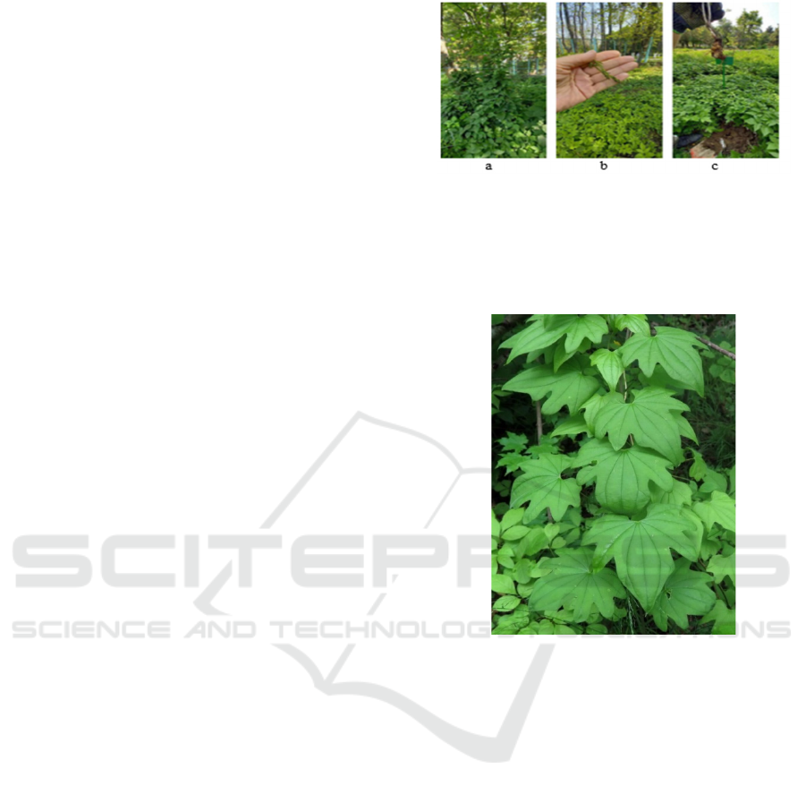
the Botanical Garden (Fig. 1.).
Figure 1: General view of Dioscorea nipponica Makino.: a
- general view,b - flowers, c – rhizome.
The leaves are three and seven-lobed. Leaves are
evenly arranged on the stem in an orderly fashion
(Fig.2).
Figure 2: Morphological structure of the leaf of Dioscorea
nipponica Makino.
Leaves are broadly heart-shaped in outline, with
strongly projecting auricles; their veining is arcuate,
most often 9 primary veins reach the leaf tip. The
marginal veins ensure the strength of these "wind"
leaves, which, obviously, is also served by
anastomoses between primary veins, formed by veins
of the 2nd order and creating a common reticulate-
nerve veining. The leaf tip is strongly elongated into
a drop-shaped tip. Such droplet spicules hanging
downward from the leaf allow water runoff from its
surface and water secreted by hydatodes. However,
they exhibit an interesting peculiarity of internal
structure. The whole length of the leaf tip is crossed
here inside by a complex glandular system of cavities
(pockets) with a slit-like exit to the surface of the drop
spicule. The glandular epithelium of the cavities
secretes mucus in them, in which nitrogen-fixing
bacteria settle. It was also found that the nitrogen
content in the drop spicules is higher than in the leaf
lamina.
Morphological and Anatomical Features of Leaf and Stem of Dioscorea Nipponica Makino
361
Features of leaf epidermis that revealed
similarities between wild-type and micropropagated
plants included amphistome state, presence of mucus,
glandular unicellular trichomes with multicellular
heads, polygonal cells with smooth walls, and type
and shape of stomata. Minor variations included a
thick cuticular wall with closed stomata in wild-type
plants compared with thin-walled open stomata in in
vitro plants. The opening of the stomata resulted in an
increase in the average size of the stomata (7.68-0.38)
μm and (6.14-0.46) μm on the adaxial side (Aina et
al., 2011).
Examined under optical microscope, scanning
electron microscope (SEM) and transmission electron
microscope (TEM) for stem, leaf, petiole, tuber, root
and flower of Dioscorea hispida Dennst. provided
detailed information on the anatomical features that
defined this species. The anatomical study showed
that the leaves of Dioscorea hispida had similar
features to eudicot plants, but the stem, tuber and
flower resembled unicotyledonous plants. The leaf
surface of Dioscorea hispida was covered with rough,
bristly and spiny trichomes or hairy surface (Bu,
2015).
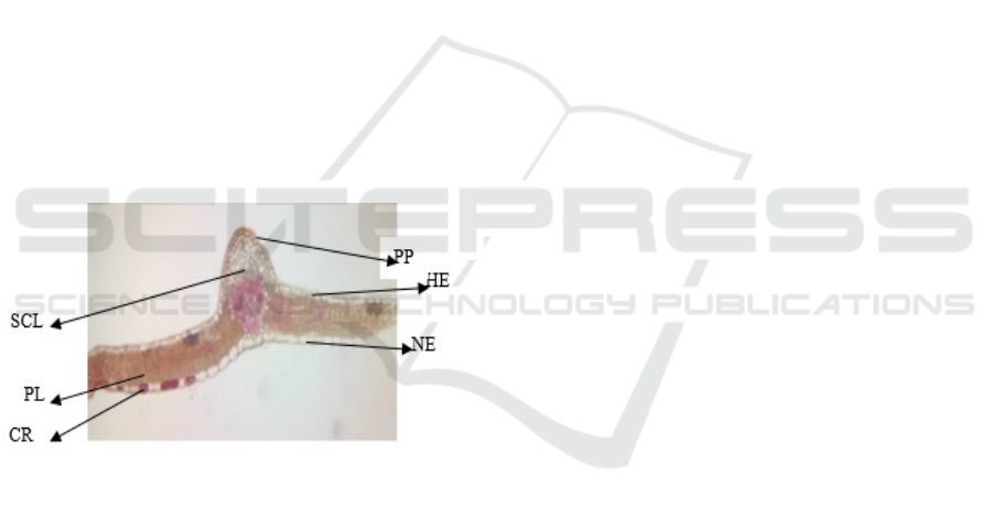
Structure of the leaf lamina of Dioscorea
nipponica (1-drawing).
Figure 3: General view of a transverse section of a
Dioscorea nipponica Makino. leaf: PP-Conducting bundle,
HE- upper epidermis, NE-lower epidermis, SCL-
sclerenchyma, PL-palisade, CR- raphid crystals.
The epidermis is a layer of cells that protects
against harmful environmental influences and
excessive water evaporation. The upper (adaxial) and
lower (abaxial) epidermis have a single layer of cells,
each covered by a cuticle. Often on top of the
epidermis, the leaf is covered with a protective layer
of waxy origin (cuticle). The stomata are restricted to
the lower surface only (hypostomal). In the epidermis
of Dioscorea nipponica leaves, anamocytic and
anisocytic stomata, as well as single pilosebaceous
trichomes with cuticular striated cell wall were found
on the adaxial and abaxial epidermis. Also, raphide
crystals are present in the epidermis.
The palisade (above, densely packed cells) and
spongy (below, loosely packed cells) portions of the
mesophyll, located between the upper and lower
epidermal layers, are shown. The mesophyll, or
parenchyma, is the inner chlorophyll-bearing tissue
that performs the main function, photosynthesis.
Network of veins formed by conducting bundles
(conducting tissue) consisting of vessels and sieve-
like tubes for the movement of water, dissolved salts,
sugars and mechanical elements.
Stomata are special complexes of cells located
mainly on the lower surface of leaves; they are used
for evaporation of excess water (transpiration) and
gas exchange. Epidermа is the outer layer of a
multilayered cell structure that covers the leaf from
all sides; the boundary area between the leaf and the
environment. The epidermis performs several
important functions: it protects the leaf from
excessive evaporation, regulates gas exchange with
the environment, excretes metabolic substances and,
in some cases, absorbs water. Most leaves have a
dorsoventral anatomy: the upper and lower surfaces
of the leaf have different structures and perform
different functions.
The epidermis is usually transparent (there are no
or few chloroplasts in its structure) and is covered on
the outside by a protective layer of waxy origin
(cuticle), which prevents evaporation. The cuticle on
the lower part of the leaf is generally thinner than on
the upper part, and thicker in biotopes with arid
climates compared to those where there is no
moisture deficit. The epidermal tissue consists of the
following cell types: epidermal (or motor) cells,
defense cells, accessory cells, and trichomes.
Epidermal cells are the most numerous, largest
(11.2±0.3, 15.4±0.4) and least adapted (9.1±0.2,
11.1±0.4). The epidermis is covered with pores called
stomata, which are part of a whole complex
consisting of a pore surrounded on all sides by
chloroplast-containing guard cells and two to four
side cells lacking chloroplast.
This complex regulates evaporation and gas
exchange of the leaf with the environment. As a rule,
the number of stomata on the lower part of the leaf.
Most of the leaf interior between the upper and
lower layers of the epidermis is parenchyma (the
main tissue), or mesophyll. Normally, the mesophyll
is formed by chlorophyll-synthesizing cells, so the
synonymous name chlororenchyma is also used. The
product of photosynthesis is called photosynthate.
Leaves are usually colored green due to
chlorophyll, a photosynthetic pigment found in
chloroplasts, the green plastids. Plants lacking or
lacking chlorophyll cannot photosynthesize. The veins
consist of xylem, the tissue used to conduct water and
I-CRAFT 2024 - 4th International Conference on Research of Agricultural and Food Technologies
362
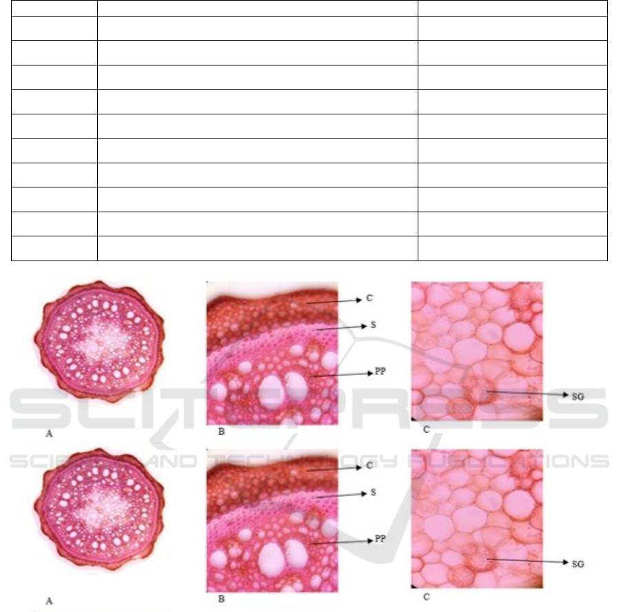
Table 1: Quantitative parameters of Dioscorea nipponica leaf mesophyll.
№ Si
g
ns Indicators, microns
1
Length of the upper epidermis
11,2±0,3
2
Width of upper epidermis
9,1±0,2
3
Length of lower epidermis
15,4±0,4
4
Width of lower epidermis
11,1±0,4
5 Palisade length 13,2±0,4
6 Palisade width 10,1±0,2
7 Sclerenchyma length 12,2±0,4
8 Sclerenchyma width 11,3±0,2
9 Parenchyma length 14,5±0,4
10 Parenchyma width 13,3±0,3
Figure 4: Transverse section of Dioscorea nipponica stem: A - general view, B - peel, C - pith: C - cortex, S - sclerenchyma,
PP - conductive bundles, SG - granular matter.
dissolved minerals, and phloem, the tissue used to
conduct organic matter synthesized by the leaves.
Usually the xylem lies on top of the phloem. Together
they form the main tissue called the leaf core. The
lamina has 3-4 protruding veins running from the base
to the apex. The cuticle of the lamina is soptically
thickened.
The leaf has a layered epidermis, the soptic wall
on the abaxial cells is convoluted and the adaxial cells
have a straight wall. Stomata with irregular
distribution and anomocytic type are found on the
abaxial surface. Palisade tissue was clearly
distinguished, elongated, compactly arranged and
oriented vertically, with a transverse section on the
adaxial side (13.2±0.4, 10.1±0.2), and spongy tissue
was arranged either loosely or compactly with
irregularly sized cells.
Dioscorea nipponica on transverse section is
round to wavy in shape, internal cells and tissues are
densely arranged (Fig. 4).
The stem is finely cellular, covered with cortex on
the outside. The structure of the cortex (containing
Morphological and Anatomical Features of Leaf and Stem of Dioscorea Nipponica Makino
363

chloroplast) consists of one row of epidermis with
cuticle and 4-5 rows of parenchyma cells under it.
The parenchyma cells are followed by a ring of 5-6
rows of thin-walled sclerenchyma cells. After the
sclerenchyma cells there are large and small
conducting bundles. The bundles have pairs of large
vessels.
The central core consists of parenchyma cells,
which increase in size as you approach the centre.
Among the parenchyma cells, granular filled cells can
be observed.
Epidermal cells are 13 µm high and 10 µm wide.
The table below summarises the size of all organoids
of the stem (Table 2).
Table 2: Anatomical parameters of stem Dioscorea nipponica.
№
S
y
mbols Indicators, µm
1
Epidermal height
13,3±0,3
2
Epidermal width
10,1±0,2
3
Sclerenchyma length
11,2±0,4
4
Sclerenchyma width
9,3±0,2
5
Parenchyma length
12,5±0,4
6
Parenchyma width
11,3±0,3
7
Height of large conductive bundles
18,8±0,5
8
Width of large conductive bundles
15,4±0,4
9
Length of smaller conductive bundles
9,4±0,3
10
Width of smaller conductive bundles
8,1±0,2
The anatomical structure of the stem of Dioscorea
nipponica is little studied than leaves, tubers, roots,
rhizomes (Martin et al., 1963), (Cunyu et al., 2022),
(Vı´tor et al., 2016), (Berdibaeva and Atabaeva,
2023).
We anatomically characterised the aerial stems of
the genus Dioscorea and evaluated the possibility of
using these anatomical characteristics to better
understand the taxonomy, systematics and diversity
of component species in the non-tropics. Air stem
fragments from 23 species were collected for
anatomical analysis using conventional
cytohistological techniques (Berdibaeva, 2021).
4 CONCLUSIONS
Dioscorea has two layers of vascular bundles, with
the outer layer containing sclerenchyma and the
medulla having sclereids, in contrast the parenchyma
had a sclerenchyma layer with vascular bundles. The
sclerenchyma layer in the stem may increase
mechanical strength.
In conclusion, we would like to report that the study
of the anatomical structure of the cross-section of the
stem Dioscorea nipponica is established, has a wavy
shape, parenchyma cells have filled with granular
substances, two-row arrangement of conductive
bundles, the presence of paired vessels.
REFERENCES
Aina OD, Atumeyi S. Foliar epidermal anatomy of four
species of Dioscorea. Adv Appl Sci Res 2011; 2(4): 21-
24.
Anthony Keith Thompson and Ibok Oduro 2021. Yams:
Botany, Production and Uses (A.K. Thompson and I.
Oduro) 129 DOI: 10.1079/9781789249279.0008
Berdibaeva D.B. Collection introductional of medicinal
plants of the Tashkent Botanical garden named after
F.N. Rusanov at the institute of botanics of republic of
Uzbekistan 2021 pp. 26-30 https://elibrary.ru/
publisher_about.asp?pubsid=23589 .
Berdibaeva Dilfuza Bazarbaevna Atabaeva Gulnoza
Shaakbar Qizi International scientific journal "Science
and innovation" Special issue: “Sustainable forestry”,
November, 2023 pp.225-229
https://cyberleninka.ru/article/n/study-of-anatomical-
structure-of-leaf-dioscorea-nipponica-makino
Bu Wei. Pharmacognostic study of individual
representatives of the genus Dioscorea L.: dissertation
... Candidate of Pharmaceutical Sciences: 04/14/02 /.-
Moscow, 2015.- 117 p.
I-CRAFT 2024 - 4th International Conference on Research of Agricultural and Food Technologies
364

Cunyu Zhou1 , Huanhuan Xiong2 , Youzhi Li2 , Chongnan
Zhao2 , Teng Li1 , Mengdi Zhang1 , Xia Zhang1 ,
Chaodong Yang1 , Zhiguo Jiang2 * Emir. J.
Anatomical and histochemical features of the
vegetative organs of Dioscorea polystachya
(Dioscoreaceae) Food Agric ● Vol 34 ● Issue 1 ● 2022
79-85.
Dospekhov B.A. Methodology of field experience (with the
basics of statistical processing of research results) —
M.: Agropromizdat, 1985. — 351 p.
Gubanov I. A. et al. Wild useful plants of the USSR / ed. by
T. A. Rabotnov. — M.: Mysl, 1976. — p. 71. — 360 p.
Harkevich S. S. Dioscorea japonica Dioscorea nipponica //
Red Book of Russia. (Verified on June 4, 2012)
Ki-Sun Park, Hye Jin Kim, Joo Tae Hwang, Byoung Seob
Ko Dioscorea nipponica extracts enhance recovery
from skeletal muscle atrophy by suppressing NF-κB
expression Journal of Functional Foods Volume 73,
October 2020, 104109
Murdakhayev Yu.M. Medicinal plants that have found their
homeland in Uzbekistan. Tashkent, 1990. 234 p.
Trankovsky D.A. Practicum on plant anatomy. – M.:
Higher School, 1979. – 221 p.
Vı´tor Tenorio 1 •Ricardo S. Couto 1 •Elaine S. B. de
Albuquerque 2 • Artur M. L. Medeiros 1 •Rafaela de
Oliveira Ferreira 4 •Joa ˜o M. A. Braga 3 • Ricardo C.
Vieira Stem anatomy of neotropical Dioscorea L.
(Dioscoreaceae) and its importance to the systematics
of the genus 4 Received: 16 March 2016 / Accepted: 18
March 2017 / Published online: 11 April 2017
W. Martin and Sonia Ortiz Origin and Anatomy of Tubers
of Dioscorea floribunda and D. spiculiflora Franklin
Botanical Gazette Vol. 124, No. 6 (Dec., 1963), pp.
416-421 (6 pages) Published By: The University of
Chicago Press https://www.jstor.org/stable/2473209
Morphological and Anatomical Features of Leaf and Stem of Dioscorea Nipponica Makino
365
