
Image Recognition of Pigmented Skin Diseases Based on Deep
Learning
Xinyu Zhao
a
College of Electronic and Information Engineering, South China University of Technology,
Guangzhou, China
Keywords: Deep Learning, Pigmented Skin Diseases, Image Recognition.
Abstract: One of the most common skin conditions is pigmentary skin disease. It is also challenging to differentiate
between the lesions of various pigmentary skin diseases with the unaided eye due to their striking similarity.
The paper wishes to investigate whether deep learning image recognition can resolve this issue because deep
learning technology has advanced significantly in recent years and has shown promise in a number of
domains. In order to help the investigation, the paper modified the weights of three pigmented skin illnesses
that have similar clinical features to help two deep learning models that paper used to identify to gain higher
accuracy. The findings demonstrate that deep learning can effectively identify many forms of pigmented skin
illnesses and is very helpful in the recognition of skin diseases.
In subsequent research, the paper will attempt
to use deep learning to determine the lesion's stage, which will be extremely beneficial for diagnosing
pigmented skin conditions.
1 INTRODUCTION
The rapid evolution of computer vision and deep
learning technologies has brought significant
advancements to medical image analysis, particularly
in the detection and evaluation of pigmented skin
diseases. Pigmented skin conditions, which are
characterized by abnormalities in skin pigmentation,
represent a common category of dermatological
disorders. (Cai, 2023) The early detection and precise
staging of these conditions are critical for effective
clinical intervention and prognosis assessment.
Traditionally, the diagnosis of skin lesions has relied
heavily on the expertise of dermatologists, because it
is challenging to recognize and categorize skin
lesions due to the wide range of pigmented skin
lesions, the high degree of resemblance between
distinct classes, and the significant differences within
the same class. (Chen, 2014) This conventional
approach, while valuable, is often subjective and can
suffer from variability in diagnostic accuracy due to
differences in clinical experience and judgment.
In recent years, the integration of deep learning
techniques, particularly Convolutional Neural
Networks (CNNs), has introduced a transformative
a
https://orcid.org/0009-0009-1026-3190
shift in the analysis of medical images. CNNs have
demonstrated exceptional performance in tasks
related to image classification and object detection,
surpassing traditional methods in accuracy and
efficiency (Chu, 2024). These advancements present
promising solutions for the automatic identification
and staging of pigmented skin lesions, offering
potential improvements in diagnostic consistency and
operational efficiency.
This paper explores the application of deep
learning technologies to the recognition and staging
of pigmented skin lesions. The paper will examine the
current state of deep learning models used in skin
lesion image analysis, highlighting their applications,
the challenges they face, and their future prospects.
The discussion will focus on several key areas: the
mainstream deep learning methods employed,
including various CNN architectures; the processes
involved in constructing and processing datasets; and
the strategies for training and optimizing models.
Furthermore, the paper will consider the practical
implications of these technologies, evaluating how
they can be integrated into clinical practice to
enhance diagnostic capabilities and improve patient
outcomes.
Zhao and X.
Image Recognition of Pigmented Skin Diseases Based on Deep Learning.
DOI: 10.5220/0013512900004619
In Proceedings of the 2nd International Conference on Data Analysis and Machine Learning (DAML 2024), pages 221-226
ISBN: 978-989-758-754-2
Copyright © 2025 by Paper published under CC license (CC BY-NC-ND 4.0)
221

By providing a thorough examination of these
aspects, this paper aims to offer insights into the
current advancements in deep learning for skin lesion
analysis and to outline the potential for future
developments in this rapidly evolving field.
2 PIGMENTED SKIN LESION
DETECTION
Pigmented skin lesions (PSLs), a common type of
skin problem, are known to show more skin coloring,
mostly happening because melanocytes start to
multiply more. Proper detection and labeling of these
lesions are important as they come in both harmless
types, like melanocytic nevi (or just nevus), and
harmful ones like melanoma. Especially melanoma, a
very serious kind of skin cancer, is usually the cause
of many deaths related to skin cancer. It becomes
quite important to detect PSLs early on and categorize
them correctly for better treatment options and
improved health results for patients.
2.1 Types of hyperpigmented lesions
Pigmented skin lesions can mainly be broken down
into either benign or malignant forms. The usual
examples include the most common ones.
Melanocytic nevus: It is where melanocytes
gather in a benign way, showing up as dark spots on
the skin. Most of the time, they do not cause harm,
but there can be rare occasions where they turn into
melanoma.
Melanoma: A very dangerous type of skin cancer
that comes from melanocytes. Melanoma has a high
level of aggression, spreading to other body parts
quickly, so detecting it early is very important. (Han,
2018)
Sunspot: often referred to as liver spot or even
senile spot, it is mostly considered as a harmless
pigmented area that is the result of prolonged
exposure to the sun. These spots do not develop into
cancer, though sometimes they can be mistaken for
dangerous lesions of a malignant kind.
Seborrheic keratosis: This is identified as a non-
cancerous growth that is wart-like in its appearance,
which can form on different parts of the body.
Though these growths pose no harm, their visual
similarity to melanomas creates certain issues during
diagnosis.
Abnormal nevus: A kind of nevus that has
irregular characteristics and may suggest early signs
of melanoma, and regular check-ups become
necessary to watch for possible changes in these skin
areas.
2.2 Limitations of traditional
recognition methods
The common ways of identifying pigmented lesions
on skin mainly depend on doctors visually inspecting,
sometimes with help from dermoscopy, which is a
tool used to magnify the skin surface for better
viewing without causing harm. (Niu, 2024) Even
though these approaches can work well, they still face
certain limits and are not always fully sufficient.
Subjectivity: The evaluation of visuals is very
subjective, depending much on the experience and
expertise of dermatologists, which introduces
variations and inconsistencies in diagnoses made.
Limited accuracy: Even with dermoscopy being
used, some lesions remain difficult to tell apart,
particularly when they are in early development or
show unusual characteristics, adding to the difficulty
in making precise distinctions.
Time being consumed: Examining several lesions
manually, particularly when a patient has many
pigmented spots, takes much time and can be not
practical in certain clinical environments due to how
long it might take to complete.
Variability among observers: Different
dermatologists might see one lesion in various ways,
which creates inconsistency in diagnoses and advice
for treatments provided across different patients and
practitioners.
2.3 Image Classification Methods
Through Deep Learning
The traditional methods have limitations that
deeplearning methods aim to overcome, especially by
using CNNS more and more for the purpose of
classifying skin lesions with pigments.
2.3.1 Convolutional Neural Network (CNN)
Basic Idea
CNNS represent a kind of deep learning models
designed to handle structured grid-like data, for
example, images. A CNN typically consists of several
layers, which might include convolutional layers,
pooling layers, and layers that are fully connected.
Convolutional layers: In these layers, convolution
operations get applied to the input, with filters
detecting various local patterns like edges or shapes,
possibly textures. What results from this is a feature
map, which highlights such patterns but does so
without strict detail. (Dong, 2017)
Pooling layer: Pooling reduces the size of the
feature map by summarizing certain features present
DAML 2024 - International Conference on Data Analysis and Machine Learning
222
in regions, which has the function of lowering
computational complexity and also reducing
overfitting risks, although the exact effect is not
always completely clear.
Fully connected layers: Each neuron in one layer
gets connected with every neuron that belongs to the
next layer, allowing predictions to be formed based
on features coming from the convolutional and
pooling layers, though these predictions can be
influenced by many factors at the same time, some of
which could change.
2.3.2 Common Deep Learning models
There have been many different deep learning models
that have managed to be applied for the classification
of skin lesions that are pigmented, which shows the
versatility of these models in handling tasks related to
this field.
ResNet: ResNet brings up the idea of residual
learning, which makes networks able to go much
deeper by reducing issues with vanishing gradients. It
has been said to perform better in tasks like image
classification, including images from medical fields,
showing better results.
EfficientNet: EfficientNet represents a group of
models that, through scaling dimensions like depth,
width, and resolution in a structured manner,
improves performance. These models manage to
reach higher accuracy levels while using fewer
parameters than what is commonly seen in older
models. The approach provides better results not just
by increasing one aspect, but by adjusting several
dimensions together. Compared to more traditional
models, which may not consider such structured
scaling, EfficientNet shows advantages in both
efficiency and accuracy, making it stand apart. (Petra,
2024) However, exact improvements can vary
depending on implementation.
3 EXPERIMENTAL PRINCIPLE
3.1 Experimental procedure
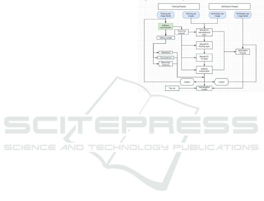
As shown in Figure 1. Images and the associated
labels (melanoma, chromatoma, seborrheic keratosis)
make up the training set. The paper applies a Softmax
loss function and modify the weights according to the
mistake.
The images are routed to the convolutional,
pooling, fully connected (FC), and Softmax output
layers of ResNet101.
The model's performance is verified using the
images and labels that make up the verification set.
Images pass through the ResNet101 layers in a
manner akin to that of training.
Following the output's progression through the
ResNet101 layers, a test set is used to optimize and
validate it. To increase classification accuracy, the
optimization model modifies the parameters in
response to the output.
Figure 1:
Flow chart of Deep learning
(Photo/Picture credit : Original)
3.2 The Role of Deep Learning in Stage
Detection
Deep learning models are becoming more significant
in how pigmented skin lesions are automatically
detected and staged, helping with:
3.2.1 Feature Extraction and Discrimination
CNNs, which are used in many cases, can find and get
important features out of dermoscopic images. These
features, which are helpful in separating the stages of
melanoma, do not only include how the lesion looks,
but also other patterns that might exist, though these
patterns may not be easily noticed by human
observers. (Wang, 2024) These patterns can
sometimes be less obvious and need more attention to
be seen clearly.
3.2.2 Semantic and Instance Segmentation
Deep learning techniques, such as the more advanced
ones including semantic segmentation and also
instance segmentation, give the possibility of a
clearer outline of lesion boundaries, as well as finding
different areas inside a lesion that might match up
with various pathological traits. Semantic
segmentation is used to assign a label to each pixel in
Image Recognition of Pigmented Skin Diseases Based on Deep Learning
223
the image, which can help with recognizing certain
areas like tumor tissue in contrast with healthy parts.
But instance segmentation goes even further to
separate different objects or lesions found in one
image, though they might still fall under the same
general category.
3.3 Current Research and Applications
Recent research shows that deep learning models
have possibilities in automatically classifying and
staging pigmented skin lesions. For instance, some
researchers have created CNN-based models, which
can sometimes reach accuracy levels close to or even
higher than experienced dermatologists when it
comes to detecting melanoma. Additionally, models
that were trained on big annotated datasets, such as
the ISIC dataset (International Skin Imaging
Collaboration), are now used to automate the staging
process for melanoma. (Niu, 2024) This offers a
useful tool to help clinicians with decisions when
making diagnoses.
4 EXPERIMENT AND RESULT
4.1 Construction and Choosing of
Datasets
The deep learning model's effectiveness when it
comes to detecting and staging pigmented skin
lesions relies a lot on the type of dataset and how
varied it is, especially for training purposes.
Important points to think about include:
Data Collection: High-resolution images must be
gathered from different groups of people and medical
settings. These images are needed for creating
reliable models. The images should cover different
phases of illness and show both frequent and unusual
types of lesions, which is important to ensure the data
is comprehensive enough for the purpose of training.
Annotation: It is necessary to annotate datasets with
accuracy. Usually, experts like dermatologists are
responsible for labeling images, and they mark them
with details like lesion categories, disease stages, and
important clinical attributes. In some situations, the
annotations may even provide pixel-wise labeling for
tasks related to segmentation.
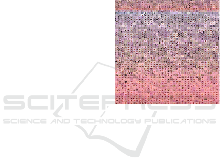
The data set used in this study was HAM10000
skin image dataset on hyperai(shows on Figure 2),
which collected 10000 skin lesions images from
different populations. The cases included several
representative types of hyperpigmented lesions,
mainly melanoma, nevus and seborrheic keratosis.
HyperAI (hyper.ai) artificial intelligence and high
performance computing community aims to help
developers and enthusiasts of data science and
artificial intelligence industry learn, understand and
practical by providing multiple services such as
accelerated download of data sets, online tutorial
demonstrations, in-depth interpretation of papers, and
integration of top conference calendar.
Figure 2: Dataset from Hyperai(Photo/Picture credit :
Original)
4.2 Model Training and Performance
Evaluation
It is very important to do model training and
performance evaluation correctly if you want deep
learning models to be used in clinical practice.
4.2.1 Choosing and Preparing the Training
data
After removing the unidentifiable bad graphs in the
dataset, the paper divided the dataset into three
independent datasets that did not cross each other to
ensure the generalization ability of the model on
unknown data. There were 6000 images in the
training set, 1800 images in the validation set, and
450 images in the test set.
The distribution of three types of tumor images
on the three datasets is shown in table 1.
DAML 2024 - International Conference on Data Analysis and Machine Learning
224
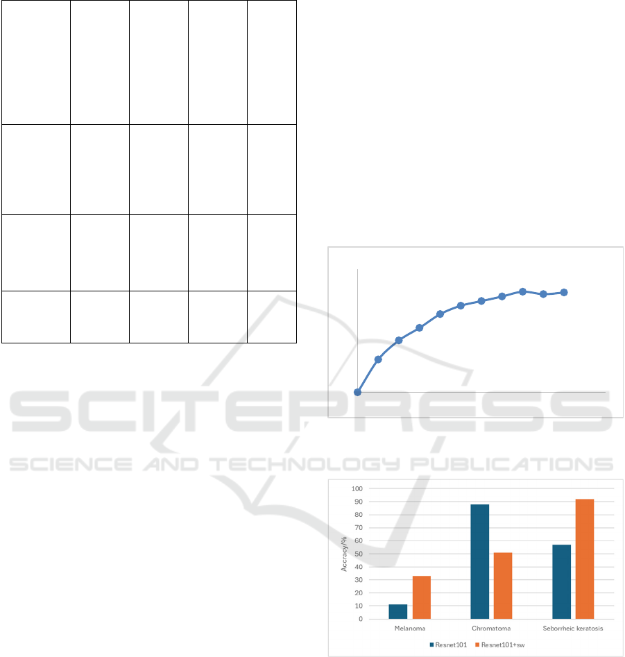
Table 1: Dataset distribution
Dataset Melan-
oma
Chrom-
atoma
Seborrh
-eic
keratosi
-s
Total
Training
set
1800 2400 1800 6000
Verificati
-on set
540 720 540 1800
Test set 145 180 135 450
4.2.2 Model Evaluation Metrics
Metrics commonly used for evaluating how models
perform include:
Accuracy: It is the ratio of correct predictions
compared to the total number of predictions that the
model has made overall.
Sensitivity (Recall): This measures how well the
model can find positive instances, like when it
identifies malignant lesions correctly.
Precision: Precision is a metric that is used in
statistical classification and information retrieval that
represents the proportion of correctly extracted
samples to total extracted samples. (Tian. 2024)
Recall, which is the ratio of the number of extracted
samples to the total number of samples, is a related
idea.
F1 Score: Precision and recall come together to
make a harmonic mean, and that gives a measure
which balances how the model performs on both
sides, providing an understanding of overall
performance.
4.3 Experimental result
During the training phase, the model's prediction
performance was assessed in real time using the
validation set, which allowed for parameter
adjustments and overall model optimization. The
final model for predicting the test set data was
determined by using the training result from the round
that had the highest accuracy of the validation set.
There were 100 training epochs in this model.
ResNet101 was used as the training model in this
experiment in order to confirm the experiment's
efficacy. By establishing category weights, the
efficacy of deep learning in recognizing photos of
pigmented skin diseases was assessed. This
experiment is trained on a Tesla V100S-PCI-32GB
GPU and is based on the Aliyun computing platform
to guarantee experiment efficiency.
It can be seen from Figure 3 that the accuracy of
diagnosing pigmentary skin illnesses improved to
65% after many training sessions, indicating that deep
learning is a viable method for doing so. The figure 4
compares the particular recognition results of each
category when the two training procedures are used
with this model.
Figure 3:
Curve of average accuracy(Photo/Picture credit :
Original)
Figure 4: Accuracy of 3 kinds of skin diseases
(Photo/Picture credit : Original)
Deep learning had generally good accuracy in
identifying chromopoma and seborrheic keratosis,
but less desirable results when it came to melanoma.
0
0.2
0.4
0.6
0.8
0 20406080100120
epochs
accuracy
Image Recognition of Pigmented Skin Diseases Based on Deep Learning
225

5 CONCLUSION
One of the most crucial responsibilities in
dermatology is the diagnosis of pigmented skin
diseases, which has a big influence on patient
outcomes. While useful, traditional diagnostic
techniques have drawbacks that deep-learning
models might be able to solve. These models are able
to correctly classify partly pigmented lesions through
the use of sophisticated techniques including
segmentation algorithms and CNN. Many obstacles
still need to be overcome, particularly in the areas of
data heterogeneity, model applicability, and
distinguishing between different kinds of pigmented
dermatoses. It will be up to continued study and
creative problem-solving to resolve these obstacles
before deep learning in dermatology can realize its
full potential.
The paper employed two distinct deep learning
models to identify three common pigmented skin
illnesses from images. By varying the weights of the
models, the paper was able to increase the recognition
accuracy. The encouraging outcomes demonstrated
that deep learning may be used to diagnose skin
illnesses, with up to 80% accuracy being able to
distinguish between chromoblastoma and seborrheic
keratosis. The paper will attempt to use deep learning
to distinguish between various phases of pigmented
skin lesions and to increase the accuracy of
diagnosing skin disorders like melanoma, which are
challenging to diagnose.
REFERENCES
Cai, Q. Y., Ma, Q., Wang, C. K. 2023 Application of
deep learning in skin OCT medical images.
Chinese Journal of Laser Biology,32(3): 193-199.
Chen, X. C. 2014. Deep Learning Algorithm and
application Research based on Convolutional neural
networks. (Doctoral dissertation, Zhejiang Gongshang
University)、
Chu, J. L., Fang, C., Wan, N., Wang, W. D., Wang,
X., Zhang, J. 2024. Application of deep learning
in classification and recognition of pneumonia CT
images. Fujian computers (07), 33-36. Doi:
10.16707 / j.carol carroll nki FJPC. 2024.07.006.
Dong, J., Jin, L. P., Zhou, F. Y. 2017. A review of
convolutional neural networks. Journal of
Computer Science, 40(6), 1229-1251.
Han, Z. Y., He, X. Y., Wei, Z. Z. 2018 Recognition
and classification of pigmented skin diseases
based on deep convolutional neural networks.
Journal of Computer Applications,38(11): 3236-
3240.
Nadia, S., Razieh, P., Sajad, S., & José L. H. 2024.
Identification of Armyworm-Infected Leaves in
Corn by Image Processing and Deep
Learning.Acta Technologica Agriculturae(2),92-
100.
Niu, H., Wang, P. Y., Yin, G. W. 2024.
Clinicopathological analysis of 15 cases of basal
cell carcinoma of the skin. Oncology
Fundamentals and Clinic (02),200-202.
Petra, R., Dorijan, R. & Goran, M. 2024. Image-
Based Leaf Disease Recognition Using Transfer
Deep Learning with a Novel Versatile
Optimization Module. Big Data and Cognitive
Computing(6),52-.
Tian, S. W., Shi, X. W., Shu, C., Yu, L. 2024.
MSMA: A multi-stage and multi-attention
algorithm for the classification of multimodal skin
lesions, Biomedical Signal Processing and
Control, Volume 93,106180, ISSN 1746-8094.
Wang, H., Wang, T. T. Wu, J. H., Yang, L. 2024.
Diagnosis of hand arthritis based on deep learning
algorithm from X-ray images. Modern Medicine
(07),1043-1049.
DAML 2024 - International Conference on Data Analysis and Machine Learning
226
