
Semantic Image Synthesis for Realistic Image Generation in Robotic
Assisted Partial Nephrectomy
Stefano Mazzocchetti
1
, Laura Cercenelli
1
, Lorenzo Bianchi
2,3
, Riccardo Schiavina
2,3
and
Emanuela Marcelli
1
1
eDIMES Lab, Laboratory of Bioengineering, Department of Medical and Surgical Sciences, University of Bologna,
Via Massarenti, 9, 40138 Bologna, Italy
2
Division of Urology, IRCCS Azienda Ospedaliero, Universitaria di Bologna, Via Massarenti, 9, 40138 Bologna, Italy
3
Department of Medical and Surgical Sciences, University of Bologna, Via Massarenti, 9, 40138 Bologna, Italy
Keywords:
Minimally Invasive Surgery, Robotic Surgery, Semantic Image Synthesis, Deep Learning, GAN,
Computer Vision.
Abstract:
With the continuous evolution of robotic-assisted surgery, the integration of advanced technologies into the
field becomes pivotal for improving surgical outcomes. The lack of labelled surgical datasets limits the range
of possible applications of deep learning techniques in the surgical field. As a matter of fact, the annotation
process to label datasets is time consuming. This paper introduces an approach for realistic image generation
in the context of Robotic Assisted Partial Nephrectomy (RAPN) using the Semantic Image Synthesis (SIS)
technique. Leveraging descriptive semantic maps, our method aims to bridge the gap between abstract scene
representation and visually compelling laparoscopic images. It is shown that our approach can effectively gen-
erate photo-realistic Minimally Invasive Surgery (MIS) synthetic images starting from a sparse set of annotated
real images. Furthermore, we demonstrate that synthetic data can be used to train a semantic segmentation
network that generalizes on real data reducing the annotation time needed.
1 INTRODUCTION
The transition from traditional open surgeries to min-
imally invasive procedures, such as laparoscopy, has
significantly reduced patient trauma and recovery
times. Concurrently, the integration of computer vi-
sion and image synthesis techniques into the surgical
domain has shown great potential for enhancing sur-
gical planning, training, and intraoperative decision-
making. As the field of robotic-assisted surgery con-
tinues to evolve, there is an increasing demand for ad-
vanced technologies that enhance both preoperative
planning and intraoperative decision-making. Data-
driven methods can develop solutions for Computer
Assisted Interventions (CAI) to support surgeons dur-
ing the procedure (Vercauteren et al., 2019). This pa-
per presents an approach to generate photo-realistic
laparoscopic images in the context of Robotic As-
sisted Partial Nephrectomy (RAPN). Our primary ob-
jective is to demonstrate that descriptive semantic
maps can serve as a bridge between abstract scene
representation and visually compelling, anatomically
accurate images. The key novelty of this paper lies
in the application of semantic image synthesis (SIS)
specifically to RAPN, showcasing that semantic maps
can effectively guide the generation of images with
high anatomical fidelity. Our work serves as an
initial step towards a comprehensive framework for
computer-assisted interventions through augmented
reality. Furthermore, we establish a foundation for in-
corporating knowledge about object positioning into
downstream tasks, such as 6-degree-of-freedom (6-
DoF) pose estimation.
In the subsequent sections of this paper, we will
delve into the related works in the field of image
generation for Minimally Invasive Surgery (MIS), the
dataset used for training and the methodology em-
ployed for semantic image synthesis in the context
of laparoscopic surgery. Additionally, we will dis-
cuss the assessment methodology and details of the
experiments undertaken, followed by concluding dis-
cussions.
Mazzocchetti, S., Cercenelli, L., Bianchi, L., Schiavina, R. and Marcelli, E.
Semantic Image Synthesis for Realistic Image Generation in Robotic Assisted Partial Nephrectomy.
DOI: 10.5220/0012611200003660
Paper published under CC license (CC BY-NC-ND 4.0)
In Proceedings of the 19th International Joint Conference on Computer Vision, Imaging and Computer Graphics Theory and Applications (VISIGRAPP 2024) - Volume 4: VISAPP, pages
645-652
ISBN: 978-989-758-679-8; ISSN: 2184-4321
Proceedings Copyright © 2024 by SCITEPRESS – Science and Technology Publications, Lda.
645

2 RELATED WORK
Image-to-image translation is a computer vision task
that involves converting an input image from one
domain to an output image in a different domain
while preserving relevant structures and features. The
goal is to learn a mapping between the two do-
mains (Zhu et al., 2017; Isola et al., 2017), allow-
ing the transformation of images from, for exam-
ple, grayscale to color, or from satellite imagery to
maps. This task is often approached using genera-
tive models, such as Generative Adversarial Networks
(GANs) (Goodfellow et al., 2020) or Variational Au-
toencoders (VAEs) (Kingma et al., 2019), to learn the
complex relationships between the input and output
domains. Image-to-image translation finds applica-
tions in various fields and recently has also been ex-
ploit in the medical domain.
(Pfeiffer et al., 2019) proposed a method based
on unpaired image to image translation (Zhu et al.,
2017) to translate simulated images taken from a 3D
software to real laparoscopic images. Those meth-
ods relies on an unpaired dataset, i.e. where there
is not a one-to-one correspondence between an im-
age in domain A and an image in domain B. The au-
thors exploited the network proposed by (Huang et al.,
2018) with an additional structural similarity loss to
preserve image content. (Rivoir et al., 2021) com-
bined unpaired image-to-image translation and neu-
ral rendering in order to transfer simulated to photo-
realistic surgical abdominal scenes with a long-term
consistency in the video. However, those methods
were proposed for liver segmentation and require a
3D scene setup from real patient-specific 3D mesh ob-
tained from medical imaging like Computed Tomog-
raphy (CT). Moreover, (Ozawa et al., 2021) employ
the cycle GANs (Zhu et al., 2017) to generate realistic
synthetic data for surgical instrument segmentation.
Another generative method is the Semantic Image
Synthesis (SIS) task which generates realistic images
starting from a semantic map. It was first introduced
by (Isola et al., 2017). Usually, it requires a paired
dataset, consisting of the coupling of the real image
to the associated semantic map. Most of the works
rely on the conditional GANs (Isola et al., 2017). In
order to augment the labelled training data for deep
learning algorithms, (Rau et al., 2019) translated en-
doscopic images into depth maps applying Image-to-
image translation (Isola et al., 2017). Recently, (Yoon
et al., 2022) released a dataset composed by real la-
belled data and synthetic images generated by seman-
tic image synthesis. They combined real data seg-
mented manually and a virtual surgery environment
created from the 3D organ meshes obtained from the
CT with different surgical instruments. They used the
SIS to minimize the semantic gap between real and
synthetic data. They showed that synthetic data are
no longer helpful when the models already achieve
high performance with the real data.
Another work that exploits SIS to generate data
is (Marzullo et al., 2021). Starting from the En-
doVis 2017 surgical instrument segmentation task
dataset (Allan et al., 2019), they added coarse seg-
mentation of fat and organ tissue to perform SIS
with (Isola et al., 2017). Different from all previ-
ous works, this approach is used to generate photo-
realistic images for the the (RAPN) surgical pro-
cedure. A dataset was collected from six surgical
procedures and labelled with more semantic infor-
mation with respect to (Marzullo et al., 2021).Even
with a limited training data availability, exploiting the
SPatially-Adaptive (DE)normalization SPADE (Park
et al., 2019) architecture, a semantic segmentation
network has been trained on the generated data
achieving good quantitative and qualitative results.
Adding more semantic features to the image let a bet-
ter mapping between semantic information and real-
istic images.
3 MATERIALS AND METHODS
In this section, the dataset and the network used for
the experiments are described.
3.1 Data
The data exploited for this work consist of 2D in-vivo
images from (RAPN) surgical procedures performed
with the da Vinci Xi robot at Division of Urology - IR-
CCS Azienda Ospedaliero-Universitaria di Bologna,
Bologna. Six clinical cases of patients with clini-
cal diagnoses of T1 renal mass extracted from data
acquired for a previous work (Bianchi et al., 2020)
have been used. Participants signed a written in-
formed consent document. The study was approved
by our Institutional Ethics Committee (IRB approval
3386/2018). A total number of 318 frames were ob-
tained from the procedures where the kidney is fully
or partially visible. The number of frames for each
clinical case (C) can be seen in Figure 1.
In order to perform the Semantic Image Syn-
thesis task, each frame has been carefully labelled
with a software tool (Wada, ). Each pixel can be
identified as one of the following 12 classes: back-
ground, kidney, Monopolar Curved Scissors, Fenes-
trated Bipolar, Bipolar Forceps, fat, abdomen tissue,
liver, blood, gauze, renal vein and others (a class con-
VISAPP 2024 - 19th International Conference on Computer Vision Theory and Applications
646
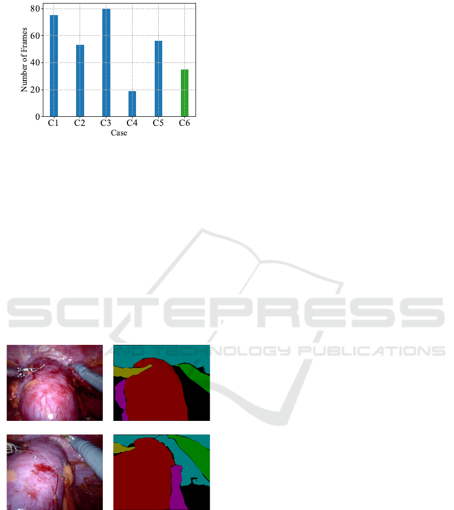
Figure 1: Number of frames extracted for each case. The
case C6 has been used as an additional test set.
taining rare surgical tools that can be seen in the pro-
cedure). Figure 2 shows two samples and the corre-
sponding semantic map. It can be noticed that differ-
ently from (Allan et al., 2019) or others Minimally In-
vasive Surgery (MIS) datasets, a part segmentation of
the surgical tools is not present. The semantic mask is
accurate only for the surgical tools, kidney and liver
(when present). In a different way from (Marzullo
et al., 2021), were the authors added only two se-
mantic information beside the manipulators to the En-
doVis (Allan et al., 2019) dataset, more information
is present in the semantic map which can help the
generative network to perform a better mapping be-
tween the mask and the real scene. All the extracted
Figure 2: Training data samples and corresponding seman-
tic segmentation map.
frames have been reshaped to a fixed size of 256x256
for memory restrictions. Five clinical cases (C1-C5)
have been used to generate the train (168 : 60% ), val-
idation (26 : 10%) and test(89 : 30%) sets. Moreover,
in order to test the ability to generalize on unseen data
of the networks, the sixth clinical case (C6) has been
used as an additional test set and is composed of 35
frames. The number of training set data has been aug-
mented to a total of 1448 frames by random vertical
or horizontal flip, elastic and affine transformation or
a combination of those.
3.2 Semantic Image Synthesis Network
Generative Adversarial Network (GAN) (Goodfellow
et al., 2020) are generative networks that the learn
probability distribution of the dataset by learning a
mapping between a random noise vector to an im-
age. It is achieved by a min-max optimization be-
tween a Generator G and a Discriminator D. Addi-
tionally, conditional GANs (Isola et al., 2017) lever-
ages on adversarial training to let a generator G learn
a mapping between a condition c and a random noise
vector z to an output image y:
G : {c, z} −→ y (1)
In contrast, the discriminator D is trained to distin-
guish between real and fake images generated by
G. Indeed, the generator G and discriminator D are
trained simultaneously and they compete to maximize
their own payoff. The supervision is achieved since
the dataset is composed by pairs {(m
i
, x
i
)}, where for
each image x
i
exists the semantic map associated m
i
.
Given a segmentation mask m
i
∈ L
H×W
with image
height H, width W and where L is a set of integers de-
noting the semantic labels, SIS networks aim to learn
a mapping function that converts input segmentation
masks to photo-realistic images. In this case the seg-
mentation mask m acts as the condition for the gener-
ative model.
In this work, the SPADE (Park et al., 2019) has
been employed to perform the image synthesis task.
The authors built the architecture starting from a pre-
vious work (Wang et al., 2018), where they added
a SPatially-Adaptive (DE)normalization. In previous
methods, the semantic map was passed directly as in-
put to the network and processed by stacks of con-
volution, normalization and activation layers. In par-
ticular, the normalization layers tend to take out the
semantic information. For this reason, (Park et al.,
2019) introduced a new conditional normalization
method. After every batch normalization, the spa-
tially adaptive modulation parameters are generated
directly from the condition (semantic map) by pro-
jecting c to an embedding space and then convolved
to produce two spatial modulation parameters that are
added and multiplied in an element-wise manner with
the batch normalization output. With this setting, the
SPADE Residual Block is composed by a stack of two
SPADE, ReLU and convolution layers. The gener-
ator is composed of several SPADE residual blocks
Semantic Image Synthesis for Realistic Image Generation in Robotic Assisted Partial Nephrectomy
647

with upsampling layers where the semantic map is
downsampled to the right dimension at each stage of
the SPADE residual block. In particular, differently
from (Wang et al., 2018), the generator G is com-
posed of only the decoding block since there is no
need to encode the segmentation map. In this work,
the deterministic version of SPADE has been used,
where the generator G starts with processing a down-
sampled version of the semantic map c. On the other
hand, the discriminator D is a multi-scale patch-based
fully convolutional network (Long et al., 2015) and
takes as inputs the concatenation of the semantic map
and the generated image and the concatenation of the
semantic map and the real image. The average predic-
tion of all patches is used to classify the whole image
as real or fake. Conditional GANs are trained with
the adversarial setting trying to model the conditional
distribution of the real image given the semantic map
to solve the adversarial min-max problem:
min
G
max
D
L
GAN
(G, D) (2)
In particular, the loss is composed by 3 terms, the
GAN loss, discriminator-based feature matching loss
and VGG perceptual loss (Wang et al., 2018). For
further details please refer to the original paper (Park
et al., 2019).
4 EXPERIMENTS
In the following section the quantitative and qualita-
tive results are presented. All experiments were per-
formed with PyTorch on a NVIDIA GeForce RTX
3070 Laptop GPU. The SPADE (Park et al., 2019)
network was trained for 50 epochs and Adam as opti-
mizer with a learning rate of 2e
−4
with a batch size of
2.
4.1 Evaluation Protocol
The evaluation of synthesized images is a challeng-
ing problem. Following previous works (Isola et al.,
2017; Park et al., 2019; Wang et al., 2018), a seg-
mentation network has been trained on the real im-
ages and tested on the synthetic data generated from
the test set and the one generated from the additional
test set. This network is a U-Net (Ronneberger et al.,
2015) architecture with a ResNet18 (He et al., 2016)
backbone pretrained on ImageNet (Deng et al., 2009).
Indeed, if the generated images reflect the distribution
of the trained data, the segmentation network should
generalize well on synthetic data.
Additionally, to demonstrate that the generated
data can be exploited to train deep learning net-
works, a segmentation network with only 3 output
classes (background, kidney and surgical tool) has
been trained on synthetic generated data and tested on
the real test set and the additional one. Both the seg-
mentation networks have been trained for 60 epochs,
Adam optimizer with learning rate of 1e
−4
and batch
size of 8. In this case, the validation set has been
used to select the best epoch. The loss function is a
weighted sum of the cross-entropy loss and the multi-
class dice loss. Semantic segmentation results are
evaluated quantitatively in terms of Dice Score, In-
tersection over Union (IoU), Precision (P) and Recall
(R):
Dice =
2T P
2T P + FN + FP
(3)
IoU =
T P
T P + FN + FP
(4)
Precision =
T P
T P + FP
(5)
Recall =
T P
T P + FN
(6)
Where, given the confusion matrix the True Positive
(TP), False Positive (FP), True Negatives (TN) and
False Negatives (FN) are defined. The Dice Score is
defined as two times the area of intersection divided
by the sum of the areas of the prediction and ground
truth mask. The IoU (or Jaccard index) is defined as
the intersection between the predicted segmentation
with respect to the ground truth divided by the area
of the union. The Precision (P) gives a measure of
how many pixels are labelled correctly over the over-
all prediction. It is a measure of the quality of the pre-
diction. Having high precision tells that the network
can accurately predict the correct pixel label with low
FP labels. On the other hand, the Recall (R) measures
the completeness of the prediction performed against
all the relevant ground truth pixels.
Table 1: Mean intersection over union (mIoU), Dice score
(Dice), precision (P) and recall (R) of the U-Net architecture
pre-trained on the real train set computed on the synthetic
test set (STS) and synthetic additional test set (SATS).
mIoU Dice P R
STS
Mean 0.78 0.82 0.85 0.90
Std 0.13 0.12 0.10 0.06
SATS
Mean 0.73 0.78 0.83 0.86
Std 0.13 0.12 0.10 0.08
4.2 Results
In Table 1 there are the results for the pre-trained seg-
mentation network on real data evaluated on the syn-
VISAPP 2024 - 19th International Conference on Computer Vision Theory and Applications
648
Real ImageSegmentation Map Synthetic Image
Figure 3: Some examples of synthetic images (centre) gen-
erated from the semantic segmentation map (left) in com-
parison with the corresponding real image (right) for the
test set.
thetic test set (STS), i.e. the synthetic images gener-
ated with the SIS starting from the real test set, and
the synthetic additional test set (SATS), i.e. the trans-
lated images from the real additional test set. The
semantic segmentation network has been trained to
label each pixel in one of the 12 classes mentioned
in 3.1. A mean IoU over all classes of 0.78 and 0.73
for the STS and the SATS, respectively, exhibits that
the SPADE (Park et al., 2019) can generate realistic
MIS images.
Moreover, in Figure 3 there are some synthetic
images generated from the ground truth segmenta-
tion map of the test set. The conditioned generative
network can effectively produce realistic laparoscopic
images starting from the semantic map. In particular,
even if some fine-grained details on the Monopolar
Curved Scissors are missing (like the da Vinci surgery
text) in the first row of Figure 3, the generated image
(central column in Figure 3) has a quality comparable
with the real one. In addition, in Figure 4 there are
samples generated starting from the semantic map of
the additional test set.
Furthermore, some label maps were generated us-
ing a painting software in order to test the network in
different conditions. As can be seen in Figure 5, the
model can generate texture maps from uniform seg-
mentation maps (first two rows) or with a more elab-
orate map (last row). Even if in the majority of the
training data there are more complex, and yet more
informative semantic maps, the network can produce
a plausible mapping from semantic segmentation la-
Real ImageSegmentation Map Synthetic Image
Figure 4: Some examples of synthetic images (centre) gen-
erated from the semantic segmentation map (left) in com-
parison with the corresponding real image (right) for the
additional test set.
bel to texture.
As said before, a U-Net architecture has been
trained on synthetic images and then tested on the real
test set (RTS) and the real additional test set (RATS).
The network was trained with only two segmentation
classes : kidney and surgical tools. In Table 2 there
are the quantitative results for this experiment for the
RTS and the RATS in terms of Dice Score mean
IoU, Precision and Recall. In particular, the metrics
are computed as mean over all the classes (O), only
for the class associated with the kidney (K) and for
the surgical tools (ST). Overall the synthetic images
provide images with informative content that lets the
model to generalize on real, previously unseen data.
In terms of mean IoU, the segmentation network
reaches an overall value of 0.83 for the RTS and 0.82
for the RATS. Even if all the training was performed
Table 2: Quantitative evaluation of the U-Net semantic seg-
mentation architecture trained on synthetic data over the
real test set (RTS) and real additional test set (RATS). There
are the average metrics and standard deviation computed
over all classes (O), only for the kidney (K) and for the sur-
gical tools (ST).
mIoU Dice P R
RTS
O
Mean 0.83 0.89 0.92 0.89
Std 0.12 0.10 0.09 0.10
K
Mean 0.84 0.90 0.91 0.91
Std 0.19 0.16 0.15 0.16
ST
Mean 0.81 0.88 0.92 0.87
Std 0.16 0.12 0.10 0.15
RATS
O
Mean 0.82 0.89 0.95 0.85
Std 0.11 0.08 0.05 0.11
K
Mean 0.90 0.94 0.98 0.92
Std 0.08 0.05 0.02 0.09
ST
Mean 0.74 0.84 0.92 0.79
Std 0.17 0.13 0.08 0.18
Semantic Image Synthesis for Realistic Image Generation in Robotic Assisted Partial Nephrectomy
649
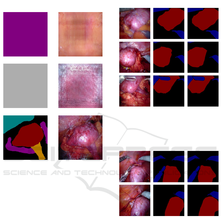
Segmentation Map Synthetic Image
Figure 5: Synthetic images (right) generated from uniform
semantic maps (first two rows) or more complex semantic
maps (last row).
on synthetic images the network generalizes over real
ones, as can be seen from qualitative results shown in
Figures 6 and 7 where there are the input image (left)
the predicted semantic map (centre) and the ground
truth map (right). In red is shown the kidney while
the surgical tools are in blue. Moreover, the inference
time is about 9ms, therefore the segmentation network
can be used for real-time applications. For example,
to mask an instrument out so that the augmented real-
ity overlay does not occlude the surgeon’s view (Allan
et al., 2019).
4.3 Discussion and Limitations
Providing the SIS network with a rich semantic map
leads to a generation of realistic laparoscopic surgery
images. Once the network is trained, it can be used
to generate synthetic data that could be useful for
the training of other networks, as shown by our ex-
periment. Some networks rely on supervision sig-
nals that are difficult to obtain for minimally inva-
sive surgeries, like the relative position of the organ
GT MapReal Image Predicted Map
Figure 6: Qualitative results for the U-Net semantic seg-
mentation architecture trained on synthetic data to predict
the kidney (red) and surgical tools (blue) semantic map for
the real test set (RTS). On the left is there is the real image,
on the centre there is the predicted semantic map and on the
right there is the ground truth (GT) semantic map.
GT MapReal Image Predicted Map
Figure 7: Qualitative results for the U-Net semantic seg-
mentation architecture trained on synthetic data to predict
the kidney (red) and surgical tools (blue) semantic map for
the real additional test set (RATS). On the left is there is
the real image, on the centre there is the predicted semantic
map and on the right there is the ground truth (GT) semantic
map.
with respect to the endoscopic camera. By leverag-
ing on the semantic image translation, segmentation
maps can be generated artificially from the patient-
specific 3D anatomical mesh in order to virtually gen-
erate a semantic mask in a given position. Moreover,
the surgical tool segmentation maps can be added
later together with other semantic information, such
as the presence of fat or abdominal tissue, in order
to generate a realistic image where the 6-DoF pose
VISAPP 2024 - 19th International Conference on Computer Vision Theory and Applications
650
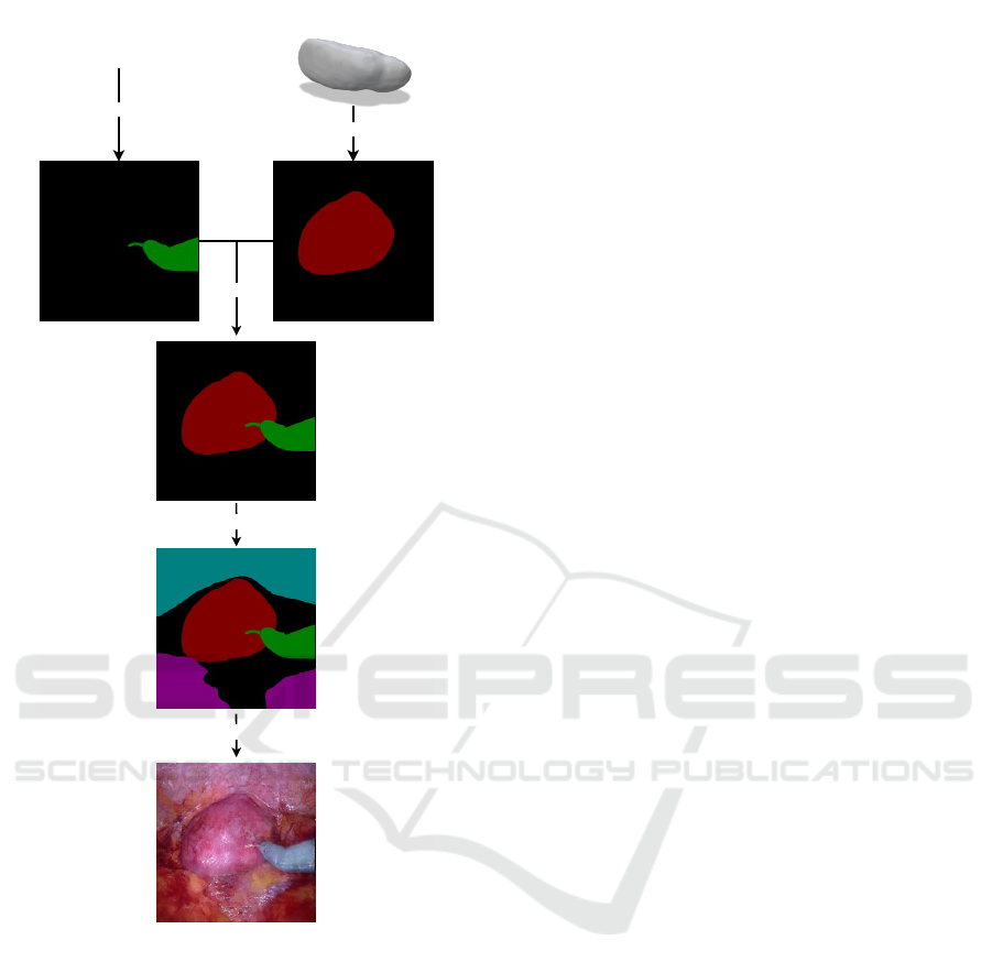
Kidney Mesh
2017 Robotic Intrument
Segmentation
Challange Dataset
Render
Overlay
Add semantic info
Generate
Surgical Tool Mask
Figure 8: Synthetic data generation pipeline.
of the organ is known. An example can be seen
in Figure 8, where the segmentation of the kidney
has been generated from a patient-specific 3D kid-
ney model. Then, the segmentation mask associ-
ated with the Monopolar Curved Scissors extracted
from the 2017 robotic instrument segmentation chal-
lenge dataset (Allan et al., 2019) has been attached
together with a rough semantic map of the fat and ab-
dominal tissue. The generated data can be used to
train networks that can retrieve the position of the
organ and be used to improve Augmented Reality
(AR) guided interventions in robotic-assisted urologic
surgery (Bianchi et al., 2021; Schiavina et al., 2021b;
Schiavina et al., 2021a; Tartarini et al., 2023).
The generative approach proposed for Minimally
Invasive Surgery (MIS) images confronts several lim-
itations. First of all, usually the images captured dur-
ing robotic surgeries have higher resolution. More-
over, the dynamic nature of MIS environments intro-
duces complexities such as smoke and motion blur,
further complicating the generation process. To ad-
dress these issues comprehensively, the dataset used
to train the generative network should encompass di-
verse and challenging scenarios, including instances
of smoke, varying degrees of motion blur, and other
intricacies characteristic of MIS procedures.
5 CONCLUSIONS
A semantic image synthesis (SIS) method has been
exploited to generate realistic Robotic Assisted Par-
tial Nephrectomy (RAPN) surgical images. Starting
from rich semantic maps we achieved highly realis-
tic synthetic images that can be used to train neural
networks. As future works, we aim to include the
powerful multi-modal generation ability of Denoising
Diffusion Probabilistic Models (DDPMs) (Ho et al.,
2020) for the conditional image generation from se-
mantic maps (Wang et al., 2022) in robotic surgery.
Moreover, we intend to generate a synthetic dataset
including 6-DoF pose estimation (Xiang et al., 2017)
starting from patient-specific organ meshes and the
presented SIS approach.
REFERENCES
Allan, M., Shvets, A., Kurmann, T., Zhang, Z., Duggal,
R., Su, Y.-H., Rieke, N., Laina, I., Kalavakonda,
N., Bodenstedt, S., et al. (2019). 2017 robotic
instrument segmentation challenge. arXiv preprint
arXiv:1902.06426.
Bianchi, L., Barbaresi, U., Cercenelli, L., Bortolani, B.,
Gaudiano, C., Chessa, F., Angiolini, A., Lodi, S., Por-
reca, A., Bianchi, F. M., et al. (2020). The impact of
3d digital reconstruction on the surgical planning of
partial nephrectomy: a case-control study. still time
for a novel surgical trend? Clinical Genitourinary
Cancer, 18(6):e669–e678.
Bianchi, L., Chessa, F., Angiolini, A., Cercenelli, L., Lodi,
S., Bortolani, B., Molinaroli, E., Casablanca, C.,
Droghetti, M., Gaudiano, C., et al. (2021). The use of
augmented reality to guide the intraoperative frozen
section during robot-assisted radical prostatectomy.
European Urology, 80(4):480–488.
Deng, J., Dong, W., Socher, R., Li, L.-J., Li, K., and Fei-
Fei, L. (2009). Imagenet: A large-scale hierarchical
image database. In 2009 IEEE conference on com-
puter vision and pattern recognition, pages 248–255.
Ieee.
Semantic Image Synthesis for Realistic Image Generation in Robotic Assisted Partial Nephrectomy
651

Goodfellow, I., Pouget-Abadie, J., Mirza, M., Xu, B.,
Warde-Farley, D., Ozair, S., Courville, A., and Ben-
gio, Y. (2020). Generative adversarial networks. Com-
munications of the ACM, 63(11):139–144.
He, K., Zhang, X., Ren, S., and Sun, J. (2016). Deep resid-
ual learning for image recognition. In Proceedings of
the IEEE conference on computer vision and pattern
recognition, pages 770–778.
Ho, J., Jain, A., and Abbeel, P. (2020). Denoising diffusion
probabilistic models. Advances in neural information
processing systems, 33:6840–6851.
Huang, X., Liu, M.-Y., Belongie, S., and Kautz, J. (2018).
Multimodal unsupervised image-to-image translation.
In Proceedings of the European conference on com-
puter vision (ECCV), pages 172–189.
Isola, P., Zhu, J.-Y., Zhou, T., and Efros, A. A. (2017).
Image-to-image translation with conditional adversar-
ial networks. In Proceedings of the IEEE conference
on computer vision and pattern recognition, pages
1125–1134.
Kingma, D. P., Welling, M., et al. (2019). An introduction
to variational autoencoders. Foundations and Trends®
in Machine Learning, 12(4):307–392.
Long, J., Shelhamer, E., and Darrell, T. (2015). Fully con-
volutional networks for semantic segmentation. In
Proceedings of the IEEE conference on computer vi-
sion and pattern recognition, pages 3431–3440.
Marzullo, A., Moccia, S., Catellani, M., Calimeri, F., and
De Momi, E. (2021). Towards realistic laparoscopic
image generation using image-domain translation.
Computer Methods and Programs in Biomedicine,
200:105834.
Ozawa, T., Hayashi, Y., Oda, H., Oda, M., Kitasaka, T.,
Takeshita, N., Ito, M., and Mori, K. (2021). Synthetic
laparoscopic video generation for machine learning-
based surgical instrument segmentation from real la-
paroscopic video and virtual surgical instruments.
Computer Methods in Biomechanics and Biomedical
Engineering: Imaging & Visualization, 9(3):225–232.
Park, T., Liu, M.-Y., Wang, T.-C., and Zhu, J.-Y. (2019).
Semantic image synthesis with spatially-adaptive nor-
malization. In Proceedings of the IEEE/CVF con-
ference on computer vision and pattern recognition,
pages 2337–2346.
Pfeiffer, M., Funke, I., Robu, M. R., Bodenstedt, S.,
Strenger, L., Engelhardt, S., Roß, T., Clarkson, M. J.,
Gurusamy, K., Davidson, B. R., et al. (2019). Gen-
erating large labeled data sets for laparoscopic im-
age processing tasks using unpaired image-to-image
translation. In Medical Image Computing and Com-
puter Assisted Intervention–MICCAI 2019: 22nd In-
ternational Conference, Shenzhen, China, October
13–17, 2019, Proceedings, Part V 22, pages 119–127.
Springer.
Rau, A., Edwards, P. E., Ahmad, O. F., Riordan, P., Janatka,
M., Lovat, L. B., and Stoyanov, D. (2019). Implicit
domain adaptation with conditional generative adver-
sarial networks for depth prediction in endoscopy.
International journal of computer assisted radiology
and surgery, 14:1167–1176.
Rivoir, D., Pfeiffer, M., Docea, R., Kolbinger, F., Riedi-
ger, C., Weitz, J., and Speidel, S. (2021). Long-term
temporally consistent unpaired video translation from
simulated surgical 3d data. In Proceedings of the
IEEE/CVF international conference on computer vi-
sion, pages 3343–3353.
Ronneberger, O., Fischer, P., and Brox, T. (2015). U-
net: Convolutional networks for biomedical im-
age segmentation. arxiv 2015. arXiv preprint
arXiv:1505.04597.
Schiavina, R., Bianchi, L., Chessa, F., Barbaresi, U.,
Cercenelli, L., Lodi, S., Gaudiano, C., Bortolani, B.,
Angiolini, A., Bianchi, F. M., et al. (2021a). Aug-
mented reality to guide selective clamping and tumor
dissection during robot-assisted partial nephrectomy:
a preliminary experience. Clinical genitourinary can-
cer, 19(3):e149–e155.
Schiavina, R., Bianchi, L., Lodi, S., Cercenelli, L., Chessa,
F., Bortolani, B., Gaudiano, C., Casablanca, C.,
Droghetti, M., Porreca, A., et al. (2021b). Real-time
augmented reality three-dimensional guided robotic
radical prostatectomy: preliminary experience and
evaluation of the impact on surgical planning. Euro-
pean Urology Focus, 7(6):1260–1267.
Tartarini, L., Riccardo, S., Bianchi, L., Lodi, S., Gaudiano,
C., Bortolani, B., Cercenelli, L., Brunocilla, E., and
Marcelli, E. (2023). Stereoscopic augmented reality
for intraoperative guidance in robotic surgery. Journal
of Mechanics in Medicine and Biology, page 2340040.
Vercauteren, T., Unberath, M., Padoy, N., and Navab, N.
(2019). Cai4cai: the rise of contextual artificial intel-
ligence in computer-assisted interventions. Proceed-
ings of the IEEE, 108(1):198–214.
Wada, K. Labelme: Image Polygonal Annotation with
Python.
Wang, T.-C., Liu, M.-Y., Zhu, J.-Y., Tao, A., Kautz, J., and
Catanzaro, B. (2018). High-resolution image synthe-
sis and semantic manipulation with conditional gans.
In Proceedings of the IEEE conference on computer
vision and pattern recognition, pages 8798–8807.
Wang, W., Bao, J., Zhou, W., Chen, D., Chen, D., Yuan,
L., and Li, H. (2022). Semantic image synthesis via
diffusion models. arXiv preprint arXiv:2207.00050.
Xiang, Y., Schmidt, T., Narayanan, V., and Fox, D. (2017).
Posecnn: A convolutional neural network for 6d ob-
ject pose estimation in cluttered scenes. arXiv preprint
arXiv:1711.00199.
Yoon, J., Hong, S., Hong, S., Lee, J., Shin, S., Park, B.,
Sung, N., Yu, H., Kim, S., Park, S., et al. (2022). Sur-
gical scene segmentation using semantic image syn-
thesis with a virtual surgery environment. In Inter-
national Conference on Medical Image Computing
and Computer-Assisted Intervention, pages 551–561.
Springer.
Zhu, J.-Y., Park, T., Isola, P., and Efros, A. A. (2017).
Unpaired image-to-image translation using cycle-
consistent adversarial networks. In Proceedings of
the IEEE international conference on computer vi-
sion, pages 2223–2232.
VISAPP 2024 - 19th International Conference on Computer Vision Theory and Applications
652
