
Decoding Visual Stimuli and Visual Imagery Information from EEG
Signals Utilizing Multi-Perspective 3D-CNN Based Hierarchical
Deep-Fusion Learning Network
Fatma Yusranur Emanet and Kazim Sekeroglu
Department of Computer Science, Southeastern Louisiana University, Hammond, LA 70402, U.S.A.
Keywords: Hierarchical Deep Learning, Brain Computer Interface, Fusion Learning, Spatiotemporal Pattern Recognition,
Multi-Perspective Learning.
Abstract: Brain-Computer Interface Systems (BCIs) facilitate communication between the brain and machines, enabling
applications such as diagnosis, understanding brain function, and cognitive augmentation. This study explores
the classification of visual stimuli and visual imagery using electroencephalographic (EEG) data. The
proposed method utilizes 3D EEG data generated by transforming 1D EEG data into 2D Spatiotemporal EEG
image mappings for feature extraction and classification. Additionally, a multi-perspective 3D CNN-based
hierarchical deep fusion learning network is employed to classify multi-dimensional spatiotemporal EEG data,
decoding brain activity for visual and visual imagery stimulation. The findings show that the suggested multi-
perspective fusion method performs better than a standalone model, indicating promising progress in using
BCIs to understand and utilize brain signals for visual and imagined stimulation.
1 INTRODUCTION
The brain-computer interface systems (BCIs) are one
of the crucial technologies in recent years that aim to
establish communication between the brain and
machines. Besides the use of BCI systems in many
scientific research areas, the main purpose of BCI
systems is to enable people to develop applications
where they can control various devices including
computers, prosthetic limbs, robots, and even video
games by using only power of human thought
(Lebedev, 2017).
BCI is a system that deals with the brain activities
of a living thing (human or animal) and turns these
activities into meaningful information about the
cognitive, perceptual, or motor processes associated
with neural activity patterns. This process is also
known as brain decoding. Meaningful knowledge
obtained thanks to brain decoding can be used for
studies such as developing brain-computer interfaces,
diagnosing disorders, understanding human brain
function, and even augmenting cognition (Tan, 2010).
In recent years, BCI technologies have started to
show their presence in fields such as medicine,
neuroscience, and gaming and are used for
revolutionary innovations in these fields. Especially
thanks to the BCI innovations made in the medical
world, many people with disabilities and limited
mobility have started to meet their needs without the
need for any physical activity (Miralles, 2015).
With many applications developed so far, this
field frequently updates itself and is very open to
developments. Therefore, it has the potential to
understand the brain and its working principles,
which is increasing day by day. This potential has
attracted the attention of scientists and it has recently
become a hot topic in the world of science and
technology.
We can give examples of these applications;
communication devices for people with disabilities
(Millán, 2010), controlling devices in hazardous
environments (Douibi, 2021), enhancing cognitive
performance (Papanastasiou, 2020), prosthetic limbs
that can be controlled by the user's thoughts (Vilela,
2020), and even brain-controlled video games
(Nijholt, 2009). In addition to many detailed and
successful studies conducted in this area, developing
reliable and robust decoding algorithms, and
obtaining consistent neural activity patterns by brain
decoding is still a challenge today due to some
reasons related to the brain such as the complexity of
the brain signals due to its nature, its dynamic
Emanet, F. and Sekeroglu, K.
Decoding Visual Stimuli and Visual Imager y Information from EEG Signals Utilizing Multi-Perspective 3D-CNN Based Hierarchical Deep-Fusion Learning Network.
DOI: 10.5220/0012568500003660
Paper published under CC license (CC BY-NC-ND 4.0)
In Proceedings of the 19th International Joint Conference on Computer Vision, Imaging and Computer Graphics Theory and Applications (VISIGRAPP 2024) - Volume 4: VISAPP, pages
381-388
ISBN: 978-989-758-679-8; ISSN: 2184-4321
Proceedings Copyright © 2024 by SCITEPRESS – Science and Technology Publications, Lda.
381

structure, and being affected by environmental
factors.
Brain decoding can be used for visual stimuli
classification. Visual stimuli classification refers to
the process of identifying the category or features of
a visual stimulus such as an image or video clip. It
uses the response of the brain which is the patterns of
neural activity that stimuli evoke in the brain for
identifying the category of a visual stimulus (Bigdely-
Shamlo, 2008).
Using various machine learning techniques,
models with high performance can be created and
successful results can be obtained to classify visual
stimuli. These models can predict the category of a
new, unseen visual stimulus based on the feature map
which is most relevant for the cognitive task at hand
by training the algorithm with known categories of
visual stimuli, such as images of faces, letters, or
simple shapes (Aggarwal, 2022).
This study aims to develop a model that utilizes
brain decoding for the purpose of classifying not only
visual stimuli but also visual imagery. Specifically,
EEG data collected from participants in an
experimental setup designed for this study will be
utilized for classification purposes.
Brain-computer interfaces are known for their
potential to provide solutions to a wide range of issues
in both scientific and everyday contexts. The primary
objective of this project was to address a research-
based problem related to the classification of EEG
signals within the context of brain-computer
interfaces. While this project was focused on
addressing this specific issue, the insights and
findings obtained through this work could be applied
to a broader range of problems in various domains.
This study contributes by analyzing EEG signals
generated by visual stimuli and visual imagery. This
involves using a 2D spatiotemporal EEG image
representation, investigating 3D EEG data for feature
extraction, and classifying based on 2D
spatiotemporal EEG (ST-EEG) maps. The approach
also incorporates a 3D convolutional neural network
(CNN) based multi-perspective hierarchical deep
fusion model for the classification of 3D EEG
representations of visually evoked and visual imagery
signals.
1.1 Literature Review
Brain-computer interface has been a hot topic in the
world of science and technology in recent years.
Although it is a topic that is widely talked about
today, it is known that studies on BCI were first
studied with animals in the 1970s (Kawala-Sterniuk,
2021). The latest studies in BCI have concentrated on
how to improve the accuracy and speed of brain
signal decoding. With many machine learning
methods, particularly deep learning, brain signals are
analyzed and examined. As seen in a study by Zhang
et al., the use of deep learning has noticeably
increased the accuracy in classifying different types
of brain signals (Sun, 2020).
Another option provided by BCI systems is the
classification of brain signals obtained using visual
stimuli. Allison, B. Z., et al. (Jin, 2012) focused on
the use of a BCI system to change frequency bands in
EEG signals via visual stimuli. The scientists
discovered that, with their claimed approach,
participants were able to effectively change their
brain signals. In another study that used visual
stimuli, Kavasidis et al. (Kavasidis, 2017) studied the
translation of visually evoked EEG signals into
meaningful images. The approach they proposed is
based on generating images using visually evoked
brain signals recorded through an
electroencephalograph (EEG). They implemented a
deep learning framework consisting of an LSTM
stacked with a generative method. They pointed out
that GAN, in general, outperforms VAE and
recommended that the study should continue by
combining these two. They also recommended
acquiring fMRI data to complement EEG data.
Similarly, Hayashi and Kawata (Hayashi, 2018)
proposed a methodology to reconstruct the images
which had been recorded from the monkey brain.
They implemented a linear decoder that predicts
visual features of viewed images at a higher-order
layer of a deep convolutional neural network, so
called CaffeNet (Jia, 2014). They refined the images
to photorealistic images through a deep generator
network (Dosovitskiy, 2016). Their approach lacks
efficient choosing critical visual features for the
subject for image reconstruction within a reasonable
time frame.
Liu, Shuang, et al. (Liu, 2014) explored the
individual identification through the extraction of
features from both resting EEG and visual evoked
potential signals. The features in this identification
consisted of fourth-order AR parameters, power
spectrum in the time and frequency domain, and
phase locking value. For the classification, the
extracted features were fed into an SVM. Thanks to
this study, as a result of the identifications made with
features, they come with the result that visually
evoked tasks show better results in identifying
individuals compared to relax tasks. Tirupattur and
Rawat (Tirupattur, 2018) introduced a GAN
architecture to generate class-specific images from
VISAPP 2024 - 19th International Conference on Computer Vision Theory and Applications
382

brain activities, achieving good results with small
datasets and they emphasize the potential for
visualizing brain signals as a video stream.
Additionally, Zhang et al. (Zhang, 2019) also used
GANs to reconstruct shapes evoked by EEG signals,
focusing on simple geometrical shapes and
suggesting future exploration of more complex
shapes. They also performed a feature extraction from
EEG data using CNN.
In another GAN-based study, Fares and Zhong
(Fares, 2020) proposed a novel DCLS-GAN
framework to integrate brain and visual features,
transforming EEG descriptions into class-relevant
images. Similarly, Wang et al. (Wang, 2021)
introduce the α-GAN approach that combines
standard GAN structure with variational auto-
encoder to reconstruct images from the EEG
framework and the fMRI framework. Rashkov et al.
(Rashkov, 2019) offered closed-loop BCI system that
reconstructs the observed or imagined stimuli images
from the co-occurring brain wave parameters. This
paradigm contains the visual-based cognitive test for
individual stimuli set selection as well as state-of- art
deep learning-based image reconstruction model for
native feedback presentation. Additionally, Qu et al.
(Qu, 2021) suggested an algorithmic idea of
extracting, selecting, and decoding the EEG features
related with the stimuli based on the supervision of
the decoding feature of the original stimulus image.
In this study, they pointed out the lack of clear
evidence to prove that humans are in visual
processing tasks.
Similarly, Palazzo et al. (Palazzo, 2020) proposes
a model, EEG-ChannelNet, to learn a brain manifold
for EEG classification. And that, they introduce a
multimodal approach that uses deep mage and EEG
encoders, trained in a Siamese configuration, to learn
a joint feature space for images and EEG signals
recorded while users look at pictures on a screen.
They trained two encoders in a siamese configuration
and maximize the compatibility score between the
corresponding images and EEGs. They also pointed
out that identifying different responses in brain
activity corresponding to different objects, patterns,
or categories is a field for future study in their study.
For EEG-based brain imaging classification,
Jiang et al. (Jiang, 2019) proposed a novel deep
framework. The proposed framework provides
multimodal brain imaging classification by using not
only the strength of integrated multiple modalities but
also the advantages of the added consistency test.
Additionally, Spampinato et al. (Spampinato, 2017)
introduce a deep learning approach to classify EEG
data as well as propose the first automated
classification approach employing visual descriptors
extracted directly from human neural processes
involved in visual scene analysis. They emphasize
that there will be a greater need for very complex deep
learning networks in the future and that the studies
conducted in this direction will be used to distinguish
brain signals produced from many image classes.
Fares et al. (Fares, 2019) create a novel region-level
stacked bi-directional deep learning framework for
visual object classification. In this framework, there
are 3 stages including the region-level information
extraction stage, the feature encoding stage, and the
classification stage. In their study, they predict that
multimedia content information can be reconstructed
through the proposed EEG representations for future
studies.
2 METHODOLOGY
2.1 Dataset
In this study, EEG was used to capture brain activity
patterns. A new dataset was created by collecting
EEG data from seven volunteer participants using
"Enobio 32" device by "NEUROELECTRICS," a
medical company specializing in non-invasive brain
stimulation ((n.d.), 2023). This device offers 32
electrodes and the option of dry electrodes for quick
setup. The sampling rate is fixed at 500. A 10-10
international electrode placement system is used in
the device (Krol, 2020).
For data collection, we gathered data from 7
participants, following a specific data collection
procedure. Participants were seated in a well-lit, quiet
room, ensuring minimal distractions. Positioned
approximately 50 cm away from an LCD computer
screen, the participants were instructed to remain
seated throughout the duration of the experiment,
with an EEG cap placed on their heads to record brain
waves. The experiment encompassed two sections
conducted on the same day. During each section, the
EEG recorder captured the participants' brain waves
while a slideshow randomly displayed stimulus.
Throughout the experiment, the EEG cap remained
on the participants' heads for consistency between the
two sections. The stimuli presented in both sections
consisted of the letters A, B, and C displayed in
random order. In the first section, participants were
directed to focus on the computer screen with their
eyes open for 30 seconds, followed by 30 seconds of
eyes-closed rest. Subsequently, the letters A, B, and
C appeared on the screen for 10 seconds each,
preceded by a 1.5-second interval of a black screen to
Decoding Visual Stimuli and Visual Imagery Information from EEG Signals Utilizing Multi-Perspective 3D-CNN Based Hierarchical
Deep-Fusion Learning Network
383
minimize the influence of the previous letter. This
sequence was repeated 20 times for each letter in
random order. In the second section, participants were
presented with a blank white screen throughout. An
auditory cue prompted participants to mentally
visualize a specific letter, which they maintained for
10 seconds. They were then instructed to imagine
another randomly selected letter for another 10
seconds. This process was repeated 20 times for each
of the letters, A, B, and C, resulting in multiple
instances of imagined letters for each participant. The
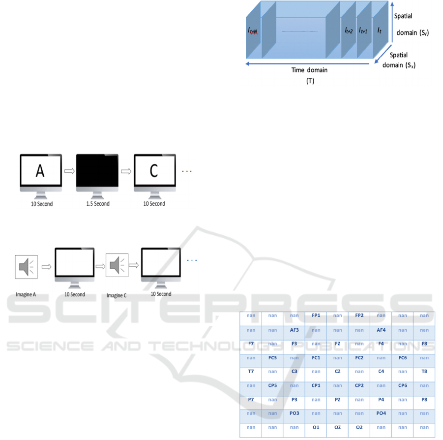
process of each experiment section is depicted in Fig.
1 and Fig. 2.
Figure 1: The First Experiment Section-Visual Stimuli
Phase.
Figure 2: The Second Experiment Section-Visual Imagery
Phase.
2.2 Data Transformation
The raw EEG data comprises one-dimensional time
series data for each channel, which reflects the
electrical activity in specific locations (referred to as
channels) of the brain over time. The recording device
utilized for gathering brain signals features 32 distinct
electrodes, making the complete raw data manifest as
a 2D matrix. This matrix encompasses 1D time series
data encapsulating the electrical activities for 32
different locations. Refer to Fig. 5 for visual
representation. In this context, the matrix 'S'
constitutes a two-dimensional array that encompasses
all the EEG data collected from 'n' channels over a
duration of 't+N' time. Each row within the matrix
corresponds to a specific channel for the entire
duration, while the columns signify the EEG data
recorded from all channels at a specific time, denoted
as 't'.
In this proposed method, we took into
consideration the potential impact of channel
proximity and neighbourhood relations on the
spatiotemporal plane. To achieve this, we
transformed the data shape into 2D spatiotemporal
Figure 3: Creation of 2D Spatiotemporal EEG image
sequence.
EEG mappings. This transformation essentially
depicts each signal as if a top-down image of the brain
was captured. Consequently, 2D spatiotemporal EEG
maps were generated for each signal, effectively
producing a three-dimensional data set. This was
achieved by arranging these generated maps
consecutively along the temporal plane. In essence,
the altered EEG data is now represented as 2D
spatiotemporal EEG maps, culminating in a 3D
dataset with two dimensions in the spatial domain and
one dimension in the time domain. Refer to Fig. 3 for
a visual depiction of this representation. EEG data
from all channels was mapped to a 9x9 matrix based
on the precise locations of the electrodes on the scalp
where the data were recorded. Channel locations in
the 9X9 matrix are illustrated in Fig. 4 below.
Figure 4: Channel Locations in the 9X9 Matrix.
The parts shown as nan in the 9x9 matrix
represent the places where the electrodes don’t exist.
In order to capture the neighborhood relation, cubic
interpolation was made for the empty ones between
the neighboring electrodes, except for the corners.
This transformation process and the transformed 2D
ST-EEG map I at time stamp t, I
t
, are illustrated in
Fig. 5 below.
2.3 Proposed Model
All 3 different perspectives of 3D EEG data were
taken into consideration and a 3D CNN model was
VISAPP 2024 - 19th International Conference on Computer Vision Theory and Applications
384
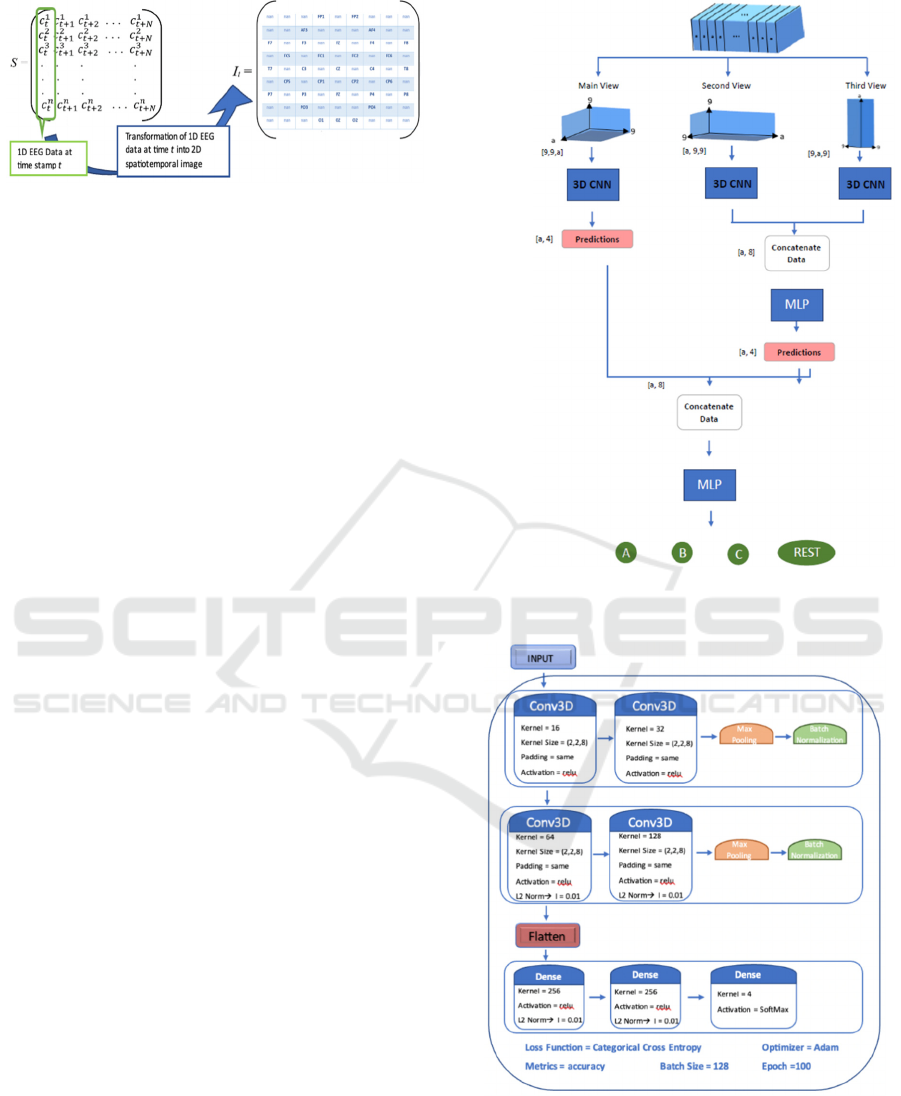
Figure 5: Transformation Process of 1D Temporal EEG
Data into 2D Spatiotemporal Image.
created for each perspective. As given in Fig. 3, two
dimensions of 3D EEG data correspond to the spatial
domain, and the third dimension pertains to the
temporal domain. The SxSy plane view offers
insights into the collected data from all channels at
time t, while the TSx and TSy planes provide
information regarding the collected data from specific
channels over a time period. Hence, this study
explores three distinct views: the main view based on
the SxSy plane, the second view based on the TSx
plane, and the third view based on the TSy plane.
In the initial phase of the proposed fusion
architecture, 3D CNN networks are employed to
identify patterns from the specific viewpoint of the
multidimensional spatiotemporal EEG data.
Subsequently, at the second stage of the architecture,
the identified patterns from the 3D CNN networks for
the second and third viewpoints are merged and
forwarded to the subsequent layer for further fusion.
The central concept is to integrate the patterns of EEG
data that share temporal information derived from the
second and third viewpoints. Finally, at the
concluding layer of the fusion architecture, the
extracted pattern from the primary perspective and
the temporal fusion layer are consolidated and
merged via the output layer to achieve the final
spatiotemporal fusion. Fig. 6 below provides a block
diagram of the proposed multi-view hierarchical deep
learning model.
As seen in Fig. 6 above, there are three 3D CNN
models in the first layer of the proposed hierarchical
model: main view, side view-1 and side view-2.
Detailed architecture of the 3D CNN model used in
main view and side views are shown in Fig. 7 below.
Whereas Fig. 8 shows the architecture of the temporal
fusion model and spatiotemporal fusion model. In this
study, we used multi-layer perceptron for the temporal
and spatiotemporal fusion, However, different
supervised machine learning models such as Support
Vector Machine, Bayesian Network, Decision Tree, or
a multidimensional classification model can be used.
Figure 6: Block diagram of the proposed multi-perspective
hierarchical deep learning model.
Figure 7: Main view and Side view model blog diagram.
Decoding Visual Stimuli and Visual Imagery Information from EEG Signals Utilizing Multi-Perspective 3D-CNN Based Hierarchical
Deep-Fusion Learning Network
385
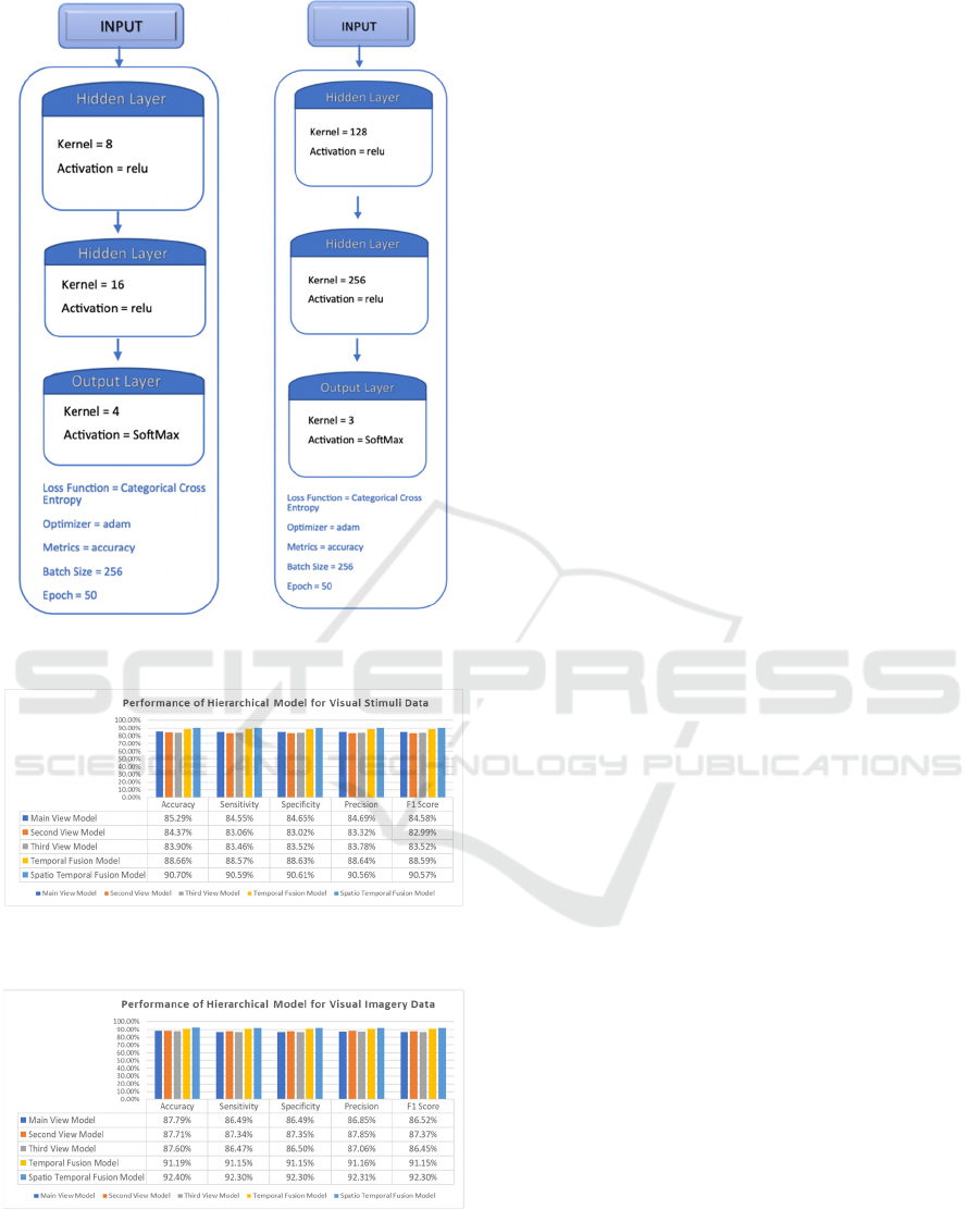
Figure 8: Temporal Fusion Model (left), Spatiotemporal
Fusion Model (right).
Figure 9: Performance measures of Hierarchical Model for
Visual Stimuli Data.
Figure 10: Performance measures of Hierarchical Model for
Visual Imagery Data.
3 RESULTS AND DISCUSSION
In our initial experimentation, we evaluated the
proposed modular fusion learning architecture,
employing a 3D CNN-based network to extract
patterns from 3D spatiotemporal EEG data. The
outcomes of our assessment are depicted in Figure 9
which presents the classification results for visual
stimuli, and Figure 10, illustrating the classification
outcomes for the imagined visual stimuli. For the
visual stimuli classification, we utilized simple
geometric representations of the letters A, B, and C,
while for the imagined visual stimuli, we instructed
the subjects to mentally visualize the corresponding
letters. As evident from the results, the Multi-
perspective and hierarchical fusion learning approach
substantially enhanced the classification accuracy.
In our preliminary findings, the utilization of the
main view alone yielded an accuracy of 85.29% for
visual stimuli and 87.79% for the imagined visual
stimuli. However, the integration of the multi-
perspective and hierarchical fusion model led to a
notable increase in accuracy, reaching 90.7% for
visual stimuli and 92.4% for the imagined visual
stimuli. These results suggest that the proposed model
is proficient in extracting patterns from
multidimensional spatiotemporal EEG data, making
it suitable for classification purposes and the
generation of both provided and imagined visual
information from EEG data.
4 CONCLUSION AND FUTURE
WORK
The current research introduces hierarchical deep
learning models that were trained to recognize
patterns in 2D spatiotemporal EEG images, for the
purpose of classifying visual stimuli and visual
imagery data. The results of the study show that the
proposed model achieved strong performance in
multi-class classification of 3D EEG data.
The investigation placed a significant emphasis on
analyzing visually evoked EEG signals and visual
imagery EEG signals, utilizing a 2D spatiotemporal
EEG image representation, and aimed to extract
features and perform classification based on these
representations.
The study also aimed to explore the benefits of a
fusion architecture and a multi-view approach in
learning the 2D ST-EEG maps. The experimental
results indicate that, in general, a fusion architecture
outperforms a stand-alone model.
VISAPP 2024 - 19th International Conference on Computer Vision Theory and Applications
386

While BCI technology has gained a lot of attention
in recent years, there are still many unanswered
questions and areas that require further research. This
study can be seen as a preliminary study for many
potential studies to be conducted in the future. By
addressing the gaps in the current literature,
researchers can build upon the findings of this study
and expand our knowledge of BCI technology,
potentially leading to new and innovative
applications in the future.
Initially, the focus of this study was on identifying
four specific classes, namely A, B, C, and Rest.
However, in future studies, the potential exists to
expand the number of classes to encompass the
entirety of the alphabet.
Moreover, we employed a common dataset that
was divided into training and testing sets in our study.
However, there is potential for further exploration on
how to improve the detection performance of models
trained on data collected at different times when
tested on data gathered at a later point. By doing so,
real-time applications can be developed, particularly
for individuals with communication difficulties who
may lack the ability to speak and could benefit from
a system that allows them to communicate their
words through BCI technology.
ACKNOWLEDGEMENTS
Institutional Development Award (IDeA) from the
National Institute of General Medical Sciences of the
National Institutes of Health under grant number
P20GM103424-21.
REFERENCES
Lebedev, M. A., & Nicolelis, M. A. (2017). Brain-machine
interfaces: From basic science to neuroprostheses and
neurorehabilitation. Physiological reviews, 97(2), 767-
837.
Tan, D., & Nijholt, A. (2010). Brain-computer interfaces
and human-computer interaction (pp. 3-19). Springer
London.
Miralles, F., Vargiu, E., Dauwalder, S., Solà, M., Müller-
Putz, G., Wriessnegger, S. C., ... & Lowish, H. (2015).
Brain computer interface on track to home. The
Scientific World Journal, 2015.
Millán, J. D. R., Rupp, R., Mueller-Putz, G., Murray-Smith,
R., Giugliemma, C., Tangermann, M., ... & Mattia, D.
(2010). Combining brain–computer interfaces and
assistive technologies: state-of-the-art and challenges.
Frontiers in neuroscience, 161.
Douibi, K., Le Bars, S., Lemontey, A., Nag, L., Balp, R., &
Breda, G. (2021). Toward EEG-based BCI applications
for industry 4.0: challenges and possible applications.
Frontiers in Human Neuroscience, 15, 705064.
Papanastasiou, G., Drigas, A., Skianis, C., & Lytras, M.
(2020). Brain computer interface based applications for
training and rehabilitation of students with
neurodevelopmental disorders. A literature review.
Heliyon, 6(9), e04250.
Vilela, M., & Hochberg, L. R. (2020). Applications of
brain-computer interfaces to the control of robotic and
prosthetic arms. Handbook of clinical neurology, 168,
87-99
Nijholt, A. (2009). BCI for games: A ‘state of the
art’survey. In Entertainment Computing-ICEC 2008:
7th International Conference, Pittsburgh, PA, USA,
September 25-27, 2008. Proceedings 7 (pp. 225-228).
Springer Berlin Heidelberg.
Bigdely-Shamlo, N., Vankov, A., Ramirez, R. R., &
Makeig, S. (2008). Brain activity- based image
classification from rapid serial visual presentation.
IEEE Transactions on Neural Systems and
Rehabilitation Engineering, 16(5), 432-441.
Aggarwal, S., & Chugh, N. (2022). Review of machine
learning techniques for EEG based brain computer
interface. Archives of Computational Methods in
Engineering, 1-20.
(n.d.). Enobio 32. Neuroelectrics. Retrieved April 16, 2023,
from https://www.neuroelectrics.com/solutions/
enobio/32.
Krol, L. R. (2020, November 25). EEG 10-10 system with
additional information. Wikimedia. https://commons.
wikimedia.org/wiki/File:EEG_10- 10_system_with_
additional_information.svg
Kawala-Sterniuk, A., Browarska, N., Al-Bakri, A., Pelc,
M., Zygarlicki, J., Sidikova, M., ... & Gorzelanczyk, E.
J. (2021). Summary of over fifty years with brain-
computer interfaces—a review. Brain Sciences, 11(1),
43.
Sun, B., Zhao, X., Zhang, H., Bai, R., & Li, T. (2020). EEG
motor imagery classification with sparse
spectrotemporal decomposition and deep learning.
IEEE Transactions on Automation Science and
Engineering, 18(2), 541-551.
Jin, J., Allison, B. Z., Wang, X., & Neuper, C. (2012). A
combined brain–computer interface based on P300
potentials and motion-onset visual evoked
potentials. Journal of neuroscience methods, 205(2),
265-276.
Kavasidis, I., Palazzo, S., Spampinato, C., Giordano, D., &
Shah, M. (2017, October). Brain2image: Converting
brain signals into images. In Proceedings of the 25th
ACM international conference on Multimedia (pp.
1809-1817).
Jia, Y., Shelhamer, E., Donahue, J., Karayev, S., Long, J.,
Girshick, R., ... & Darrell, T. (2014, November). Caffe:
Convolutional architecture for fast feature embedding.
In Proceedings of the 22nd ACM international
conference on Multimedia (pp. 675- 678).
Decoding Visual Stimuli and Visual Imagery Information from EEG Signals Utilizing Multi-Perspective 3D-CNN Based Hierarchical
Deep-Fusion Learning Network
387

Dosovitskiy, A., & Brox, T. (2016). Generating images
with perceptual similarity metrics based on deep
networks. Advances in neural information processing
systems, 29, 658-666.
Hayashi, R., & Kawata, H. (2018, October). Image
reconstruction from neural activity recorded from
monkey inferior temporal cortex using generative
adversarial networks. In 2018 IEEE International
Conference on Systems, Man, and Cybernetics (SMC)
(pp. 105-109). IEEE
Liu, S., Bai, Y., Liu, J., Qi, H., Li, P., Zhao, X., ... & Ming,
D. (2014). Individual feature extraction and
identification on EEG signals in relax and visual evoked
tasks. In Biomedical Informatics and Technology: First
International Conference, ACBIT 2013, Aizu-
Wakamatsu, Japan, September 16-17, 2013. Revised
Selected Papers (pp. 305-318). Springer Berlin
Heidelberg.
Tirupattur, P., Rawat, Y. S., Spampinato, C., & Shah, M.
(2018, October). Thoughtviz: Visualizing human
thoughts using generative adversarial network. In
Proceedings of the 26th ACM international conference
on Multimedia (pp. 950-958).
Zhang, X., Chen, X., Dong, M., Liu, H., Ge, C., & Yao, L.
(2019). Multi-task Generative Adversarial Learning on
Geometrical Shape Reconstruction from EEG Brain
Signals. arXiv preprint arXiv:1907.13351.
Fares, A., Zhong, S. H., & Jiang, J. (2020, October). Brain-
media: A Dual Conditioned and Lateralization
Supported GAN (DCLS-GAN) towards Visualization
of Image- evoked Brain Activities. In Proceedings of
the 28th ACM International Conference on Multimedia
(pp. 1764-1772).
Wang, P., Zhou, R., Wang, S., Li, L., Bai, W., Fan, J., ... &
Guo, Y. (2021). A General Framework for Revealing
Human Mind with auto-encoding GANs. arXiv preprint
arXiv:2102.05236.
Rashkov, G., Bobe, A., Fastovets, D., & Komarova, M.
(2019). Natural image reconstruction from brain waves:
a novel visual BCI system with native feedback.
bioRxiv, 787101.
Qu, L., Chen, D., & Yin, K. (2021, June). Research on EEG
Feature Decoding Based on Stimulus Image. In 2021
IEEE 4th Advanced Information Management,
Communicates, Electronic and Automation Control
Conference (IMCEC) (Vol. 4, pp. 467-470). IEEE.
Palazzo, S., Spampinato, C., Kavasidis, I., Giordano, D.,
Schmidt, J., & Shah, M. (2020). Decoding brain
representations by multimodal learning of neural
activity and visual features. IEEE Transactions on
Pattern Analysis and Machine Intelligence.
Jiang, J., Fares, A., & Zhong, S. H. (2019). A context-
supported deep learning framework for multimodal
brain imaging classification. IEEE Transactions on
Human- Machine Systems, 49(6), 611-622.
Spampinato, C., Palazzo, S., Kavasidis, I., Giordano, D.,
Souly, N., & Shah, M. (2017). Deep learning human
mind for automated visual classification. In
Proceedings of the IEEE conference on computer vision
and pattern recognition (pp. 6809-6817).
Fares, A., Zhong, S. H., & Jiang, J. (2019). EEG-based
image classification via a region- level stacked bi-
directional deep learning framework. BMC medical
informatics and decision making, 19(6), 1-11.
VISAPP 2024 - 19th International Conference on Computer Vision Theory and Applications
388
