
Instance Selection Framework for Alzheimer’s Disease Classification
Using Multiple Regions of Interest and Atlas Integration
Juan A. Castro-Silva
1,2
, Maria N. Moreno-Garcia
1
, Lorena Guachi-Guachi
3
and
Diego H. Peluffo-Ordo
˜
nez
4,5
1
Universidad de Salamanca, Salamanca, Spain
2
Universidad Surcolombiana, Neiva, Colombia
3
Department of Mechatronics, International University of Ecuador, Simon Bolivar Avenue 170411, Quito, Ecuador
4
College of Computing, Mohammed VI Polytechnic University, Lot 660, Hay Moulay Rachid Ben Guerir, 43150, Morocco
5
SDAS Research Group
Keywords:
Alzheimer’s Disease, Swin Transformer, Weighted Ensemble, Instance Selection, Multiple Region of Interest.
Abstract:
Optimal selection of informative instances from a dataset is critical for constructing accurate predictive mod-
els. As databases expand, leveraging instance selection techniques becomes imperative to condense data into a
more manageable size. This research unveils a novel framework designed to strategically identify and choose
the most informative 2D brain image slices for Alzheimer’s disease classification. Such a framework integrates
annotations from multiple regions of interest across multiple atlases. The proposed framework consists of six
core components: 1) Atlas merging for ROI annotation and hemisphere separation. 2) Image preprocessing
to extract informative slices. 3) Dataset construction to prevent data leakage, select subjects, and split data.
4) Data generation for memory-efficient batches. 5) Model construction for diverse classification training and
testing. 6) Weighted ensemble for combining predictions from multiple models with a single learning algo-
rithm. Our instance selection framework was applied to construct Transformer-based classification models,
demonstrating an overall accuracy of approximately 98.33% in distinguishing between Cognitively Normal
and Alzheimer’s cases at the subject level. It exhibited enhancements of 3.68%, 3.01%, 3.62% for sagittal,
coronal, and axial planes respectively in comparison with the percentile technique.
1 INTRODUCTION
Alzheimer’s disease (AD) is the leading cause of de-
mentia in older adults. It is a progressive brain dis-
order that causes nerve cells to die, leading to sig-
nificant brain volume reduction and affecting almost
all brain functions (WHO, 2023). AD affects crucial
brain regions, such as the Entorhinal Cortex, Fornix,
Hippocampus, Frontal lobe, Temporal lobe, and Pari-
etal lobe, impacting spatiotemporal orientation, cog-
nition, memory, intelligence, judgment, behavior, and
language (Yu and Lee, 2020).
Medical imaging, including modalities like MRI,
PET, and DTI, aids in diagnosis and treatment by vi-
sualizing brain structures. The performance of AD
classification models is influenced by data quality and
quantity. Instance selection methods, varying in strat-
egy and selected slice numbers, may involve remov-
ing slices based on position or informative content
(Castro-Silva. et al., 2022).
The mentioned instance selection methods have
limitations. Including less informative content can in-
crease computational time and introduce noise, harm-
ing model performance. Selecting a low or fixed num-
ber of slices per volume may not guarantee MRI rep-
resentativeness and may exclude AD-related or infor-
mative instances.
Data leakage occurs when test data is used in the
training process, leading to bias. An incorrect split
dataset, lack of independent testing data, or biased
transfer learning can cause it (Wen et al., 2020).
Deep learning in computer vision excels at detect-
ing brain structural changes via MRI, using 2D or 3D
models for image or ROI analysis. Originally de-
signed for sequence-to-sequence tasks like machine
translation, the widely adopted Transformer (Vaswani
et al., 2017) is now applied in diverse domains, in-
cluding NLP, CV, and speech processing. The Swin
Transformer (Liu et al., 2021), with its novel hierar-
chical architecture and Shifted windows, is known for
Castro-Silva, J., Moreno-Garcia, M., Guachi-Guachi, L. and Peluffo-Ordoñez, D.
Instance Selection Framework for Alzheimer’s Disease Classification Using Multiple Regions of Interest and Atlas Integration.
DOI: 10.5220/0012469600003654
Paper published under CC license (CC BY-NC-ND 4.0)
In Proceedings of the 13th International Conference on Pattern Recognition Applications and Methods (ICPRAM 2024), pages 453-460
ISBN: 978-989-758-684-2; ISSN: 2184-4313
Proceedings Copyright © 2024 by SCITEPRESS – Science and Technology Publications, Lda.
453

efficient computation, making it suitable for tasks like
image classification.
Ensemble methods can outperform single classi-
fiers for AD diagnosis (Young et al., 2018) Homoge-
neous ensembles use one base classifier with diverse
training data, and weighted ensembles assign weights
based on member performance.
To overcome the limitations mentioned above, in
this paper, we propose a novel framework for select-
ing the most informative 2D image slices based on
the annotations fusion of multiple Regions of Inter-
est (ROIs) such as the entorhinal cortex, fornix, hip-
pocampus, frontal, parietal, and temporal lobes. The
ROI analysis involves using multiple atlases or maps
to divide the brain into distinct regions, leading to a
method called Multiple ROI and Multiple Atlas-based
Instance Selection, which classifies Cognitively Nor-
mal (CN) and AD cases.
The proposal consists of several components:
• A volume dataset builder to prevent data leakage,
conducting an early subject dataset split for creat-
ing independent training, validation, and test sets.
• A data generator in charge of producing multiple-
view instances from a volume belonging to a pre-
processed dataset with skull-stripping and regis-
tration.
• A weighted ensemble builder that combines ho-
mogeneous methods. This ensemble employs
a single base classifier 2D Swin Transformer
model, trained on various datasets encompassing
ROIs, planes, and hemispheres.
The major contributions of this novel proposal can
be summarized as follows.
1. To capture the most informative images, we pro-
pose an ROI content extraction method. Using the
mode, it identifies the centroid position (x, y) and
crops the 2D slice image accordingly.
2. To enhance diagnostic accuracy in AD tasks, we
propose Multiple ROI and Multiple Atlas-based
Instance Selection. It combines ROI annotations
from multiple atlases to select informative ROI
slice images and remove useless instances.
3. To tackle insufficient sample utilization and lim-
itations of a single classifier, we propose a ho-
mogeneous weighted ensemble using a 2D Swin
Transformer model. This method uses multiple-
view samples in a single classifier with different
weights according to their accuracy performance.
4. The experimental results demonstrate that the pro-
posed method’s accuracy outperforms the state-
of-the-art related works.
The remainder of this paper is structured as fol-
lows: Section 2 presents some related works. The
materials and methods used for preprocessing and in-
stance selection are included in Section 3. Section
4 provides a detailed description of the experiments
conducted in this work and the parameter settings
used. The results of the experiments are discussed
in Section 5. Finally, Section 6 summarizes the con-
cluding remarks of this work.
2 RELATED WORKS
Most proposed approaches for image instance selec-
tion for AD classification differ in the number of
slices selected, and the technique used to obtain the
most representative or discard the least informative
slices.
In previous studies (Choi and Lee, 2020), a com-
mon method involves computing entropy values for
slices, sorting by descending entropy, and selecting
a fixed number (typically 8 to 32) of top slices. In
(Qin et al., 2022), a pre-trained U-Net is used for skull
stripping, resulting in 3D MRI images (64 × 64 × 64).
In (Hu et al., 2023; Altay et al., 2021), 96 axial slices
are selected after skull stripping and volume registra-
tion. In (Lyu et al., 2022), images are resized and
down-sampled (70×75×50) after preprocessing. Fi-
nally, in (Castro-Silva. et al., 2022), instance selec-
tion uses percentile positions for 32 slices.
ROI extraction, a crucial image processing task,
varies in approaches. In (Zaabi et al., 2020), the MRI
scan is divided into 32*32-pixel blocks, extracting
only those containing the hippocampus as ROIs. In
(Bae et al., 2020), the complete hippocampus, ana-
lyzed with 30 MRI coronal slices. Other studies, such
as (Pan et al., 2022; Li et al., 2022), create an ensem-
ble classifier by extracting ROI-based patches from
different brain regions, including the hippocampus,
amygdala, and insulae.
The previously mentioned instance selection
methods have some limitations. The model’s perfor-
mance greatly depends on the number of slices per
volume (Castro-Silva. et al., 2022). Adding more
image slices with less informative content can result
in redundant or less representative information, in-
creases the computational cost (time) of training, in-
troduce noise, and deteriorates model performance.
On the other hand, selecting a fixed number of slice
images could exclude AD-related or more informative
instances. A low number of slices per volume, for ex-
ample (1, 8), does not ensure the representativeness
of the 170-256 slice instances that comprise an MRI
volume.
ICPRAM 2024 - 13th International Conference on Pattern Recognition Applications and Methods
454

Classification algorithms must be evaluated with-
out bias to ensure clinical relevance. Biased evalua-
tions caused by data leakage issues, such as incorrect
dataset splitting, absence of an independent test set,
delayed splitting, and biased transfer can lead to mis-
leading results by inflating model performance (Wen
et al., 2020).
Vision and Swin Transformers are applied to AD
classification using MRI data. Examples include the
hybrid 2D model Conv-Swinformer in (Hu et al.,
2023), merging a CNN module (VGGNet-16) with
a Swin Vision Transformer. In (Lyu et al., 2022),
a ViT model pre-trained on ImageNet-21K is used
for AD/CN classification. Additionally, (Zhang and
Khalvati, 2022) proposes a Convolutional Voxel Vi-
sion Transformer (CVVT) for 3D MRI scans, and
(Altay et al., 2021) presents a 3D Recurrent Visual At-
tention Model and an Attention Transformer. Finally,
(Xin et al., 2023) introduces ECSnet, a two-stream
model combining CNN and Swin Transformer using
the 2.5D-subject approach.
This work proposes an innovative framework for
selecting informative 2D image slices. It merges
annotations from various ROIs (entorhinal cortex,
fornix, hippocampus, frontal, parietal, and temporal
lobes) from several atlases. To prevent data leak-
age, the dataset is separated early on at both the sub-
ject (volume) and slice levels. A data generator pro-
duces multiple perspectives from a volume, using pre-
processed datasets with skull-stripping and registra-
tion. The network’s input diversity is accomplished
by combining 2D perspectives, utilizing the cropped
ROI. A weighted classification ensemble employs a
single Swin Transformer base classifier trained on
various datasets to optimize performance and model
reliability.
3 MATERIALS AND METHODS
This section presents the dataset used and the frame-
work proposed for building classification models. It
also describes the image preprocessing techniques,
instance selection, data generator, swin model of
transformer classifiers, ensembles, and model perfor-
mance evaluation used in this work.
3.1 Datasets
This study employs T1-weighted structural MRI im-
ages, merging diverse datasets—Alzheimer’s Dis-
ease Neuroimaging Initiative (ADNI, 2023), Aus-
tralian Imaging, Biomarker & Lifestyle Flagship
Study of Ageing (AIBL, 2023), and Open Access
Series of Imaging Studies (OASIS, 2023)—for en-
hanced model robustness and generalization. The
multicenter dataset characterizes subjects using the
Clinical Dementia Rating (CDR) scale (ranging from
0 to 3) to determine dementia severity. Cases with
CDR zero are Cognitively Normal (CN), while those
with CDR one or greater are identified as Alzheimer’s
Disease (AD) cases. Demographic information is
summarized in Table 1.
Table 1: Summary of participant demographics and global
clinical dementia rating (CDR) scores of all the study
datasets.
Dataset Class Subjects Age Gender Total
F / M Subjects
ADNI CN 70 78.63 ± 5.82 34/36 140
AD 70 78.63±6.50 31/39
AIBL CN 70 74.56 ± 5.81 37/33 140
AD 70 74.87±7.57 43/27
OASIS CN 70 69.89 ± 9.38 39/31 140
AD 70 76.36±9.15 34/36
ALL-420 CN 210 74.36 ± 8.01 110/100 420
AD 210 76.62 ± 7.93 108/102
3.2 Proposed Framework
This framework involves six components: 1) An at-
las merging in charge of fusing ROI annotations from
multiple atlases; 2) An image preprocessor responsi-
ble for obtaining the most informative content of each
slice; 3) A dataset builder in charge of avoiding data
leakage by early selecting subjects, volumes, and im-
age slice; splitting the data into train, validation, and
test sets; 4) A data generator that provides batch-by-
batch data to fit in memory; 5) A model builder to
train and test different classification models; and 6)
A weighted ensemble builder that combines the pre-
dictions from two or more models trained on multiple
datasets. The proposed framework is shown in Figure
1.
3.2.1 Atlas Merging
In this study, various regions, including the Entorhi-
nal Cortex, Fornix, Hippocampus, and the Frontal,
Parietal, and Temporal Lobes, which are associated
with cognitive decline in Alzheimer’s Disease, are
employed. However, due to variations in voxel con-
tent within a given ROI across different atlases, our
proposal involves the fusion of multiple atlas annota-
tions to address this issue.
The instance selection proposal, which is based on
the fusion of ROI annotations from multiple atlases,
involves selecting appropriate atlases, merging them,
and separating left and right hemisphere structures,
Instance Selection Framework for Alzheimer’s Disease Classification Using Multiple Regions of Interest and Atlas Integration
455
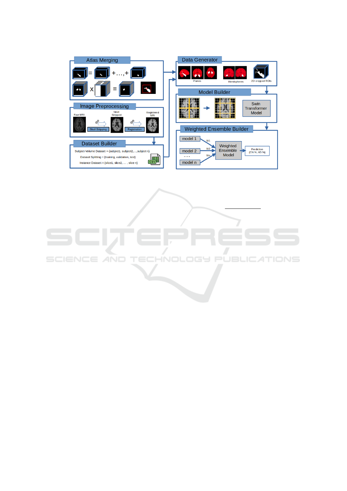
Figure 1: Proposed framework.
outlined as follows:
[Step-1] Atlas Fusion. In this research, a set of at-
lases (A
A
A) has been integrated, including JHU DTI-
based white-matter, J
¨
ulich histological, Talairach, and
Harvard-Oxford cortical and subcortical structural at-
lases (FSL, 2023). These atlases, featuring ROI an-
notations for both hemispheres, are registered into
MNI152 space. The (n) selected atlases (A
A
A) contain-
ing a specific region of interest are merged into a sin-
gle mean values map (M
M
M
∗
∈ R
w×h×d
).
The merged atlas (M
M
M
∗
) is binarized, converting to
a “1” any voxel having a numerical value greater than
zero, as follows: Let (M
M
M ∈ R
w×h×d
) the binarized
merged atlas given by:
∀
{
i, j, k ∈ N, 0 ≤ i < h, 0 ≤ j < w, 0 ≤ k < d
}
,
M
i jk
=
(
1, ifM
∗
i jk
> 0,
0, otherwise,
(1)
where (h) is the slice image height, (w) is the slice
image width, and (d) is the number of slices of the
(M
M
M) volume.
[Step-2] Atlas Volume Bounding. The volume
(M
M
M
whd
) is traversed by each plane in ascending order
to obtain the initial slice numbers (x
1
, y
1
, and z
1
) and
in descending order to obtain the final slice numbers
(x
2
, y
2
, and z
2
) for sagittal, coronal, and axial planes
respectively.
The slice number (i) of the image
(M
M
M
i
) with a mean greater than zero is in-
cluded in the boundaries list, (A
A
A) for initial
slices and (Ω
Ω
Ω) for finals, using Equation 2.
A
A
A
n+1
= i, ∀
{
i ∈ N, 0 ≤ i < d
}
, and
Ω
Ω
Ω
m+1
= i, ∀
{
i ∈ N, d > i ≥ 0
}
,
if
∑
w
j=1
∑
h
k=1
M
M
M
i jk
w × h
!
> 0,
with x
1
= A
A
A
1
and x
2
= Ω
Ω
Ω
1
,
(2)
where (d) is the number of slice images for a particu-
lar plane. The (n) and (m) variables are the number of
elements in the A
A
A and Ω
Ω
Ω lists. (x
1
) is the initial slice
number and (x
2
) the final.
3.2.2 Image Preprocessing
The raw volumes undergo preprocessing, involving
skull stripping and registration. The skull-stripped
dataset volumes are then registered to the MNI152 T1
template MRI scan, ensuring uniformity in shape, po-
sition, and alignment. The resulting scans have nor-
malized intensity, dimensions of 182 × 218 × 182, and
a resolution of 1 mm.
3.2.3 Dataset Builder
[Step-1] Subject - Volume Dataset Building. The
volumes for each subject are arranged in chronologi-
cal order based on their visit dates, and the filename
of the T1-weighted MRI volume from the last visit is
incorporated into the subjects’ dataset. This approach
guarantees the inclusion of only one volume per sub-
ject in the dataset. The subject dataset is balanced
using a simple random sampling, including the same
(k) number of subjects per class, where (k) is less than
or equal to the number of samples from the minority
class, thus avoiding class imbalance problems.
ICPRAM 2024 - 13th International Conference on Pattern Recognition Applications and Methods
456

[Step-2] Dataset Splitting. The dataset-splitting
process ensures reproducible testing and prevents data
leakage by first splitting the subject dataset to create
independent training, validation, and test sets. The
datasets are split randomly, ensuring that an MRI vol-
ume per subject is included in only one distribution
(training, validation, or test).
[Step-3] Dataset Metadata. Metadata information
is generated for each slice containing the ROI, cat-
egorized by plane and hemisphere. This metadata
includes details such as the group (ADNI, AIBL, or
OASIS), volume filename, label, and the position of
the ROI’s center. The ROI position is represented as
a voxel (x, y, z), which includes the slice number (z)
and the centroid position (x, y). This centroid position
is crucial in cropping the 2D slice image to extract the
most informative content from the ROI.
The process involves several steps, including the
creation of a list of slice numbers containing the ROI
(S
S
S), the generation of lists for pixel positions along
the x and y axes, and the determination of the centroid
position (x, y) based on the mode. The ROI center po-
sition (Φ
Φ
Φ
i
, Λ
Λ
Λ
i
), is calculated for each slice (i) included
in the ROI slice list S
S
S, using a mode-based approach.
The function ( f (χ)) obtains the mode from the X
X
X and
ϒ
ϒ
ϒ list of pixel positions, as follows:
∀
{
i ∈ N, i ∈ S
S
S
}
,
Φ
Φ
Φ
i
= f (X
X
X
i
),
Λ
Λ
Λ
i
= f (ϒ
ϒ
ϒ
i
)
(3)
where the f (χ) function gets the mode of the χ list.
(Φ
Φ
Φ
i
, Λ
Λ
Λ
i
) represent the x and y mode values for each
slice in the ROI slice list (S
S
S).
3.2.4 Data Generator
This component prepares training data for batch load-
ing by extracting metadata from the instance dataset.
It uses the image preprocessor to create an instance
image batch in memory based on specified output
preferences. The dataset batch size is limited by com-
putational resources like GPU and memory.
3.2.5 Model Builder
This component builds diverse Transformer classi-
fication models using datasets that include ROIs,
planes, and hemispheres. This task also includes eval-
uating model performance, as follows:
Swin Transformers. The 2D Swin Transformer
(Liu et al., 2021) serves as a weak learner, boosting
training speed and diagnostic accuracy. It augments
the number of instances and facilitates the use of pre-
trained models through transfer learning.
Model Performance Evaluation. The Transformer
model’s performance in Alzheimer’s case classifi-
cation is evaluated through average accuracy and
its standard deviation. Assessment is conducted at
the subject-patient level by combining classifications
from a subject’s slice level using majority voting.
3.2.6 Weighted Ensemble Builder
This component selects the highest-accuracy single
base classifiers (2D Swin Transformer), trained on di-
verse datasets, to form a homogeneous weighted en-
semble model. The goal is to improve performance,
robustness, and reliability in AD classification.
4 EXPERIMENTAL SETUP
The proposed Multiple ROI and Multiple Atlas-Based
Instance Selection method is rigorously examined
through four experiments.
4.1 Instance Selection
This experiment compares our framework to existing
instance selection methods, using a single base classi-
fier (Swin Transformer). The 2D slice image datasets
are uniformly derived from the same volumes.
[Method 1] - Percentile Fixed Number. Following
(Altay et al., 2021) and (Hu et al., 2023), 96 MRI
slices from the middle of all anatomical planes, pre-
cisely at the 50th percentile, are chosen to evaluate the
instance selection method.
[Method 2] - Multiple ROI and Multiple
Atlas-Based Instance Selection (Our Proposal).
Atlas annotations were amalgamated to form merged
ROIs. The position and the number of slice instances
(n) for this experiment vary based on the ROI,
anatomical plane, and hemisphere.
4.2 Regions of Interest Datasets
Diverse perspectives are attained by consolidating
information from various sources, including ROIs,
brain hemispheres, and anatomical planes. This ex-
periment aims to identify the most informative ROI
datasets using the proposed framework.
Instance Selection Framework for Alzheimer’s Disease Classification Using Multiple Regions of Interest and Atlas Integration
457

4.3 Weighted Ensemble
Diverse instance datasets, combining data from var-
ious sources like ROIs, brain hemispheres, and
anatomical planes, train a single 2D Swin Trans-
former classifier for enhanced diversity. The weighted
ensemble model in this experiment selects the most
accurate and diverse models, employing a weighted
approach for final classification based on each mem-
ber’s performance.
4.4 Performance Comparison
This experiment compares the model performance
obtained using the proposed Multiple ROI and
Multiple Atlas-based Instance Selection framework
with that of state-of-the-art methods. The related
works analyzed in this experiment use diverse
datasets (ADNI, AIBL, OASIS), input types (2D
and 3D), model architectures (Vision Transformer,
Swin Transformer, and mixed models combining
Convolutional Neural Networks with Transformers),
ROIs, and instance selection techniques (Cropping,
ROI extraction).
All experiments are reported at the subject level.
The volumetric dataset comprises 420 subjects,
divided into 70% (300) for training, 15% (60) for
validation, and an additional 15% (60) for testing.
This subject-volume dataset has been previously
randomly selected and partitioned. The instance
datasets exclusively contain slices of specific ROIs,
planes, and hemispheres. Cropped ROI images
consistently maintain a size of 32 x 32 x 3 (width,
height, channels). The number of slices per subject
varies depending on the specific ROI, plane, and
hemisphere.
A single base classifier model, trained on various
datasets, is utilized to evaluate their impact on the pro-
posed framework. The 2D Swin Transformer (Lyu
et al., 2022) was selected for its effectiveness in im-
age classification.
Hyperparameter optimization was conducted us-
ing Hyperband. The values employed to train the pro-
posed framework are as follows: Optimizer Name:
Adam, Learning Rate: 1e − 04, Clip Value Rate: 0.5,
Dropout: 0.15, Batch Size: 10, and Epochs: 100.
Python libraries NiBabel, TorchIO, PIL, and
NumPy preprocess the images. The FreeSurfer tools
are used for skull stripping and MRI registration us-
ing the MNI152 template. The Keras library is used
to build the classification models. All experiments are
repeated three times. We carry out the experiments
using ten workstations with an Intel Core i9 9900K
processor, 32 GB RAM, and 11 GB NVIDIA RTX
2080Ti GPU.
5 RESULTS AND DISCUSSION
Four experiments are conducted to test the pro-
posed framework: I) Compares state-of-the-art in-
stance selection techniques with the proposed Mul-
tiple ROI and Multiple Atlas-Based Instance Selec-
tion; II) Evaluates the effect of diverse datasets trained
with the same base classifier; III) Tests the weighted
ensemble model, combining a single base classifier
trained on multiple-view datasets; and IV) Compares
the models’ performance of the proposed method
with state-of-the-art related works. The presented ex-
perimental results correspond to the model accuracy
mean.
5.1 Instance Selection
Since selecting the most informative slices from the
original dataset may improve the overall performance
of the prediction model (Khan et al., 2019), this exper-
iment compares the proposed instance selection based
on a multiple region of interest and multiple atlas with
techniques based on percentiles, as shown in Table 2.
Table 2: Accuracy summary from different instance se-
lection techniques, using the same subject-volume dataset
(ALL-420). Multi-ROI-Atlas is our proposal.
Technique Sagittal % Coronal % Axial %
Percentile 92.99 ± 0.96 91.43 ± 0.79 91.941 ± 0.79
Multi-ROI-Atlas 96.67 ± 0.00 94.44 ± 0.79 95.56 ± 0.79
Hippocampus(Right)
Table 2 shows that all instance selection tech-
niques achieve the highest average accuracy values
for the sagittal plane, capturing the most critical in-
formation about the regions affected by AD. On the
other hand, the average accuracy values for each tech-
nique show that our proposed Multiple ROI and Mul-
tiple Atlas-Based Instance Selection technique en-
sures higher accuracy for all three planes (sagittal,
coronal, and axial).
5.2 Region of Interest Datasets
In homogeneous ensembles, the main difficulty is
generating diversity, despite using the same learning
algorithm (Sabzevari et al., 2022). This work experi-
mentally evaluates different MRI datasets trained us-
ing a single base classifier.
Table 3 demonstrate that the most informative
ICPRAM 2024 - 13th International Conference on Pattern Recognition Applications and Methods
458

ROIs are as follows: a) The Parietal Lobe and the
Hippocampus exhibit the highest accuracies, with val-
ues of 94.63% and 94.35%, respectively. This is
attributed to these ROIs capturing crucial informa-
tion from regions affected by AD. b) The right hemi-
sphere (93.70%) and the left hemisphere (92.81%)
display a marginal difference of 0.89% in their mean
accuracy across all planes. This disparity indicates
structural asymmetry between the left and right brain
hemispheres. Furthermore, c) the sagittal right plane
achieves the highest accuracy at 94.26%. This can
be attributed to the sagittal plane effectively capturing
critical information from regions affected by AD.
These multiple-view dataset models provide vari-
ety to build the weighted ensemble model.
Table 3: Summary of accuracy from different ROIs, planes,
and hemispheres.
Left Right
ROI Sagittal Coronal Axial Sagittal Coronal Axial Mean
Entorhinal Cortex 92.78 93.89 95.00 93.89 90.00 95.00 93.43
Fornix 94.44 96.11 93.33 93.33 93.89 92.78 93.98
Frontal Lobe 91.11 93.33 91.67 91.11 90.56 91.67 91.58
Hippocampus 91.67 93.89 93.89 96.67 94.44 95.56 94.35
Parietal Lobe 95.56 97.22 91.67 95.56 95.56 92.22 94.63
Temporal Lobe 88.33 88.33 88.33 95.00 94.44 95.00 91.57
Mean 92.32 93.80 92.32 94.26 93.15 93.71 93.26
5.3 Weighted Ensemble
The optimal ensemble composition is problem-
dependent, and determining the number of classifiers
for each type remains an open question (Sabzevari
et al., 2022). Models from the previous experiments
(II) with the highest accuracy and variety are used to
create a weighted ensemble, as shown in Table 4.
The contribution of each ensemble member is
weighted proportionally to the member’s performance
to obtain the final classification, creating a weighted
ensemble.
Finally, Table 4 shows that combining homoge-
neous ensemble methods produces accuracy perfor-
mance results significantly higher (98.33%) than a
single learning classification model, providing variety
to the weighted ensemble classifier.
5.4 Performance Comparison
Table 5 presents a comparison of the proposed method
with the state-of-the-art related works in terms of CN
versus AD classification performance.
The experimental results indicate that the pro-
posed Multiple ROI and Multiple Atlas-based In-
stance Selection method using a weighted ensemble
(98.33%) and a single base classifier, such as 2D Swin
Table 4: Summary of accuracy from different model mem-
bers of the weighted ensemble.
Ensemble model members accuracy
Model (ROI) Plane Hemisphere Accuracy % Weight %
Hippocampus Sagittal Right 96.67 8.32
Parietal Lobe Sagittal Right 96.67 8.32
Temporal Lobe Sagittal Left 96.67 8.32
Temporal Lobe Sagittal Right 96.67 8.32
Entorhinal Cortex Coronal Left 96.67 8.32
Fornix Coronal Left 96.67 8.32
Hippocampus Coronal Left 96.67 8.32
Parietal Lobe Coronal Left 98.33 8.46
Parietal Lobe Coronal Right 96.67 8.32
Entorhinal Cortex Axial Right 96.67 8.32
Hippocampus Axial Right 96.67 8.32
Temporal Lobe Axial Right 96.67 8.32
Ensemble 98.33
Transformer, slightly outperforms the state-of-the-art
instance selection methods regarding overall results.
This behavior can be attributed to the careful as-
sembly of subject and slice distribution sets, optimal
selection of the most significant slice instances, and
the most informative content from the ROIs.
Table 5: Performance comparison of the proposed method
with other related works for the classification tasks (AD vs.
CN).
Author Model Dataset Accuracy (%)
(Lyu et al., 2022) Vision Transformer ADNI 96.80
(Hu et al., 2023) 2D CNN+Transformer ADNI, OASIS 93.56
(Altay et al., 2021) Vision Transformer OASIS 91.18
(Xin et al., 2023) CNN+Swin-Transformer ADNI, AIBL 93.90
(Huang and Li, 2023) Swin Transformer ADNI+AIBL 94.05
(Mora-Rubio et al., 2023) Vision Transformer ADNI+OASIS 89.02
Hybrid Ensemble Swin Transformer ADNI+AIBL 98.33
Our Proposal +OASIS
6 CONCLUSIONS AND FUTURE
WORK
This work introduces a novel framework for strate-
gically identifying and selecting the most informa-
tive 2D image slices based on the fusion of multiple
regions of interest annotations from multiple atlases
(Multiple ROI and Multiple Atlas-based Instance Se-
lection).
The proposed framework’s impact on
Transformer-based classification models is ex-
perimentally explored. The performance of the 2D
Swin Transformer model varies with the dataset
(region of interest, plane, and hemisphere), and
using 2D slices increases instances, allowing for
training with transfer learning or from scratch. The
classifications obtained at the slice level are fused to
obtain a classification at the subject level. Finally,
the weighted ensemble improves the classification
model performance and reliability by combining
Instance Selection Framework for Alzheimer’s Disease Classification Using Multiple Regions of Interest and Atlas Integration
459

homogeneous ensemble methods.
For future work, researchers should consider us-
ing multiple inputs, mixed data, and 3D transformer
model ensembles based on multiple ROIs to enhance
the classification model performance and reliability.
REFERENCES
ADNI (2023). Alzheimer’s Disease Neuroimaging Initia-
tive. http://adni.loni.usc.edu.
AIBL (2023). Australian Imaging, Biomarker & Lifestyle
Flagship Study of Ageing. https://aibl.csiro.au.
Altay, F., S
´
anchez, G. R., James, Y., Faraone, S. V., Veli-
pasalar, S., and Salekin, A. (2021). Preclinical stage
alzheimer’s disease detection using magnetic reso-
nance image scans. Proceedings of the AAAI Confer-
ence on Artificial Intelligence, 35(17):15088–15097.
Bae, J. B., Lee, S., Jung, W., Park, S., Kim, W., Oh, H., Han,
J. W., Kim, G. E., Kim, J. S., Kim, J. H., and Kim,
K. W. (2020). Identification of Alzheimer’s disease
using a convolutional neural network model based on
T1-weighted magnetic resonance imaging. Scientific
Reports, 10(1):1–10.
Castro-Silva., J. A., Moreno-Garc
´
ıa., M. N., Guachi-
Guachi., L., and Peluffo-Ord
´
o
˜
nez., D. H. (2022). In-
stance selection on cnns for alzheimer’s disease clas-
sification from mri. In Proceedings of the 11th Inter-
national Conference on Pattern Recognition Applica-
tions and Methods - ICPRAM,, pages 330–337. IN-
STICC, SciTePress.
Choi, J. Y. and Lee, B. (2020). Combining of multiple
deep networks via ensemble generalization loss, based
on mri images, for alzheimer’s disease classification.
IEEE Signal Processing Letters, 27:206–210.
FSL (2023). Templates and Atlases included with FSL),
https://fsl.fmrib.ox.ac.uk/fsl/fslwiki/Atlases.
Hu, Z., Li, Y., Wang, Z., Zhang, S., and Hou, W. (2023).
Conv-swinformer: Integration of cnn and shift win-
dow attention for alzheimer’s disease classification.
Computers in Biology and Medicine, 164.
Huang, Y. and Li, W. (2023). Resizer swin transformer-
based classification using smri for alzheimer’s dis-
ease. Applied Sciences (Switzerland), 13.
Khan, N. M., Abraham, N., and Hon, M. (2019). Trans-
fer Learning with Intelligent Training Data Selection
for Prediction of Alzheimer’s Disease. IEEE Access,
7:72726–72735.
Li, C., Cui, Y., Luo, N., Liu, Y., Bourgeat, P., Fripp, J., and
Jiang, T. (2022). Trans-resnet: Integrating transform-
ers and cnns for alzheimer’s disease classification. In
2022 IEEE 19th International Symposium on Biomed-
ical Imaging (ISBI), pages 1–5.
Liu, Z., Lin, Y., Cao, Y., Hu, H., Wei, Y., Zhang, Z., Lin,
S., and Guo, B. (2021). Swin transformer: Hierarchi-
cal vision transformer using shifted windows. In 2021
IEEE/CVF International Conference on Computer Vi-
sion (ICCV), pages 9992–10002.
Lyu, Y., Yu, X., Zhu, D., and Zhang, L. (2022). Classifi-
cation of alzheimer’s disease via vision transformer:
Classification of alzheimer’s disease via vision trans-
former. ACM International Conference Proceeding
Series, pages 463–468.
Mora-Rubio, A., Bravo-Ort
´
ız, M. A., Arredondo, S. Q., Tor-
res, J. M. S., Ruz, G. A., and Tabares-Soto, R. (2023).
Classification of alzheimer’s disease stages from mag-
netic resonance images using deep learning. PeerJ
Computer Science, 9.
OASIS (2023). Open Access Series of Imaging Studies.
http://www.oasis-brains.org.
Pan, D., Luo, G., Zeng, A., Zou, C., Liang, H., Wang,
J., Zhang, T., Yang, B., and the Alzheimer’s Dis-
ease Neuroimaging Initiative (2022). Adaptive 3dcnn-
based interpretable ensemble model for early diag-
nosis of alzheimer’s disease. IEEE Transactions on
Computational Social Systems, pages 1–20.
Qin, Z., Liu, Z., Guo, Q., and Zhu, P. (2022). 3d
convolutional neural networks with hybrid attention
mechanism for early diagnosis of alzheimer’s dis-
ease. Biomedical Signal Processing and Control,
77:103828.
Sabzevari, M., Mart
´
ınez-Mu
˜
noz, G., and Su
´
arez, A. (2022).
Building heterogeneous ensembles by pooling homo-
geneous ensembles. International Journal of Machine
Learning and Cybernetics, 13(2):551–558.
Vaswani, A., Shazeer, N., Parmar, N., Uszkoreit, J., Jones,
L., Gomez, A. N., Kaiser, L., and Polosukhin, I.
(2017). Attention is all you need.
Wen, J., Thibeau-Sutre, E., Diaz-Melo, M., Samper-
Gonz
´
alez, J., Routier, A., Bottani, S., Dormont, D.,
Durrleman, S., Burgos, N., and Colliot, O. (2020).
Overview of classification of Alzheimer’s disease.
Medical Image Analysis, 63.
WHO (2023). World Health Organization. https://www.wh
o.int/news-room/fact-sheets/detail/dementia.
Xin, J., Wang, A., Guo, R., Liu, W., and Tang, X. (2023).
Cnn and swin-transformer based efficient model for
alzheimer’s disease diagnosis with smri. Biomedical
Signal Processing and Control, 86.
Young, S., Abdou, T., and Bener, A. (2018). Deep super
learner: A deep ensemble for classification problems.
Lecture Notes in Computer Science (including sub-
series Lecture Notes in Artificial Intelligence and Lec-
ture Notes in Bioinformatics), 10832 LNAI:84–95.
Yu, J. and Lee, T. M. (2020). Verbal memory and hippocam-
pal volume predict subsequent fornix microstructure
in those at risk for alzheimer’s disease. Brain Imaging
and Behavior, 14:2311–2322.
Zaabi, M., Smaoui, N., Derbel, H., and Hariri, W. (2020).
Alzheimer’s disease detection using convolutional
neural networks and transfer learning based methods.
In 2020 17th International Multi-Conference on Sys-
tems, Signals & Devices (SSD), pages 939–943.
Zhang, Z. and Khalvati, F. (2022). Introducing vision trans-
former for alzheimer’s disease classification task with
3d input.
ICPRAM 2024 - 13th International Conference on Pattern Recognition Applications and Methods
460
