
Concentric Ring Tattoo Electrodes for Biosignal Recordings
Gema Prats-Boluda
1a
, Eduardo Garcia-Breijo
2b
, José L. Martinez-de-Juan
1c
,
Javier Garcia- Casado
1d
, Yiyao Ye-Lin
1e
, Oleksandr Makeyev
3f
and Piero Cossedu
4
1
Centro de Investigación e Innovación en Bioingenieria, Universitat Politècnica de València, 46022, Spain
2
Instituto Interuniversitario de Investigación de Reconocimiento Molecular y Desarrollo Tecnológico, 46022, Spain
3
School of STEM, Diné College, Tsaile, AZ 86556, U.S.A.
4
Department of Electrical and Electronic Engineering, University of Cagliari, via Marengo, Cagliari, 09123, Italy
piero.cosseddu@diee.unica.it
Keywords: Tattoo Electrodes, Concentric Ring Electrodes, Electrocardiogram, Inkjet.
Abstract: Non-invasive bioelectrical recordings utilize monopolar or bipolar disc electrodes. However, these electrodes
suffer from poor spatial resolution, leading to susceptibility to physiological interferences. Concentric ring
electrodes have been implemented on rigid and flexible substrates to enhance spatial resolution. The present
work aims to develop an ultra-flexible and ergonomic concentric ring tattoo electrode based on PEDOT: PSS
ink and check its feasibility of picking up surface bioelectric signals such as the electrocardiogram. Results
reveal that it is possible to capture good quality bioelectric signals with tattoo electrodes implemented through
inkjet techniques on tattoo paper substrate using PEDOT: PSS as ink. The main problem associated with this
option is the cost in time of the machine for manufacturing the electrodes.
1 INTRODUCTION
The recording of electrophysiological signals in its
simplest form, that is, through contact electrodes
attached to the skin, is subject to continuous studies
both to optimize these recordings and to search for
new technologies that improve the manufacturing
process.
Today, diagnosis, therapy, and health monitoring
are largely based on the recording of
encephalographic, cardiac, and muscle (myoelectric)
signals. Even so, most recording systems for these
signals continue to have a traditional approach, using
monopolar disk electrodes (mainly Ag or AgCl). The
use of conventional electrodes entails important
limitations. On the one hand, they have limited
ergonomics and comfort, and the costs of the
a
https://orcid.org/0000-0002-9362-5055
b
https://orcid.org/0000-0002-9745-8485
c
https://orcid.org/0000-0001-9133-3123
d
https://orcid.org/0000-0003-1410-2721
e
https://orcid.org/0000-0003-2929-181X
f
https://orcid.org/0000-0003-2648-0500
manufacturing techniques used are also high. On the
other hand, the records obtained show a low spatial
resolution, which originates mainly from the
smearing effect due to the different conductivities of
the body volume conductor (Bradshaw et al., 2001).
This fact limits the ability to localize the sources of
bioelectric potential, which translates, for example,
into the difficulty of using conventional
electrocardiography (ECG) for the diagnosis of
pathologies associated with localized alterations in
the electrical conduction of the heart, as occurs in the
case of ventricular ischemia, atrial hypertrophy
(Macias et al., 2019), or difficulty in accurately
locating epileptic foci using conventional
electroencephalographic recordings (Aghaei-Lasboo
et al., 2020). Addressing these issues often
necessitates invasive electrophysiology, a procedure
Prats-Boluda, G., Garcia-Breijo, E., Martinez-de-Juan, J., Garcia-Casado, J., Ye-Lin, Y., Makeyev, O. and Cossedu, P.
Concentric Ring Tattoo Electrodes for Biosignal Recordings.
DOI: 10.5220/0012427800003657
Paper published under CC license (CC BY-NC-ND 4.0)
In Proceedings of the 17th Inter national Joint Conference on Biomedical Engineering Systems and Technologies (BIOSTEC 2024) - Volume 1, pages 159-163
ISBN: 978-989-758-688-0; ISSN: 2184-4305
Proceedings Copyright © 2024 by SCITEPRESS – Science and Technology Publications, Lda.
159

associated with considerable risks for the patient, and
an extended diagnosis time.
In recent years, an effort has been made to search
for alternative geometries and new technologies for
the manufacture of contact electrodes that allow
obtaining signals of better quality and/or with
improved spatial resolution. In addition, the
integration of the electrodes into clothing is sought,
which would lead to medical control beyond the
clinical work environment. To this end, the
development of electrodes on flexible biocompatible
substrates that provide characteristics such as
lightness, high flexibility, and adaptation to the
surface is sought out (Trung & Lee, 2016). Of these,
screen printing is the most widely used and mature
technology that has been used for decades in the
manufacturing of electronic systems and, more
recently, in the manufacturing of bioelectrodes.
To overcome the limited spatial resolution of
bioelectric recordings obtained from conventional
disk electrodes, the recording of the surface Laplacian
potential has been proposed (Lu & Tarjan, 2002)(Liu
et al., 2020) (Wang et al., 2023). The literature has
confirmed that Laplacian recordings are capable of
providing better spatial resolution of surface potential
recordings, that is, they can improve the detection of
bioelectric potential sources closest to the recording
electrodes, rejecting the contribution of sources of
bioelectric potential that are further away.
First, the surface Laplacian potential was
estimated using monopolar disc electrodes and
applying discretization techniques to estimate the
Laplacian potential from them (Hjorth,
1975)(Tandonnet et al., 2005)(Prats-Boluda et al.,
2007). Subsequently, concentric annular electrodes
were designed, whose configurations (bipolar, quasi-
bipolar, and tripolar) allowed obtaining a direct
estimate of the Laplacian of the potential captured on
the body surface. Subsequently, Laplacian potentials
of ECG, electrohysterographic, and
electroenterographic signals were obtained with
concentric ring electrodes (CRE) implemented on
rigid substrates, mainly using printed circuit boards
(Lu & Tarjan, 2002) (Prats-Boluda et al., 2011) (Mas-
Cabo et al., 2017)
The next step was the development of new
flexible CREs (fCREs). Different printing
technologies (screen printing, inkjet, gravure) and
materials such as Melinex or Ultem, MEMS) were
studied and compared for the development of fCREs
(Wang et al., 2023)(Trung & Lee, 2016),(Wei et al.,
2016). These fCREs allowed the capture of more
robust signals against respiratory interference than
conventional disc electrode recordings. Still, they
presented greater low-frequency interference, this last
observation being attributable to the fact that the
recordings with fCRE were carried out dry.
Therefore, despite the improvements introduced by
the implementation of CREs on flexible substrates,
the use of fCREs has not yet been transferred to the
clinical setting.
The objective of this work was to create an ultra-
flexible and ergonomic concentric ring tattoo
electrode using PEDOT: PSS ink assessing its
capability to capture surface bioelectric signals,
specifically the ECG. With this type of recording,
tools could be developed that would bring
electromyography closer to clinical practice in
different areas such as cardiology or obstetrics,
improving and/or complementing the information
provided by the non-invasive recording systems
common in clinical settings.
2 MATERIALS AND METHODS
A concentric ring electrode was designed using the
AutoCAD 2017 software (see Figure 1) and
considering the results of previous works (Prats-
Boluda et al., 2016).
Figure 1: Diagram of the concentric ring electrode design.
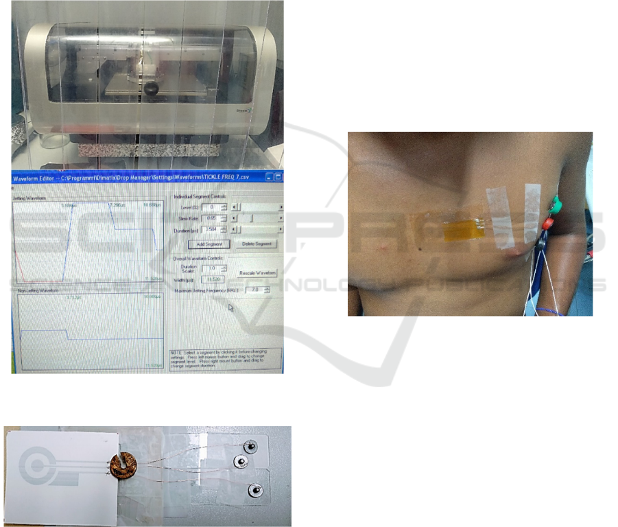
The technology tested in the manufacturing of
CRE tattoo prototypes was inkjet printing using the
Dimatix printer available at DEALAB (Figure 2).
Tattoo paper (tattoo 2.1) and commercial PEDOT:
PSS conductive ink GSD9011 (Heraeus®) have been
used as a substrate. Before printing the electrodes, the
tattoo paper is activated by plasma (30', 100W).
Likewise, the printing machine was programmed with
the configuration parameters shown in Figure 2.
To ensure correct deposition of the material, a 5-
layer print was made. The next step in the
manufacturing of these prototypes has been allowing
BIODEVICES 2024 - 17th International Conference on Biomedical Electronics and Devices
160
the extraction of signals from the tattoo electrodes. To
do this, two cables have been glued to the conductive
exit tracks of the electrodes prepared for this purpose,
using the CW 2400 conductive glue, and the joint has
been insulated and protected with Kapton adhesive.
In addition, snap connectors are glued and crimped on
the opposite end of the electrodes to allow their
connection to the biosignal capture system available
in the DEALAB, TMSI Porti 7®, see Figure 3. To
cure the conductive glue it is necessary to put it in the
electrode in the oven for 10 minutes at a temperature
of 50º.
Figure 2: Inkjet printer (top panel) and the setup screen
(bottom panel).
Figure 3: Output cables and connectors (at right) glued to
the CRE tattoo.
Both, concentric bipolar (CRE-ECG; outer ring
minus inner disc) and Lead II ECG (Lead II-ECG)
signals were simultaneously recorded with the TMSI
Porti7®, from DC to 500Hz, and acquired at 2048 Hz.
3 RESULTS
The capacity of this prototype of fCRE for the capture
of bioelectric signals was tested. Specifically, an ECG
signal was recorded. First, the skin surface where the
fCRE was positioned, comparable to precordial lead
V1 (CMV1), was shaved to minimize contact
impedance and minimally exfoliated using Nuprep
from Weaver and Company, USA. Following the
subjects' skin was cleaned with alcohol.
Subsequently, tattoo paper where the fCRE was
printed was placed over the area to be recorded,
CMV1, eliminating the “sacrificial layer”, a top
transparent film that covers the printed electrode. It
protects the electrode and helps transfer it onto the
skin. Figure 4 shows the arrangement of the
developed tattoo electrode on the chest to test its
ability to record the CRE-ECG signal. Together with
the CRE electrode, the Lead II-ECG was
simultaneously recorded.
Figure 4: Placement of the CRE tattoo of PEDOT: PSS, for
recording ECG signal.
Figure 5 corresponds to the simultaneous raw
recordings of the Lead II-ECG with conventional
commercial disc electrodes and with the PEDOT:
PSS tattoo electrode printed with the Dimatix on
tattoo paper. Recordings in Figure 5 demonstrate the
feasibility of capturing the ECG signal with the
PEDOT: PSS CRE tattoo electrodes on tattoo paper.
As expected, the amplitude of the signal captured
with the CRE (ECG-CRE) is much weaker than that
associated with Lead II-ECG recordings made with
conventional disc electrodes. In this regard, it should
be pointed out that one of the advantages of the
designed CRE is the possibility of picking up the
cardiac signal without using an external reference
electrode located on the hip or extremities. Still,
rather the tattoo electrode itself incorporates its own
reference. Also, the presence of powerline noise is
Concentric Ring Tattoo Electrodes for Biosignal Recordings
161
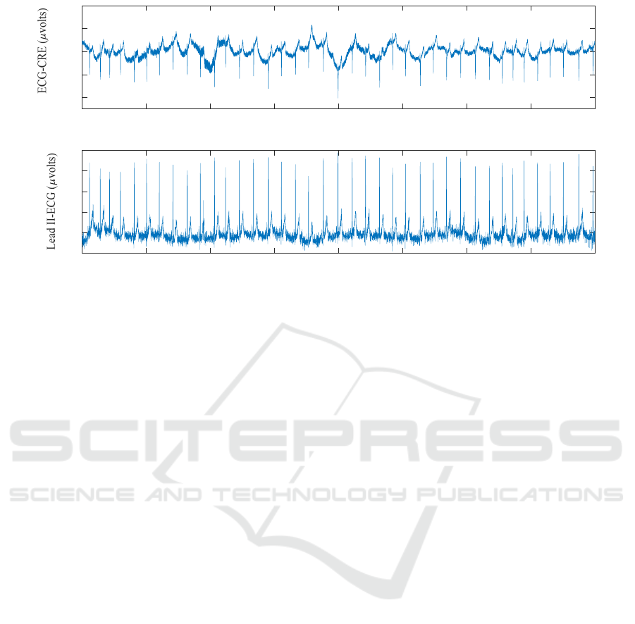
Figure 5: 40 seconds or raw signals corresponding to simultaneous recording of ECG-CRE bipolar signal captured with CRE
tattoo of PEDOT: PSS printed by inkjet using the Dimatix printer on plasma-activated tattoo paper (top panel) and Lead II-
ECG recording obtained with conventional disc electrodes (bottom panel).
noticeable in the recorded signals, both in the ECG-
CRE recording and in the Lead II-ECG signal.
Concerning this aspect, tests to check the ECG signal
capture capacity with the CRE tattoos printed with the
Dimatix were carried out in the laboratory, where a
large number of machines and wiring are located, that
is, in a very unfavorable environment for the
recording of bioelectric signals.
One of the main problems with this technology is
the slowness of printing, requiring 3.5 hours of
machine use per electrode. That is why it is proposed
in the future to carry out a study about developing
tattoo electrodes using screen printing or gravure
techniques, that enable the large-scale production of
this type of electrodes at an industrial scale.
Additionally, to enhance signal quality and facilitate
extensive use, it would be advisable to incorporate
additional elements such as electrolytic gel, adhesive,
and connectors commonly used in the biomedical
industry.
Another important aspect that needs to be
addressed to bring the use of CRE electrodes into the
clinical setting is to establish standard recording
positions on the torso and extract patterns of
normality/abnormality from the morphology of
cardiac waves. In this regard, preliminary studies
have been conducted using flexible concentric multi-
ring electrodes in precordial positions (Prats-Boluda
et al., 2016) or even mapping the torso (Besio &
Chen, 2007) (Prats-Boluda et al., 2018). However,
comprehensive studies are needed to validate the
results and, above all, to determine biomarkers
associated with pathological conditions.
As for the applicability of the developed tattoo
electrode, in the present work, we have focused on
ECG recording for the electrode design. Similar
electrodes could be used for picking up other
bioelectric signals (Estrada-Petrocelli et al., 2021)
(Ye-Lin et al., 2022). The optimal TCRE dimensions
will depend on the depth of the bioelectric source (the
deeper the higher the electrode diameter), the
required signal-to-noise ratio, or spatial resolution
(Makeyev et al., 2021).
4 CONCLUSIONS
It is possible to capture good quality bioelectric
signals with tattoo electrodes implemented through
inkjet techniques on tattoo paper substrate using
PEDOT: PSS as ink. The main problem associated
with this option is the cost in time of the machine for
manufacturing the electrodes. Future works will be
carried out to develop tattoo electrodes by screen-
printing or gravure techniques.
ACKNOWLEDGMENTS
This work was supported by the Generalitat
Valenciana (Spain) BEST/2019/168.
0 5 10 15 20 25 30 35 40
Time (seconds)
-200
-100
0
100
20
0
0 5 10 15 20 25 30 35 40
Time (seconds)
-200
0
200
400
600
800
BIODEVICES 2024 - 17th International Conference on Biomedical Electronics and Devices
162

REFERENCES
Aghaei-Lasboo, A., Inoyama, K., Fogarty, A. S., Kuo, J.,
Meador, K. J., Walter, J. J., Le, S. T., Graber, K. D.,
Razavi, B., & Fisher, R. S. (2020). Tripolar concentric
EEG electrodes reduce noise. Clinical
Neurophysiology, 131(1), 193–198. https://doi.org/
10.1016/j.clinph.2019.10.022
Besio, W., & Chen, T. (2007). Tripolar Laplacian
electrocardiogram and moment of activation isochronal
mapping. Physiol Meas., 28(5), 515–529.
https://doi.org/10.1088/0967-3334/28/5/006
Bradshaw, L. A., Richards, W. O., & Wikswo, J. P. (2001).
Volume conductor effects on the spatial
resolution of magnetic fields and electric
potentials from gastrointestinal electrical activity.
Med.Biol.Eng.Comput., 39(1), 35–43.
Estrada-Petrocelli, L., Torres, A., Sarlabous, L., Rafols-De-
Urquia, M., Ye-Lin, Y., Prats-Boluda, G., Jane, R., &
Garcia-Casado, J. (2021). Evaluation of Respiratory
Muscle Activity by Means of Concentric Ring
Electrodes. IEEE Transactions on Biomedical
Engineering, 68(3), 1005–1014. https://doi.org/10.11
09/TBME.2020.3012385
Hjorth, B. (1975). An on-line transformation of EEG scalp
potentials into orthogonal source derivations.
Electroencephalogr.Clin.Neurophysiol., 39(5), 526–
530.
Liu, X., Makeyev, O., & Besio, W. (2020). Improved
Spatial Resolution of Electroencephalogram Using
Tripolar Concentric Ring Electrode Sensors. Journal of
Sensors, 2020. https://doi.org/10.1155/2020/6269394
Lu, C. C., & Tarjan, P. P. (2002). Pasteless, Active,
Concentric Ring Sensors for Directly Obtained
Laplacian Cardiac Electrograms. J.Med.Biol.Eng., 22,
199–203.
Macias, C., Khakpour, H., Buch, E., Shivkumar, K., &
Bradfield, J. S. (2019). Limitations of 12-lead
electrocardiogram wide complex tachycardia
algorithms in a patient with left atrial flutter and large
myocardial infarction. HeartRhythm Case Reports,
5(2). https://doi.org/10.1016/j.hrcr.2018.04.001
Makeyev, O., Ye-Lin, Y., Prats-Boluda, G., & Garcia-
Casado, J. (2021). Comprehensive optimization of the
tripolar concentric ring electrode based on its finite
dimensions model and confirmed by finite element
method modeling. Sensors, 21(17). https://doi.org/
10.3390/s21175881
Mas-Cabo, J., Ye-Lin, Y., Benalcazar-Parra, C., Alberola-
Rubio, J., Perales, A., Garcia-Casado, J., & Prats-
Boluda, G. (2017). Electrohysterogram Signals from
Patients with Threatened Preterm Labor: Concentric
Ring Elecctrode vs. Disk Electrode Recordings.
BIOSIGNALS 2017 - 10th International Conference on
Bio-Inspired Systems and Signal Processing,
Proceedings; Part of 10th International Joint
Conference on Biomedical Engineering Systems and
Technologies, BIOSTEC 2017, 5, 78–83.
https://doi.org/10.5220/0006155000780083
Prats-Boluda, G., Garcia-Casado, J., Martinez-de-Juan, J.
L. L., & Ponce, J. L. L. (2007). Identification of the
slow wave component of the electroenterogram from
laplacian abdomianl surface recording in Humans.
Physiological Measurement, 28(9), 1115–1133.
https://doi.org/10.1088/0967-3334/28/9/012
Prats-Boluda, G., Garcia-Casado, J., Martinez-de-Juan, J.
L., & Ye-Lin, Y. (2011). Active concentric ring
electrode for non-invasive detection of intestinal
myoelectric signals. Medical Engineering and Physics,
33(4), 446–455. https://doi.org/10.1016/j.medengphy.
2010.11.009
Prats-Boluda, G., Ye-Lin, Y., Bueno-Barrachina, J. M.,
Rodriguez De Sanabria, R., & Garcia-Casado, J.
(2016). Towards the clinical use of concentric
electrodes in ECG recordings: Influence of ring
dimensions and electrode position. Measurement
Science and Technology, 27(2). https://doi.org/
10.1088/0957-0233/27/2/025705
Prats-Boluda, G., Ye-Lin, Y., Pradas-Novella, F., Garcia-
Breijo, E., & Garcia-Casado, J. (2018). Textile
Concentric Ring Electrodes: Influence of Position and
Electrode Size on Cardiac Activity Monitoring. Journal
of Sensors, 2018. https://doi.org/10.1155/2018/7290
867
Tandonnet, C., Burle, B., Hasbroucq, T., & Vidal, F.
(2005). Spatial enhancement of EEG traces by surface
Laplacian estimation: comparison between local and
global methods. Clin.Neurophysiol., 116(1), 18–24.
Trung, T. Q., & Lee, N.-E. (2016). Flexible and Stretchable
Physical Sensor Integrated Platforms for Wearable
Human-Activity Monitoringand Personal Healthcare.
Advanced Materials, 28(22), 4338–4372.
https://doi.org/10.1002/adma.201504244
Wang, Z., Zhao, N., Shen, G., Jiang, C., & Liu, J. (2023).
MEMS-Based Flexible Wearable Tri-Polar Concentric
Ring Electrode Array With Self-Adhesive Graphene
Gel for EEG Monitoring. IEEE Sensors Journal, 23(3).
https://doi.org/10.1109/JSEN.2022.3230679
Wei, Y., Torah, R., Li, Y., & Tudor, J. (2016). Dispenser
printed capacitive proximity sensor on fabric for
applications in the creative industries. Sensors and
Actuators A: Physical, 247, 239–246. https://doi.org/
10.1016/J.SNA.2016.06.005
Ye-Lin, Y., Martinez-De-Juan, J. L., Jareño-Silvestre, A.,
& Prats-Boluda, G. (2022). Concentric ring electrodes
for non-invasive recording of gastric myoelectric
activity. Measurement: Journal of the International
Measurement Confederation, 188(110607), 1–9.
https://doi.org/10.1016/j.measurement.2021.110607
Concentric Ring Tattoo Electrodes for Biosignal Recordings
163
