
Visual Insights in Human Cancer Mutational Patterns: Similarity-Based
Cancer Classification Using Siamese Networks
Rocco Zaccagnino, Clelia De Felice, Marco Russo and Rosalba Zizza
Dip. di Informatica, University of Salerno, Salerno, Italy
Keywords:
Cancer Detection, Siamese Neural Networks, Mutational Signature, Explainable AI, Information
Visualization.
Abstract:
In recent years, a number of innovations concerning the diagnosis and treatment of diseases through the ap-
plication of genomics have opened the door to the detailed analysis of somatic mutation patterns in human
cancers. Several AI-based systems have been proposed to identify correlations between mutations and type of
cancer. However, the use of AI in Bioinformatics still presents two main limitations: (i) the explainability, i.e.,
the ability of the methods to partially explain and motivate their behavior, and (ii) the usability, i.e., about the
strong limitations that are found in the actual use of such methods in real bio-medical contexts and scenarios.
In this work, we propose a novel ML-based cancer-type detection system which integrates explainability and
usability techniques. To this aim, we first formulate the cancer-type detection problem using the similarity-
based classification paradigm. Then, given a cancer sample, we assume to have a set of somatic mutation
features available which can be interpreted as cancer mutational view of the sample itself. Finally, we propose
the use of a special Machine Learning model defined for learning similarity functions, namely the Siamese
Neural Network (SNN). The proposed SNN learns to take a pair of cancer mutational views as input, and to
compute a similarity score that can be used to verify whether such samples are similar or not.
Preliminary experiments carried out to assess the effectiveness of the proposed system show high performance
reaching f1 score 97.61%, and highlight how the similarity-based classification paradigm could be more suit-
able than the commonly used classification paradigm for the formulation of the cancer-type detection problem.
1 INTRODUCTION
1.1 Cancer and Somatic Mutations
In recent years, a number of technical innovations
have been developed regarding the diagnosis and
treatment of diseases through the application of ge-
nomics. The most evident result is the standardiza-
tion of tumor profiling techniques based on recur-
rent targeted mutations analysis. This has led to an
evident efficacy of molecularly targeted therapies on
distinct types of tumor by exploiting information re-
garding shared genetic features. Today, based on re-
cent large-scale exome and genome-sequencing stud-
ies, we know that major tumour types present specific
patterns of somatic mutations (Kandoth et al., 2013;
Lawrence et al., 2013; Ciriello et al., 2013).
In this direction, several research initiatives have
developed recently. As an example, at Memorial
Sloan Kettering Cancer Center
1
, a NGS panel named
msk-impact has been developed to show the feasi-
bility and utility of large-scale prospective clinical
sequencing of tumors to guide clinical management.
msk-impact has been used to detect all protein-
coding mutations, copy number alterations, and se-
lected promoter mutations and structural rearrange-
ments in 410 cancer-associated genes, for a total of
62 sequenced principal tumors from more than 10,000
patients. The result is a comprehensive and detailed
catalog of somatic mutations for every tumor se-
quenced, publicly available online
2
.
1.2 Contribution of this Work
Explainable and Usable AI. Artificial Intelligence
(AI) and in particular Machine Learning (ML) sys-
tems are increasingly used in Bioinformatics. This
because the massive amounts of bio-medical data, in-
1
https://www.mskcc.org/
2
http://cbioportal.org/msk-impact
462
Zaccagnino, R., De Felice, C., Russo, M. and Zizza, R.
Visual Insights in Human Cancer Mutational Patterns: Similarity-Based Cancer Classification Using Siamese Networks.
DOI: 10.5220/0012399600003657
Paper published under CC license (CC BY-NC-ND 4.0)
In Proceedings of the 17th International Joint Conference on Biomedical Engineering Systems and Technologies (BIOSTEC 2024) - Volume 1, pages 462-470
ISBN: 978-989-758-688-0; ISSN: 2184-4305
Proceedings Copyright © 2024 by SCITEPRESS – Science and Technology Publications, Lda.

cluding heterogeneous high-dimensional data, intro-
duce challenges to existing ML methods (Karim et al.,
2021), which are increasingly being used successfully
for data analysis and interpretation.
To date, the use of AI techniques in Bioinformat-
ics has two main limitations. The first is the so-called
explainable AI (XAI), i.e., the ability of the methods
to partially explain or motivate their behavior, while
the second is about the usable AI, i.e., the actual use
of such systems in real-world scenarios.
While ML models are able to address complex
problems, their “black-box” nature raises concerns
about transparency and accountability, which also
overshadow their ability to solve the problems them-
selves. The field of XAI aims to make AI systems
more transparent by explaining how they make deci-
sions and so to enhance the human-comprehensibility,
reasoning, transparency, and accountability.
As mentioned earlier, another strong limitation of
the use of AI in Bioinformatics is about the actual “us-
ability” of such systems in real-world scenarios. Ad-
vanced ML models facing really complex problems
often suffer from scalability problems. In some cases,
the motivation could be found in the “classification”
paradigm used to formulate the problem faced: there
are n classes of samples, and the model is trained on a
training set to classify a new sample in one of such n
classes. This approach, especially in Bioinformatics,
could suffers from some issues, including the enor-
mous amount of data on which the model must be
trained, the strong imbalance of the classes that can
arise when working on real data, and above all the
problem of scaling the model when new classes of
samples must be classified. In this case, the model
must be retrained on the whole set of data, with se-
vere impact on the computational effort, but also in
contexts where a timely response can be crucial.
Proposed Strategy. We propose a novel ML-based
cancer-type detection system with the the aim of in-
tegrating it with explainability and usability tech-
niques. We first formulate such a problem in terms
of similarity-based classification (Chen et al., 2009).
Given a cancer sample, we assume to have a set
of somatic mutation features available which can be
interpreted as a cancer mutational view of the sam-
ple itself. Then, according to the central idea of the
similarity-based classification paradigm, we define a
model which does not simply learn to classify a can-
cer sample by observing its cancer mutational view,
but which is able to learn, starting from a set of sam-
ple pairs, a similarity function and which therefore is
able to tell whether two samples are similar or not.
Clearly, the more the starting set of samples is repre-
sentative of the problem, the more accurate the func-
tion is. The advantage of this approach is that once
the similarity function has been calculated, the model
can also be used on new samples (even of a cancer-
type never seen during the training) of which to find
out which classes are more similar to. Furthermore,
to make the system scalable on large amounts of data,
we keep track, for each cancer-type class, of one sin-
gle representative view, and using them to find out
which classes are more similar to a test view, with
great benefits both in terms of memory and privacy.
There are numerous examples of works in Bioin-
formatics based on the similarity-based classification
paradigm (Mathai and Kirchmair, 2020). In this pa-
per, we propose the usage of special ML models de-
fined for learning similarity functions, i.e., Siamese
Neural Networks (SNN). We define a novel SNN
which given a pair of cancer mutational views out-
puts a similarity score that can be used to verify that
they are similar. The proposed solution is based on the
following two main ideas that, in our opinion, could
limitate the issues discussed above. First, the somatic
mutation features of a cancer sample could be used
as “similarity view” that can be exploited as effec-
tive feature embedding for ML methods. Second, we
show that the SNN increases the level of discrimina-
tion strength within the proposed cancer mutational
views (Bell and Bala, 2015).
Several studies have been proposed in the litera-
ture to face the problem of using ML techniques to
determine tumour organ of origin and histology using
the patterns of somatic mutation identified by whole
genome DNA sequencing, such as (Jiao et al., 2020).
However, most of these are based on the classification
paradigm. Furthermore, several works use SNNs in
Bioinformatics (Bechar et al., 2023; Narmatha et al.,
2023), but to the best of our knowledge this is the
fist attempt to propose a similarity-based classifica-
tion paradigm based on SNNs exploiting somatic mu-
tation features for the cancer-type detection problem.
Our Contributions:
• A novel cancer-type detector integrating explain-
ability and usability techniques, and based on can-
cer mutational views for training SNNs at verify-
ing the similarity between cancer samples.
• Preliminary experiments to assess the effective-
ness of the proposed method; results obtained on
a dataset of somatic mutation features show ac-
curacy 89.25%, precision 97.60%, recall 97.63%,
and f1 score 97.63%, highlighting the advantages
of the similarity-based classification paradigm.
Visual Insights in Human Cancer Mutational Patterns: Similarity-Based Cancer Classification Using Siamese Networks
463
Source code and files are available online
3
.
2 THE PROPOSED SYSTEM
In this section, we describe a novel ML-based cancer-
type detection system. We assume that the reader is
familiar with ML notions. For further details, refer
to (Tan et al., 2016).
2.1 Overview
Here, we provide an overview of the scenario in which
the proposed system can be placed (Figure 1).
• Usability. The system must be designed to be
able to manage views in a scalable and efficient
way. To this aim, the typical scenario in which we
imagine it could be used is the one in which it is
used to store cancer mutational views to be com-
pared from time to time with new test cancer sam-
ples that are analyzed to find out their type. More
in detail, at every moment it has in memory a rep-
resentative view of each type of cancer analyzed
up to that moment. Each time a new cancer sam-
ple c
t
must be detected, the corresponding cancer
mutational view s
t
, named test view, is provided
to the system; during the search, s
t
is compared
with every stored enrollment view; then, the sys-
tem returns the type of cancer corresponding to
the enrollment view s
e
(corresponding to a spe-
cific cancer sample c
e
) which is most similar to s
t
,
formally denoted with c
t
∼ c
e
. We assume that if
this level of similarity does not exceed a threshold
(established during the training of the S ), then s
t
is a sample of a new type of cancer and therefore
will be memorized as a view of this new type.
The advantages of such a system are numerous.
First, there is no need to keep in memory a huge
amount of data relating to samples to be used for
a re-training of the ML model, but for each type
of cancer only the view of a representative sample
is stored. Furthermore, a significant implication is
that of data privacy, which in this case must focus
on the privacy of a very small set of data.
• Explainability. S has been designed to integrate
the attemption mechanism, through special lay-
ers. “Attention” was first used in computer vision,
inspired by the idea to mimic the attention abil-
ity of the human brains to deal with the massive
amount of visual input. Attention layers mainly
3
https://github.com/FLaTNNBio/few-shot-learning-f
or-cancer-detection/tree/master
consist in a weighted mean reduction, where each
element is weighted in proportion to its contribu-
tion to the mean. One way to interpret the atten-
tion weights is to plot them as a feature heatmap,
where each row corresponds to an output item and
each column corresponds to an input feature, and
the color or intensity of each cell indicates the
level of the attention weight. This can help you
visualize which parts of the input are more im-
portant for each output. Thus, by showing the vi-
sualization of the feature heatmap of the attemp-
tion layer we can interpret the relation between
the features and better understand the key issues
which affect the performance of S . As we will
see in Section 3, such heatmaps can be used to
highlight the most relevant somatic mutations in
the several cancer types.
2.2 Cancer Mutational View
The dataset used for our experiments is extracted from
the msk-impact (Kübler et al., 2019), a genomic pro-
filing dataset generated by Memorial Sloan Kettering
Cancer Center. It contains molecular profiling data of
10,945 successfully sequenced tumor samples from
10,336 individuals, for 62 principal tumor types. The
dataset, generated using NGS technologies, includes
molecular features that are relevant for cancer diagno-
sis, prognosis, and treatment, such as protein-coding
mutations, copy number alterations (CNAs), and se-
lected promoter mutations and structural rearrange-
ments in 410 cancer-associated genes.
To extract the data used for our experiments, first
we downloaded such a dataset
4
, and then we merged
the following files to into a csv file: data_cna.txt,
data_sv.txt, data_clinical_sample.txt,
data_clinical_patient.txt. The dataset ob-
tained consists of 433 features, organized into:
• Clinical info (13): Sample ID, Cancer Type,
Mutation Count, Sex, Sample Type, DNA
Input, Matched Status, Oncotree Code,
Overall Survival Status, Patient’s
Vital Status, Sample Collection Source,
Smoking History, Somatic Status.
• Structural variations info (10): Site1 Chr,
Site1 Region, Site1 Hugo Symbol, Site2
Chr, Site2 Region, Site2 Hugo Symbol,
Class, Connection Type, Tumor Variant
Count, Breakpoint Type.
• Copy Numbers (410).
4
https://www.cbioportal.org/study/summary?id=msk_
impact_2017
BIOINFORMATICS 2024 - 15th International Conference on Bioinformatics Models, Methods and Algorithms
464
view
view
view
view
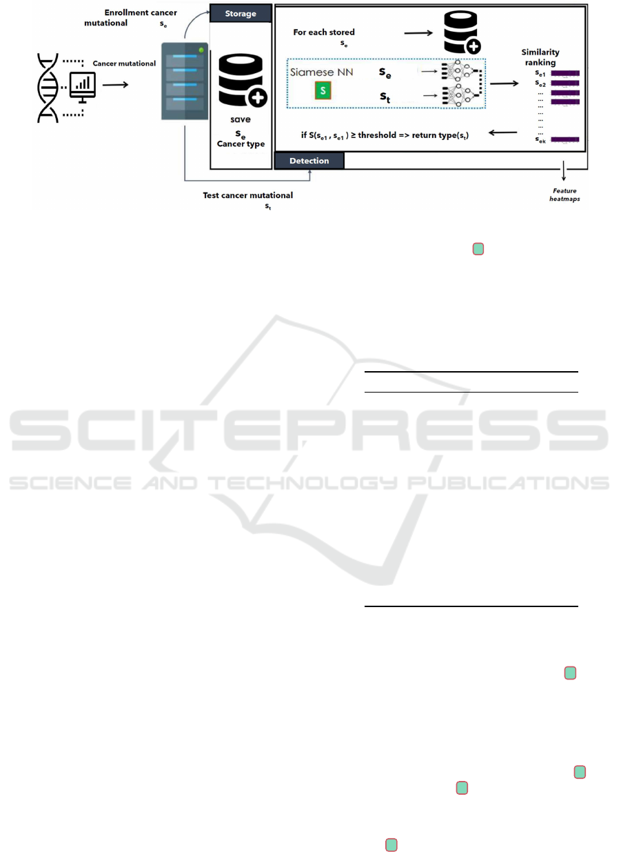
Figure 1: The overall scenario. The system manages cancer mutational views. To store a view s
e
, it must be entered, and if
the corresponding cancer type is not in the system then to save it together with the cancer type; to detect a view s
t
, it must
compared with all the stored views; a similarity ranking s
e1
, . . . , s
ek
is built by using the SNN S , and if the similarity score
between s
t
and s
e1
is greater or equal to a threshold, then the cancer-type of s
t
is the same of s
e
.
At the end of this extraction phase, each cancer
sample was represented by a set of 433 features.
Then, the dataset underwent a normalization pro-
cedure for numeric features, and a one-hot encoding
for non-numeric features, so reaching a number of
features equal to 2181. The reason for the increased
number of features is due to the use of one-hot coding
which notoriously could generate huge vectors since
the size of a generated feature vector is equal to the
number of possible values. To reduce the high di-
mensionality of the input data, several techniques in
the literature can be applied, such as the feature se-
lection procedure. However, in order to limit the loss
of information that could occur by choosing which
input features to keep and which to discard, in this
work we have decided to use the Principal Compo-
nent Analysis with several values for the number of
components parameter. Results showed that best re-
sults have been obtained with 1403 components. For
each cancer sample, this set of 1403 components is
the cancer mutational view.
Finally, since one of the goals of this work is
to compare detection by classification with that by
similarity-based classification, we tried to maintain,
of the 62 types of cancer managed in the starting
dataset, only those that have a minimum number of
instances that maximize the capability of classifica-
tion models. This is because, as is known, a strong
class imbalance is a problem when training classifi-
cation models. From empirical observations and pre-
liminary experiments, we have observed that by guar-
anteeing a minimum number of instances equal to 30,
this allows us to obtain a classification model, with
which we will compare ourselves, with excellent per-
formance (see Section 3) for further details).
Table 1 reports the 16 types of cancer, i.e., the
classes of our problem, which have at least 30 in-
stances, by indicating for each of them the exact num-
ber of instances (#instances).
Table 1: Number of instances for each cancer-type class.
Cancer type #instances
Prostate Cancer 336
Non-Small Cell Lung Cancer 313
Breast Cancer 242
Soft Tissue Sarcoma 104
Colorectal Cancer 100
Glioma 158
Hepatobiliary Cancer 70
Melanoma 66
Esophagogastric Cancer 63
Pancreatic Cancer 56
Bone Cancer 54
Cancer of Unknown Primary 43
Bladder Cancer 42
Ovarian Cancer 40
Head and Neck Cancer 35
Endometrial Cancer 32
2.3 The Proposed Siamese NN
In this section, we first describe the SNN S trained
to compute the similarity between two cancer muta-
tional views, and then details of the pseudo-code.
Siamese Architecture and One-Shot Learning.
Given a pair of cancer mutational views s(c
i
) and
s(c
j
), where c
i
and c
j
are cancer samples, S com-
putes a similarity score S (s(c
i
), s(c
j
)). Then, to ver-
ify that c
i
and c
j
are of the same type, the following
rule is used by the system:
S (s(c
i
), s(c
j
)) ≥ δ =⇒ c
i
∼ c
j
Visual Insights in Human Cancer Mutational Patterns: Similarity-Based Cancer Classification Using Siamese Networks
465
where δ ∈ [0, 1] is the cancer mutational view thresh-
old empirically estimated during the training of S . In
the following, we provide details about the architec-
ture and the training of S . S consists of three sec-
tions: the branches, the info, and the similarity.
The branches section consists of two identical
subnetworks, each one defined as follows. It starts
with a Linear layer using ReLu activation, which
takes as input the cancer mutational view and returns
a vector of size 1754. Such a layer is then followed by
5 blocks each one consisting of: (i) one Linear with
ReLu activation function and returning a vector of size
750, (ii) one Dropout layer with probability 0.1, and
(iii) one BatchNormalization layer. The info section
essentially consists of two layers, each taking as input
the concatenation of the outputs o
1
and o
2
of the two
identical subnetworks described above: one Attention
layer used to integrate S with the attention mecha-
nism described in Section 2.1, and one Lambda layer
used to compute the Euclidean distance between o
1
and o
2
. As for the similarity section, the concatena-
tion of the Attention layer output and of the Lambda
layer output is given as input to 3 blocks where each
block consists of: (i) one Linear layer with ReLu ac-
tivation function and returning a vector of size 320,
(ii) one Drouput layer with probability 0.1, (iii) one
BatchNormalization layer. Then, the blocks are fol-
lowed by Linear layer with output of size 1 (“similar
or not similar”) and Sigmoid activation function.
One of the most interesting advantages of using
SNNs is the ability to adopt the One-Shot Learn-
ing strategy, shown to be effective in identifying new
classes based on one (or only a few) examples. The
idea is to learn patterns and similarities on previously
seen classes instead of fitting the ML model to fixed
classes, in order to be able of classifying previously
unseen classes using one instance. This strategy is
very helpful in the scenario described in Section 2.1.
Indeed, it allows us to define a detection system “cal-
ibrated” on a significant initial set of cancer-types,
i.e, with a SNN trained on an initial set of cancer
mutational views corresponding to a “representative”
set of cancer-types; a new cancer-type can be added
to the system without having to retrain the network,
but simply by saving a reference cancer mutational
view, used every time during the detection tasks. S is
trained using the One-Shot learning (Algorithm 1).
Pseudocode:
• One-Shot Learning (Algorithm 1).
It takes as input the dataset S of cancer samples
organized into N cancer-type classes, and the cho-
sen cancer mutational view similarity threshold.
First, the algorithm initializes the weights of S
(line 1), and an empty list one-shot-accuracy
which will contain the accuracy obtained at each
evaluation step (line 2). Then, for each cancer-
type class t
i
∈ C, t
i
is split in t
l
i
(labelled samples),
and t
u
i
(unlabelled samples) (line 5). Each of the
remaining N − 1 classes is split into two balanced
subsets (line 11): the first one using the methods
GetSimilarPairs and GetDissimilarPairs to
generate the training set of similar and dissimi-
lar pairs, while the second one used as evaluation
pool (lines 12 and 13). Thus, the training pro-
cess (line 17) and the testing process (line 18)
run, by excluding s
i
. For the evaluation, the
method GetOtherPairs (line 17) is used to build
a set of evaluation pairs P
i
. Then, using the
method Voting, each instance e
i
∈ P
i
is classi-
fied using the class with the highest votes. Fi-
nally, the trained S and the average accuracy
one-shot-accuracy are returned.
• The overall detection system (Algorithm 2). It
takes as input a cancer_sample, and the type of
request (“storage” or “detect”). At the begin-
ning, the type of request is checked. If a “stor-
age” is required, then the system first check if
a the cancer-type of the sample is already stored
in the database using the method GetCancerType
(line 4). If a cancer type has been found, then the
system communicates a cancer mutational view
for the cancer-type of the input sample is already
stored. Otherwise, this means that the the cancer-
typer of the input sample is not stored. Then,
the system saves the cancer mutational view of
cancer_sample as enrollment view through the
method SaveCancerView (line 8). Instead, if
a “detect” is required, the most similar view is
searched within the system (line 12).
3 PRELIMINARY EXPERIMENTS
Here, we report the results obtained during prelimi-
nary experiments carried out to assess the effective-
ness of the proposed detection system. To this aim,
we have compared the performance obtained by the
proposed SNN S described in Section 2.3, with that
obtained by a baseline Deep Neural Network (DNN)
trained for classify the cancer-type of cancer samples.
In these experiments, such a baseline DNN has been
obtained by extracting only one of the subnetworks of
the branches section of S .
BIOINFORMATICS 2024 - 15th International Conference on Bioinformatics Models, Methods and Algorithms
466
Algorithm 1: S One-Shot Learning.
Input : C = {t
1
, . . . , t
N
}, threshold
Output: ⟨ S ,one-shot-accuracy⟩
1 S ← InitializeSiamese( S );
2 one-shot-accuracy ← [];
3 for i = 1 to N do
4 /
*
Select “new” speaker s
i
5 ⟨t
l
i
, t
u
i
⟩ ←
SplitSamplesByCancerType(t
i
,0.5);
6 training_set
i
←
/
0;
7 testing_set
i
←
/
0;
8 /
*
Build training/testing sets
without t
i
9 for j = 1 to N do
10 if j ̸= i then
11 ⟨t
l
j
, t
u
j
⟩ ←
SplitSamplesByCancerType(t
j
,0.5);
12 training_set
i
← training_set
i
∪ t
l
j
;
13 testing_set
i
← testing_set
i
∪
t
u
j
;
14 P
t
← GetSimilarPairs(training_set
i
);
15 P
d
←
GetDissimilarPairs(training_set
i
);
16 /
*
Train and Test Siamese NN
17 S ←
Train( S ,P
t
,P
d
,SV_threshold,“Triplet
Loss”);
18 accuracy ←
Test( S ,testing_set
i
,SV_threshold);
19 /
*
One-Shot Evaluation
20 P
i
←
GetOtherPairs(t
u
i
,{t
1
, . . . , t
i−1
, t
l
i
, t
i+1
, . . . , t
N
};
21 correct ← 0;
22 for k = 1 to |P
i
| do
23 x ← Voting(P
i
[k], S );
24 if x == i then
25 /
*
Correct classification
26 correct ← correct +1;
27 accuracy
i
←
correct
100
;
28 one-shot-accuracy.append(accuracy
i
);
29 return ⟨ S ,Average(one-shot-accuracy)⟩;
3.1 Results
We have split, using a stratified approach, the dataset
into training set, consisting of the 70% of cancer sam-
ples of the dataset (1,403 samples), and testing set
consisting of the 30% (351 samples). Then, the train-
ing set has been split into two subsets: (i) the first
one consisting of the 80% (1,122 samples) and used
to train both S and the baseline DNN, and (ii) the sec-
ond one consisting of the 20% (281 samples) and used
to validate both S and the baseline DNN. Finally, the
testing set has been used to test the two networks.
Algorithm 2: The proposed detection system.
Input : cancer_sample, request
Output: outcome
1 /
*
Check type of request
2 if request == “storage” then
3 /
*
Storage request
4 test-view ←
GetCancerType(cancer_sample);
5 if test-view != null then
6 return “cancer-type already
exists!”;
7 else
8 SaveCancerView(cancer_sample);
9 return “cancer-type stored!”;
10 else
11 /
*
Detection request
12 most_similar_view ←
Back-End(cancer-sample);
13 if most_similar_view != None then
14 return most_similar_view.type();
15 else
16 return “Cancer type not
found!”;
Table 2 (resp. Table 3) reports the average per-
formance achieved during the testing of the baseline
DNN (resp. S ). As we can see, the average perfor-
mances achieved by S are evidently superior to those
achieved by the baseline DNN.
Table 2: Baseline DNN average testing performance.
Accuracy Precision Recall F1 score
0.7380 0.8499 0.7977 0.7879
Table 3: S average testing performance.
Accuracy Precision Recall F1 score
0.8925 0.9760 0.9763 0.9761
This is even more evident if we look at the data
reported in Table 4, which the accuracy achieved by
both the models for each of the 16 cancer-type class.
Notice that for 6 classes (Bladder Cancer, Bone
Cancer, Breast Cancer, Cancer of Unknown
Primary, Hepatobiliary Cancer, Non-Small
Cell Lung Cancer) the baseline DNN shows
performances superior to those achieved by S , while
for the remaining 10 classes S proves to be more
efficient. However, the maximum gap between
the performance by the baseline DNN and that by
S when the baseline DNN is better than S , i.e,
1.0000 − 0.9069 = 0.0931 for the class Bladder
Cancer, is lower of the the gap calculated in the
opposite case, i.e., 0.6949 − 0.1818 = 0.5131 for the
Visual Insights in Human Cancer Mutational Patterns: Similarity-Based Cancer Classification Using Siamese Networks
467
class Endometrial Cancer.
Furthermore, the baseline DNN tends to overfit for
the classes that have a higher number of instances,
while the S network has a more stable behavior, try-
ing to distribute the accuracy more uniformly among
the various classes. This can be deduced from the
performances achieved in the worst cases, which are
much lower for the baseline DNN (0.1818 for the
Endometrial Cancer) than for S (0.6949 for the
Endometrial Cancer).
Table 4: Accuracy achieved by the baseline DNN and S for
each of the 16 cancer-type class.
Cancer type DNN accuracy S accuracy
Prostate Cancer 0.8513 0.9605
Non-Small Cell Lung Cancer 0.9824 0.9524
Breast Cancer 1.0000 0.9392
Soft Tissue Sarcoma 0.3030 0.7037
Colorectal Cancer 0.8823 0.9581
Glioma 0.7391 0.8280
Hepatobiliary Cancer 0.9230 0.8666
Melanoma 0.8666 0.9318
Esophagogastric Cancer 0.7000 0.7900
Pancreatic Cancer 0.7000 0.8333
Bone Cancer 0.9166 0.9537
Cancer of Unknown Primary 0.8333 0.8314
Bladder Cancer 1.0000 0.9069
Ovarian Cancer 0.4285 0.7984
Head and Neck Cancer 0.5000 0.9296
Endometrial Cancer 0.1818 0.6949
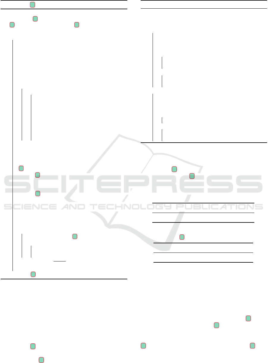
3.2 Attention Feature Heatmaps
As explained in Section 2.1, one of the main goals of
this work is to design a cancer-type detection system
one that is explainability oriented. To this aim, in the
structure of S has been integrated an attention layer
used to produce special feature heatmaps which can
help to visualize which parts of the input are more
important for the detection. Figure 2 shows the fea-
ture heatmaps generated using the attention layer of
S
5
. We remark that to facilitate the viewing and in-
terpretation of the heatmaps, we have we have super-
imposed special dotted rectangles whose color is that
indicated by the heatmap and the size is proportional
to the intensity of the highlighted areas.
As we can see, for each cancer-type class C
i
, the
corresponding heatmap has size 750 ×|C
i
| where 750
is the size of the input vector of the attention layer,
and |C
i
| indicates the number of instances of C
i
. The
most evident aspect that emerges from the visualiza-
tion of the heatmaps is that each class activates a spe-
cific set of features of the vector given in input to
the attention layer. This allows to identify a sort of
5
https://github.com/FLaTNNBio/few-shot-learning-f
or-cancer-detection/tree/master
visual pattern extracted from the cancer mutational
views given in input. However, it is important to un-
derline that, in this preliminary version of the work,
this explainability component still needs a lot of work
so that it can be profitably used for analysis. What is
missing at the moment is a correspondence between
the areas highlighted in the heatmaps and the corre-
sponding features in the view which in fact determine
the activation of the various areas.
In the same way, however, it is important to under-
line how the production of visual information to sup-
port the analysis of this type of problem, as well as
orienting the system towards the question of explain-
ability, makes it open to the possibility of integrating
Information Visualization (IV) techniques. IV tech-
niques consist in computerized methods that involve
selecting, transforming and representing data in a vi-
sual form that facilitates human interaction for ana-
lyzing and understanding the data (Tao et al., 2004).
IV techniques have been used in many areas of Bioin-
formatics. Although they have been successfully used
in many biological domains, such as structure visual-
ization, expression profile analysis, sequence analy-
sis, visualization of genome, pathway and hierarchi-
cal data, in our opinion the study of the specific pat-
terns of somatic mutations in the major cancer types
is still challenging. We believe that a system such as
the one proposed in this paper, i.e., oriented towards
an explainable and usable approach, although still in-
complete and in a preliminary form, can provide inter-
esting starting points for future work in this direction.
4 DISCUSSION AND
CONCLUSION
Although the obtained results are interesting, there are
some obvious limitations that need to be addressed.
The proposed method is a preliminary attempt
to simultaneously satisfy explainability and usabil-
ity needs when applying AI techniques in Bioinfor-
matics. In our opinion, the potential in the use of
feature maps, on which however to date there is in-
sufficient evidence to demonstrate their effectiveness
in terms of explainability, is amplified by the use of
SNNs whose advantage in terms of usability is evi-
dent. However, we plan to use explainability tech-
niques that can return a heatmap with respect to the
input sequence, which is easier to interpret.
We used the term “view” and not “signature” as
the latter was already introduced in the literature, and
there are different methods to calculate them. We can-
not consider what we obtained as a real “signature”
since on the downloaded data set we only considered
BIOINFORMATICS 2024 - 15th International Conference on Bioinformatics Models, Methods and Algorithms
468
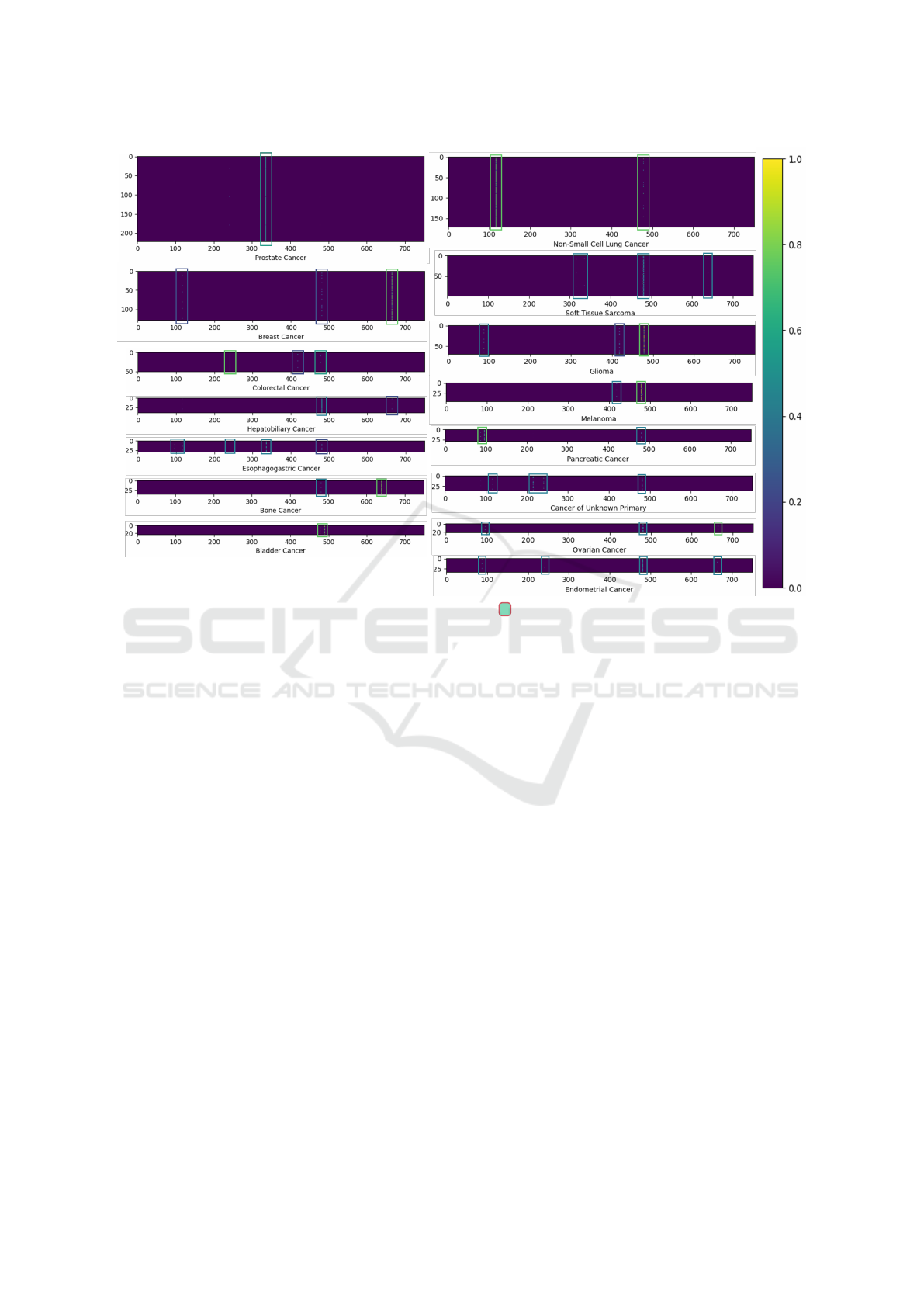
Figure 2: Attention feature heatmaps generated by S for each of the cancer-type classes.
molecular features (of which 13 variables with clini-
cal information) and performed PCA.
Further investigations will be carried out with the
aim to collect larger datasets to evaluate the perfor-
mance of the model in a wider range of contexts.
REFERENCES
Bechar, M. E. A., Guyader, J.-M., El Bouz, M., Douet-
Guilbert, N., Al Falou, A., and Troadec, M.-B. (2023).
Highly performing automatic detection of structural
chromosomal abnormalities using siamese architec-
ture. Journal of Molecular Biology, 435(8):168045.
Bell, S. and Bala, K. (2015). Learning visual similarity for
product design with convolutional neural networks.
ACM transactions on graphics (TOG), 34(4):1–10.
Chen, Y., Garcia, E. K., Gupta, M. R., Rahimi, A., and Caz-
zanti, L. (2009). Similarity-based classification: Con-
cepts and algorithms. Journal of Machine Learning
Research, 10(3).
Ciriello, G., Miller, M. L., Aksoy, B. A., Senbabaoglu, Y.,
Schultz, N., and Sander, C. (2013). Emerging land-
scape of oncogenic signatures across human cancers.
Nature genetics, 45(10):1127–1133.
Jiao, W., Atwal, G., Polak, P., Karlic, R., Cuppen, E.,
Danyi, A., De Ridder, J., van Herpen, C., Lolkema,
M. P., et al. (2020). A deep learning system accu-
rately classifies primary and metastatic cancers using
passenger mutation patterns. Nature communications,
11(1):728.
Kandoth, C., McLellan, M. D., Vandin, F., Ye, K., Niu, B.,
Lu, C., Xie, M., Zhang, Q., McMichael, J. F., Wycza-
lkowski, M. A., et al. (2013). Mutational landscape
and significance across 12 major cancer types. Na-
ture, 502(7471):333–339.
Karim, M. R., Beyan, O., Zappa, A., Costa, I. G., Rebholz-
Schuhmann, D., Cochez, M., and Decker, S. (2021).
Deep learning-based clustering approaches for bioin-
formatics. Briefings in bioinformatics, 22(1):393–
415.
Kübler, K., Karli
´
c, R., Haradhvala, N. J., Ha, K., Kim, J.,
Kuzman, M., Jiao, W., Gakkhar, S., Mouw, K. W.,
Braunstein, L. Z., et al. (2019). Tumor mutational
landscape is a record of the pre-malignant state.
BioRxiv, page 517565.
Lawrence, M. S., Stojanov, P., Polak, P., Kryukov, G. V.,
Cibulskis, K., Sivachenko, A., Carter, S. L., Stewart,
C., Mermel, C. H., Roberts, S. A., et al. (2013). Muta-
tional heterogeneity in cancer and the search for new
cancer-associated genes. Nature, 499(7457):214–218.
Mathai, N. and Kirchmair, J. (2020). Similarity-based
methods and machine learning approaches for target
prediction in early drug discovery: performance and
scope. International Journal of Molecular Sciences,
21(10):3585.
Narmatha, P., Gupta, S., Lakshmi, T. V., and Manikavelan,
D. (2023). Skin cancer detection from dermoscopic
images using deep siamese domain adaptation convo-
Visual Insights in Human Cancer Mutational Patterns: Similarity-Based Cancer Classification Using Siamese Networks
469

lutional neural network optimized with honey badger
algorithm. Biomedical Signal Processing and Control,
86:105264.
Tan, P.-N., Steinbach, M., and Kumar, V. (2016). Introduc-
tion to data mining. Pearson Education India.
Tao, Y., Liu, Y., Friedman, C., and Lussier, Y. A. (2004). In-
formation visualization techniques in bioinformatics
during the postgenomic era. Drug Discovery Today:
BIOSILICO, 2(6):237–245.
BIOINFORMATICS 2024 - 15th International Conference on Bioinformatics Models, Methods and Algorithms
470
