
Can Electromyography Alone Reveal Facial Action Units? A Pilot
EMG-Based Action Unit Recognition Study with Real-Time Validation
Abhinav Veldanda
a
, Hui Liu
b
, Rainer Koschke
c
, Tanja Schultz
d
and Dennis K
¨
uster
e
Cognitive Systems Lab, University of Bremen, Germany
Keywords:
Action Units, Electromyography, Facial Action Coding System, EMG, sEMG, fEMG, Pattern Recognition,
Machine Learning.
Abstract:
Facial expressions play a crucial role in non-verbal and visual communication, often observed in everyday
life. The facial action coding system (FACS) is a prominent framework for categorizing facial expressions as
action units (AUs), which reflect the activity of facial muscles. This paper presents a proof-of-concept study
for upper face action unit recognition (AUR) using electromyography (EMG) data. The study recorded facial
EMG data of a subject over four sessions, who imitated facial expressions corresponding to four different
AUs. The subject-dependent models that were trained achieved high accuracy in near-real time and were able
to classify AUs not directly underneath the recording sites.
1 INTRODUCTION
A large part of human communication is believed to
be nonverbal and visual in nature, with facial expres-
sions playing a key role (Kappas et al., 2013). We
may notice this in everyday life, when we cannot see
someone’s face (e.g., on the phone), or when facial
expressions are partially obscured – for example, due
to a face mask (Giovanelli et al., 2021), or when we
interact with someone wearing a virtual reality (VR)
headset (Oh Kruzic et al., 2020).
Considerable work has been done on facial ex-
pression analysis since the early 1970s. Perhaps most
prominent among these is the Facial Action Coding
System (FACS), which was developed by Paul Ek-
man and Wallace Friesen (Ekman et al., 2002), and
based on prior work by (Hjortsj
¨
o, 1969), a Swedish
anatomist who had catalogued the facial configura-
tions (Barrett et al., 2019) depicted by Duchenne
(Duchenne and Cuthbertson, 1990). FACS provides
a framework for categorizing all possible facial ex-
pressions into constituent action units (AUs), which
reflect the activity of facial muscles that can be con-
a
https://orcid.org/0009-0007-3749-4971
b
https://orcid.org/0000-0002-6850-9570
c
https://orcid.org/0000-0003-4094-3444
d
https://orcid.org/0000-0002-9809-7028
e
https://orcid.org/0000-0001-8992-5648
trolled independently. In contrast to discrete or “basic
emotions” (Ekman, 1999), AUs are purely descriptive
for movements of certain muscles, and do not pro-
vide any inferential labels (Zhi et al., 2020). There-
fore, accurate tracking of AUs provides an objective
basis for behavioral research into facial emotional ex-
pressions, as well as for 3D-modelling of emotions
(van der Struijk et al., 2018). In total, the FACS (Ek-
man et al., 2002) provides coding instructions for 44
AUs.
1.1 Automatic Action Unit Recogntion
Action unit recognition (AUR) is an important re-
search direction within facial expression analysis,
which aims to automatically identify the activation
of AUs that correspond to specific emotions, expres-
sions, and actions. This approach analyzes the dy-
namics of subtle changes in the face, such as wrin-
kling of the nose, raising of the eyebrows, or lip cor-
ner pulling. For decades, AUR had to rely exclu-
sively on costly and time-consuming manual recog-
nition by certified FACS experts, with a ratio of more
than one hour to manually label one minute of video
data (Bartlett et al., 2006; Zhi et al., 2020). Today,
automatic affect recognition tools allow for a much
more cost-effective consideration of facial activity in
most experimental research paradigms (K
¨
uster et al.,
2020). From early classifiers, such as the Computer
142
Veldanda, A., Liu, H., Koschke, R., Schultz, T. and Küster, D.
Can Electromyography Alone Reveal Facial Action Units? A Pilot EMG-Based Action Unit Recognition Study with Real-Time Validation.
DOI: 10.5220/0012399100003657
Paper published under CC license (CC BY-NC-ND 4.0)
In Proceedings of the 17th International Joint Conference on Biomedical Engineering Systems and Technologies (BIOSTEC 2024) - Volume 1, pages 142-151
ISBN: 978-989-758-688-0; ISSN: 2184-4305
Proceedings Copyright © 2024 by SCITEPRESS – Science and Technology Publications, Lda.

Expression Recognition Toolbox (Littlewort et al.,
2011)) to current open-source tools, e.g., OpenFace
(Baltrusaitis et al., 2018), researchers can now rely on
a wide range of out-of-the-box software for camera-
based automatic affect recognition. Due to the pop-
ularity of basic emotion theories (BETs) (Ortony,
2022), many of these tools have traditionally aimed
to distinguish prototypical patterns of expressions be-
lieved to reflect discrete emotional states such as hap-
piness, anger, or sadness (Dupr
´
e et al., 2020). More
recently, reliable assessment and validation of facial
AUs has been gaining more attention because they can
be measured objectively without requiring the lens of
BET (K
¨
uster et al., 2020).
Although some work has previously tested their
own database without comparative evaluations be-
tween different platforms (Krumhuber et al., 2021),
machine learning (ML) models for camera-based
AUR have been the focus of a number of recent
challenges in facial expression recognition and anal-
ysis (Zhi et al., 2020). Additionally, a few works
have studied the performance of freely available pre-
trained AUR systems such as OpenFace (Namba
et al., 2021a; Namba et al., 2021b; Lewinski et al.,
2014). Overall, these works have demonstrated
the usefulness and reliability of camera-based AUR.
However, there still remain methodological and con-
ceptual challenges, including the nearly exclusive re-
liance of facial AUR on visual data. Here, EMG re-
search and other recent approaches such as the use
of inertial measurement units (IMUs) (Verma et al.,
2021) may contribute towards improving the conver-
gent validity of ML-based AUR.
1.2 Methodological Challenges
Perhaps unsurprisingly, camera-based AUR perfor-
mance still varies depending on factors such as the
specific AU (Namba et al., 2021a), viewing angle
(Namba et al., 2021b), and database (Zhi et al., 2020)
in question. Cross-database evaluations and chal-
lenges for discrete and AU-based affect recognition
have also generally been based on a limited number
of well-known databases of mostly posed expressions
(K
¨
uster et al., 2020; Zhi et al., 2020). Compared
to well-controlled posed datasets, spontaneous facial
expressions in the wild are likely to be more subtle
(Zhi et al., 2020) and involve more complex dynam-
ics (Krumhuber et al., 2023), as well as other cues
such as head movements (e.g., nodding) (Zhi et al.,
2020). Spontaneous facial behavior also includes the
possibility of co-occurring AUs, e.g., smiling with the
eyes and the mouth, which can potentially yield thou-
sands of distinct classes (Zhi et al., 2020). Together,
these considerations raise the question of how well
the said classifiers will perform for completely new
and less standardized data. Finally, including a cam-
era may sometimes interfere with the phenomenon to
be measured. For example, the feeling of being ob-
served has been shown to eliminate facial feedback
phenomena that were once believed to be robust and
well-established (Noah et al., 2018). These factors
still pose significant challenges to the vibrant field of
camera-based AUR.
1.3 Conceptual Challenges
Conceptually, facial expression research still faces
substantial challenges relating to a lack of cohesion
between measures of emotion (Kappas et al., 2013),
as well as the interpretation of AUs as part of their
physical and social context (Kuester and Kappas,
2013).
As demonstrated by earlier reviews, agreement
between physiological measures of emotion and sub-
jective self-report has often been surprisingly low
(Mauss and Robinson, 2009). Furthermore, while
theories of emotion have generally assumed biosig-
nals, cognitive, and behavioral components of emo-
tions to be synchronized and/or coordinated, empiri-
cal data has repeatedly challenged notions of strong
concordance (Hollenstein and Lanteigne, 2014).
Here, novel approaches in ML combining different
modalities may yield more stable predictions than
previous psychological models, as well as eventu-
ally provide some further insights into the ways in
which the different components of the emotional re-
sponse may synchronize and relate to each other.
In consequence, leveraging easily obtainable data,
such as jointly recorded audio-visual emotional re-
sponses together has been a core aim of a series of
multimodal emotion recognition challenges for over
a decade (Schuller et al., 2012). More recently,
such approaches have proven to be fruitful across a
wide range of subject areas, e.g., recognition of emo-
tional engagement of people suffering from dementia
(Steinert et al., 2021). Perhaps surprisingly, however,
fEMG has thus far rarely been included in such ap-
proaches.
Apart from the practical challenges of recording
fEMG as a high-quality and high-resolution signal of
facial activity, a second major conceptual challenge
relates to the interpretation of facial muscle activ-
ity beyond a FACS-based categorization. Here, an
increasing number of works have demonstrated that
notions such as Ekman’s “basic emotions” (Ekman,
1999) may no longer be tenable (Ortony, 2022; Criv-
elli and Fridlund, 2018; Crivelli and Fridlund, 2019).
Can Electromyography Alone Reveal Facial Action Units? A Pilot EMG-Based Action Unit Recognition Study with Real-Time Validation
143

However, while we are aware of this ongoing debate,
the present work is focused on a methodological con-
tribution. I.e., by demonstrating the possibility of
a reliable automatic recognition of facial AUs from
EMG, we aim to help pave the way towards provid-
ing a more sensitive and high-resolution measure of
facial activity compared to the now commonly used
webcam data.
1.4 Facial Electromyography for
Automatic Action Unit Recognition
In this paper, we solely focus on the use of fa-
cial electromyography (fEMG) as our basis for AUR.
While most research on facial expressions today has
been conducted on the basis of video data or mainly
video supplemented by electromyography (EMG)
data. Nevertheless, the use of fEMG has been the
true gold standard for the high-precision recording of
facial expressions in the psychophysiological labora-
tory for decades (Fridlund and Cacioppo, 1986; Win-
genbach, 2023). In particular, facial surface EMG is
capable of detecting very subtle muscle activity, in-
cluding muscle relaxation (e.g., of the eyebrows), be-
low what would be observable with the naked eye
(Kappas et al., 2013; Larsen et al., 2003). Thirdly,
some previous studies further indicate that, beyond re-
liable detection of emotional facial expressions, these
may be leveraged to substantially improve human-
computer interaction (Gibert et al., 2009; Schultz,
2010). Last but not least, many up-to-date in-house
and external research works have confirmed the prac-
ticality, convenience, and effectiveness of EMG in
different areas of the human body and physiological
exploration (Liu et al., 2023; Cai et al., 2023; Hart-
mann et al., 2023; Liu and Schultz, 2022).
Within the scope of this paper, we use our in-
house recorded dataset of fEMG sensor data to pre-
dict a subset of AUs. To the best of our knowledge,
no work has been published yet in AUR relying only
on EMG data as its source.
2 METHODOLOGY
The proposed framework is based on a pilot dataset,
which contains synchronised video modality data
with fEMG recordings and output labels correspond-
ing to appropriate AUs. Multiple widely-applied ML
models with default hyperparameters will be trained
using the acquired data and subsequently, classifica-
tion metrics will be calculated for the same. The best-
performing model will be chosen for further analysis.
2.1 Dataset Preparation
To construct a dataset, we recorded a proof-of-
concept fEMG dataset of one subject across four ses-
sions. Each session comprised of 25 recording tri-
als, yielding a total of 100 trials. We have on average
2.12 ± 0.8 minutes of data per trial across all the ses-
sions. Within each recording trial, the subject was
asked to imitate facial expressions shown to them in
the stimulus videos through a custom-made graphical
user interface (GUI) 1(see Figure 1). These stimu-
lus videos were taken from the MPI Video Database
(Kleiner et al., 2004). These videos provide accurate
portrayals of AU activation, which have been verified
by FACs coders.
The subject was shown stimulus videos pertaining
to four different AUs (see Table 1 and asked to imi-
tate the AUs at maximum intensity and hold them for
at least five seconds while fEMG data was being par-
allely recorded. In addition to the different AUs, the
fEMG data corresponding to the neutral expression
was also recorded, which we shall refer to as AU0 for
representation purposes in the rest of the paper.
The recording setup consisted of a computer dis-
playing the stimuli, a webcam, and an fEMG record-
ing setup. The subject was seated in front of the dis-
play screen approximately 70 cm away. The fEMG
setup was a bipolar recording setup consisting of 2
channels covering the Frontalis and Corrugator Su-
percili facial muscles. These positions are defined
by the guidelines of the Society for psychophysio-
logical research (Fridlund and Cacioppo, 1986), with
slight deviations from the standard sensor positions
(see Figure 2), based on extensive pre-testing to min-
imize the amount of crosstalk, and to account for the
slightly larger size of our electrodes compared to the
original guideline paper. These deviations were based
on intensive pre-testing to optimize the quality of the
recording. It is known that fEMG signals occur in the
range of 15–500Hz (Boxtel, 2001), so the sampling
frequency was chosen to be 2000 Hz as required by
the limitations imposed by the Nquist theorem, which
states that a periodic signal must be sampled at more
than twice the highest frequency component of the
signal. We employed the Biosignal Plux
1
sensors and
hub as our acquisition system because its high-quality
EMG acquisition was confirmed by many preliminary
in-house works (Liu and Schultz, 2018; Hartmann
et al., 2022; Liu et al., 2021a).
The synchronization between the imitated actions
and the EMG sensor data is handled using the lab
streaming layer (LSL) protocol. Along with the EMG
data, we also record the video data of the participant
1
www.pluxbiosignals.com
BIODEVICES 2024 - 17th International Conference on Biomedical Electronics and Devices
144

using the standard inbuilt webcam within the host
computer. The timestamps associated with the video
stream for the imitated actions are used to extract the
relevant EMG signals and store them in a usable for-
mat for further processing.
Figure 1: Graphical User Interface (GUI) implemented for
recording trials.
Figure 2: Sensor Placement.
Table 1: Selected actions units for proof-of-concept study.
Action Unit Action
AU1 Inner Brow Raiser
AU2 Outer Brow Raiser
AU4 Brow Lowerer
AU9 Nose Wrinkler
AU0 Neutral Expression
2.2 Pre-Processing and Feature
Extraction
Raw EMG data typically includes a substantial
amount of electrical noise, which should be removed
before amplification (Tassinary et al., 2007). Tradi-
tionally, remaining noise (e.g., 50/60 Hz noise) and
artifacts are then removed via filtering prior to any sta-
tistical analyses (Fridlund and Cacioppo, 1986; Tassi-
nary et al., 2007). Within the field of biosignals-based
ML, however, this latter type of noise may be bet-
ter accounted for by the ML algorithm than by a fil-
ter, which might filter out some relevant data along
with the noise. Therefore, some recent works in this
field have suggested running their feature extractors
directly on the raw fEMG (Liu and Schultz, 2019; Ro-
drigues et al., 2022; Liu, 2021), which we adopted in
our work. The raw EMG data was first segmented
into windows of a specified length with a pre-defined
overlap percentage.
Training ML models require the proper set of fea-
tures to be fed into the model. Considering our model
orients a real-time application for interaction and con-
trol in the future, it is essential to consider the features
that can be computed quickly. Therefore, following
the windowing of signals, 16 temporal features were
extracted using a time-series feature extraction library
(TSFEL) (Barandas et al., 2020):
• Absolute energy
• Area under the curve (AUC)
• Autocorrelation
• Centroid
• Entropy
• Mean absolute difference (MAD)
• Median difference
• Negative turning points (NT)
• Neighbourhood peaks
• Peak to peak distance
• Positive turning points
• Signal distance
• Slope
• Sum absolute difference (SAD)
• Total energy
• Zero crossing rate
Most of the features listed above are frequently
used in time-series ML, like AUC, MAD, and SAD;
some features exist almost uniquely in TSFEL, such
as NT, whose effectiveness and low computational
Can Electromyography Alone Reveal Facial Action Units? A Pilot EMG-Based Action Unit Recognition Study with Real-Time Validation
145

cost have been validated in previous research (Liu
et al., 2022). The feature extraction was followed by
linear discriminant analysis (LDA) for dimensional-
ity reduction (Hartmann et al., 2020; Liu et al., 2021b;
Hartmann et al., 2021). Such an operation reduced the
feature set and wad used for the training of different
models.
2.3 Classification Models
We applied six widely-used ML and deep learning
classification models to our pilot dataset.
1. Random forest RF
2. Support vector machine SVM
3. Gaussian naive Bayes GNB
4. K nearest neighbors k-NN
5. Artificial neural networks (ANN, deep)
6. Temporal convolutional networks (TCN, deep)
Five of these models were trained on features ex-
tracted using TSFEL library while the TCN model
used the windowed data as its input. All non-deep
models were trained on default hyperparameters pro-
vided by the scikit learn library (Pedregosa et al.,
2011). The ANN model was designed as a vanilla
seven layer network with leaky rectified linear unit
(Leaky ReLU) and dropout layers stacked in between
and ending with a softmax layer at the end. The TCN
network was designed to takeinput as samples of de-
fined windowed length. The output of the TCN mod-
ule is of the same length as the input. This output is
flattened out and fed to a single neural network with
the number of output nodes same as the number of
categories, followed by a softmax activation for pre-
diction.
3 RESULTS
3.1 Cross-Validation Results
We conducted cross-validation studies using three of
the acquired data sessions as our training set. The
training data was first segmented into windows of 400
ms in length and 20% overlap, followed by tempo-
ral feature extraction and LDA. Subesequently, a five-
fold cross-validation was performed using stratified
sampling, and the mean accuracy results are show-
cased in Table 2.
Table 2: Accuracies for the results accumulated from five-
fold cross-validation using combinations of different ses-
sions of data.
Session(s): 1 1 and 2 1,2, and 3
RF 0.99 0.99 0.99
SVM 0.95 0.95 0.96
GNB 0.99 0.98 0.97
KNN 0.98 0.98 0.98
ANN 0.27 0.39 0.35
TCN 0.77 0.80 0.76
3.2 Leave-One-Channel-Out Testing
Considering the high performance of the models
demonstrated in Table 2. We wanted to test how our
models would perform when they were only trained
on individual data channels. Cross-validation studies
were done on the same training data but only using in-
dividual channels from our dataset. We analysed how
changing the number of channels used for training the
models would impact the performance. Table 3 indi-
cates the results for the performances of the models
on independent channels. Noticeably, there is an ex-
pected drop in the performance of the models but not
much of a clear discernible pattern in terms of one
channel performing better than the other. For exam-
ple RF seems to be better trained with Frontalis data
while SVM works better with the Corrugator data.
TCN has the most significant drop in performance,
suggesting that it may require a combination of fea-
tures from multiple channels to recognize classes.
Table 3: Accuracies for the results accumulated from five-
fold cross-validation using single channel data based on
random forest.
Frontalis Corrugator
RF 0.97 0.91
SVM 0.75 0.83
GNB 0.88 0.84
KNN 0.89 0.89
TCN 0.28 0.37
3.3 Performance of Test Set
Although validation scores present one side of the
argument on how the model performs on a similar
distribution of data on which it was performed. We
wanted to know how the model performs on an en-
tirely unseen dataset, i.e session-independent data.
We trained different models and calculated the accu-
racy metrics on the unseen test data set that we had
kept apart from the start. The results are displayed in
Figure 3. The trained models provide excellent test
BIODEVICES 2024 - 17th International Conference on Biomedical Electronics and Devices
146
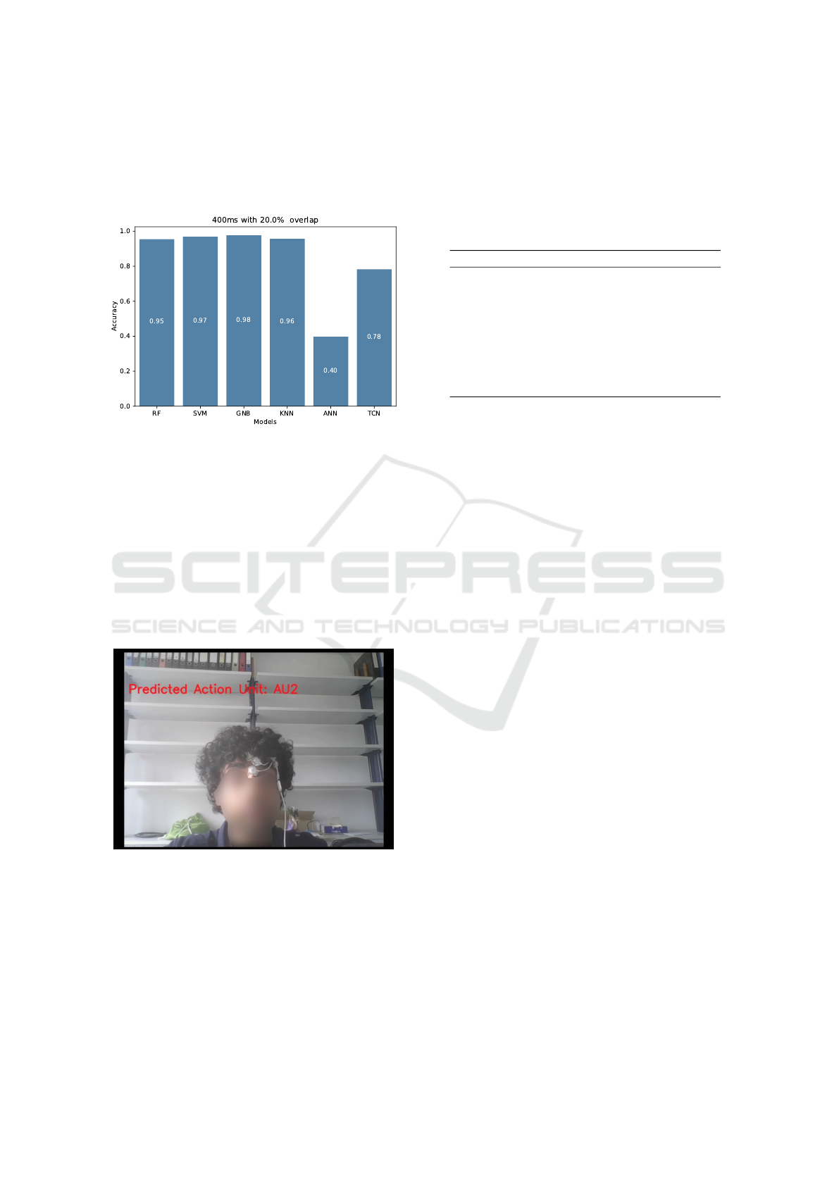
set scores. Based on the accuracy graphs obtained,
it was apparent that all models except for ANN were
showing promising results. We continue to see how
the best-performing model performs in real time.
Figure 3: Accuracies on unseen test set.
3.4 Real-Time Analysis
RF was chosen as the base model for further analy-
sis, as it outperformed the other models in our offline
investigation. Satisfactorily, RF is up to the task in
real time (see Figure 4) as it was able to recognize the
participant’s facial AU’s in near-real time with a de-
cent rate of correctness, which supports our hypoth-
esis that EMG biosignals can be used to accurately
predict AUs.
Figure 4: Real-time action unit recognition.
3.5 Joint Study of Window Length and
Overlap Percentage
One helpful comparison to showcase is how the
model performs when we change the window length
and the overlap percentage value. We again chose RF
as our basis and applied window lengths varying from
150ms to 500ms in increments of 50ms. The overlap
percentages values varied from 10% to 50% in incre-
ments of 10% (see Table 4).
Table 4: Mean accuracy results with five-fold cross-
validation for jointly studying window length and overlap
percentage.
10% 20% 30% 40% 50%
150 ms 0.97 0.97 0.97 0.97 0.97
200 ms 0.97 0.98 0.98 0.98 0.98
250 ms 0.98 0.98 0.98 0.98 0.98
300 ms 0.98 0.98 0.98 0.98 0.98
350 ms 0.98 0.98 0.98 0.98 0.99
400 ms 0.99 0.98 0.98 0.98 0.98
450 ms 0.98 0.98 0.98 0.98 0.98
500 ms 0.98 0.98 0.98 0.98 0.98
4 DISCUSSION
To the best of our knowledge, this paper is the first
to provide a proof-of-concept for upper face AU
recognition based solely on EMG data. The subject-
dependent model achieved high accuracy in near-real
time. Furthermore, we showed that our models could
also classify AUs that were not directly underneath
the respective recording sites, suggesting that future
systems may achieve adequate results from conve-
niently placed electrodes that are even more distal
from the respective facial muscles.
4.1 Evaluation and Analysis
We focused on recording EMG data from two mus-
cle sites, Frontalis and Corrugator Supercili, to cap-
ture the activity of the eyebrows and distinguish be-
tween four different AUs (AU1, AU2, AU4, AU9).
While we did not record above the levator labii (Nose
Wrinkler), we assumed that sufficient signals could
still be detected from these nearby sites also to allow
a reliable classification of AU9. As demonstrated by
our results, this classification was successful and ro-
bust, even when using only single channel data. Thus,
while previous work has pointed out the often prob-
lematic effects of crosstalk phenomena of other mus-
cles (Van Boxtel and Jessurun, 1993; van Boxtel et al.,
1998), our models appear to have been able to suc-
cessfully leverage these data to recognize the intended
AUs.
As demonstrated by our validation results, all non-
deep learning models obtained good results. As TCN
is a deep learning-based model, we did not expect
it to perform well given the relatively small amount
Can Electromyography Alone Reveal Facial Action Units? A Pilot EMG-Based Action Unit Recognition Study with Real-Time Validation
147

of data. As suggested by our training runs, the TCN
model was still unstable. Nevertheless, its results ap-
peared promising, with performance at a level simi-
lar to that of an ANN. Future work could therefore
investigate whether TCN-based models may achieve
even better and more stable results once larger EMG-
datasets become available. Contrarily, ANNs did not
provide sufficiently strong results to be considered a
good candidate compared to the other models. We
hypothesize that this could be attributed to two fac-
tors. First, the vanilla neural networks utilized for our
model training may require more extensive data. Sec-
ond, ANNs may have performed worse than TCNs
because they failed to construct adequate internal fea-
ture maps.
4.2 Comparison with State-of-the-Art
Work
To the best of our knowledge, very few prior works
have aimed to leverage EMG data for action unit
recognition. (Perusquia-Hernandez et al., 2021) re-
lied on the fusion of computer vision data along with
EMG to train their models, while (Gibert et al., 2009)
used EMG data only to predict prototypical facial ex-
pressions. Similarly, (Gruebler and Suzuki, 2014)
developed a wearable device to detect smiling and
frowning based on two electrode pairs. Some works
have relied on independent video data (Baltrusaitis
et al., 2018) or even using other modalities such as
electroencephalography (Li et al., 2020). Finally a re-
cent approach has utilised earbud IMUs (Verma et al.,
2021) and TCNs to detect and classify a large range
of AUs. While this approach has obtained promising
results in a subject-dependent setting, ear-mounted
IMU sensors may be at a disadvantage with respect
to detecting more subtle naturalistic facial activity,
the presence of movement artefacts (e.g., head move-
ments) or interference due to the sound waves pro-
duced by the earbud when it is in use. Thus, the sound
waves may themselves excite the IMUs as well as the
earable device (Verma et al., 2021).
5 CONCLUSION
We propose a novel approach to upper-face AUR us-
ing EMG data, as demonstrated by the successful
training of subject-dependent models in this initial
case study. Our results furthermore show potential for
classifying new AUs based on more distally placed
electrodes in future applications, e.g., in VR. These
results also suggest that deep learning models such
as TCN can be considered for further research in this
domain, while highlighting the limitations of using
fewer channels. Overall, this work contributes to the
emerging field of EMG-based AUR recognition and
paves the way for future research.
We believe that this approach could be comple-
mentary to the development of IMU-based earable de-
vices, as they are subject to different sources of noise
and environmental as well as practical constraints.
Thus, while EMG sensors require direct contact with
the skin, they are likely to be more robust towards
artefacts due to head movements or the sound waves
produced by the earable itself. Conversely, earbuds
are likely to be less susceptible to electrical noise
from other devices, whereas EMG should outperform
other sensor types for detecting subtle expressions.
Considering the challenges of multimodal emotion
recognition in the wild (K
¨
uster et al., 2020), we there-
fore envision a joint system comprising of both IMUs
and a few EMG sensors to be able to provide the most
robust and precise AUR performance. However, more
work is still required to develop robust and versatile
AUR from fEMG.
Despite the successful AU recognition in this pi-
lot, our present models were still limited to training
on subject-dependent data near the traditional record-
ing sites for the respective action units in the up-
per face. As demonstrated by prior work, control
of human facial muscles is complex (Cattaneo and
Pavesi, 2014) and subject to significant anatomical
differences (D’Andrea and Barbaix, 2006) as well
as variability in signal power across muscle sites
(Schultz et al., 2019). To address these challenges, we
plan to build subject-independent models to examine
whether the underlying muscle activity patterns are
sufficiently reliable. In our future work, we therefore
aim to to extend our EMG-based AUR approach also
to lower face AUs, while further examining the via-
bility of a distal electrode placement in multi-subject
studies.
REFERENCES
Baltrusaitis, T., Zadeh, A., Lim, Y. C., and Morency, L.-
P. (2018). OpenFace 2.0: Facial Behavior Analysis
Toolkit. In 2018 13th IEEE International Conference
on Automatic Face & Gesture Recognition (FG 2018),
pages 59–66, Xi’an. IEEE.
Barandas, M., Folgado, D., Fernandes, L., Santos, S.,
Abreu, M., Bota, P., Liu, H., Schultz, T., and Gamboa,
H. (2020). TSFEL: Time Series Feature Extraction
Library. SoftwareX, 11:100456.
Barrett, L. F., Adolphs, R., Marsella, S., Martinez, A. M.,
and Pollak, S. D. (2019). Emotional Expressions Re-
considered: Challenges to Inferring Emotion From
BIODEVICES 2024 - 17th International Conference on Biomedical Electronics and Devices
148

Human Facial Movements. Psychological Science in
the Public Interest: A Journal of the American Psy-
chological Society, 20(1):1–68.
Bartlett, M. S., Littlewort, G. C., Frank, M. G., Lainscsek,
C., Fasel, I. R., and Movellan, J. R. (2006). Auto-
matic Recognition of Facial Actions in Spontaneous
Expressions. Journal of Multimedia, 1(6):22–35.
Boxtel, A. (2001). Optimal signal bandwidth for the record-
ing of surface EMG activity of facial, jaw, oral, and
neck muscles. Psychophysiology, 38(1):22–34.
Cai, L., Yan, S., Ouyang, C., Zhang, T., Zhu, J., Chen,
L., Ma, X., and Liu, H. (2023). Muscle synergies
in joystick manipulation. Frontiers in Physiology,
14:1282295.
Cattaneo, L. and Pavesi, G. (2014). The facial motor sys-
tem. Neuroscience & Biobehavioral Reviews, 38:135–
159.
Crivelli, C. and Fridlund, A. J. (2018). Facial Displays Are
Tools for Social Influence. Trends in Cognitive Sci-
ences, 22(5):388–399. Publisher: Elsevier.
Crivelli, C. and Fridlund, A. J. (2019). Inside-Out: From
Basic Emotions Theory to the Behavioral Ecology
View. Journal of Nonverbal Behavior, 43(2):161–194.
Duchenne, G.-B. and Cuthbertson, R. A. (1990). The mech-
anism of human facial expression. Studies in emotion
and social interaction. Cambridge University Press
; Editions de la Maison des Sciences de l’Homme,
Cambridge [England] ; New York : Paris.
Dupr
´
e, D., Krumhuber, E. G., K
¨
uster, D., and McKeown,
G. J. (2020). A performance comparison of eight com-
mercially available automatic classifiers for facial af-
fect recognition. PLOS ONE, 15(4):e0231968. Pub-
lisher: Public Library of Science.
D’Andrea, E. and Barbaix, E. (2006). Anatomic research
on the perioral muscles, functional matrix of the max-
illary and mandibular bones. Surgical and Radiologic
Anatomy, 28(3):261–266.
Ekman, P. (1999). Basic emotions. In Handbook of cog-
nition and emotion, pages 45–60. John Wiley & Sons
Ltd, Hoboken, NJ, US.
Ekman, P., Friesen, W. V., and Hager, J. C. (2002). Facial
action coding system: the manual. Research Nexus,
Salt Lake City, Utah.
Fridlund, A. J. and Cacioppo, J. T. (1986). Guidelines for
Human Electromyographic Research. Psychophysiol-
ogy, 23(5):567–589.
Gibert, G., Pruzinec, M., Schultz, T., and Stevens, C.
(2009). Enhancement of human computer interaction
with facial electromyographic sensors. In Proceed-
ings of the 21st Annual Conference of the Australian
Computer-Human Interaction Special Interest Group:
Design: Open 24/7, pages 421–424, Melbourne Aus-
tralia. ACM.
Giovanelli, E., Valzolgher, C., Gessa, E., Todes-
chini, M., and Pavani, F. (2021). Unmasking
the difficulty of listening to talkers with masks:
lessons from the covid-19 pandemic. i-Perception,
12(2):2041669521998393.
Gruebler, A. and Suzuki, K. (2014). Design of a Wear-
able Device for Reading Positive Expressions from
Facial EMG Signals. IEEE Transactions on Affective
Computing, 5(3):227–237. Conference Name: IEEE
Transactions on Affective Computing.
Hartmann, Y., Liu, H., Lahrberg, S., and Schultz, T. (2022).
Interpretable high-level features for human activity
recognition. In Proceedings of the 15th Interna-
tional Joint Conference on Biomedical Engineering
Systems and Technologies (BIOSTEC 2022) - Volume
4: BIOSIGNALS, pages 40–49.
Hartmann, Y., Liu, H., and Schultz, T. (2020). Feature space
reduction for multimodal human activity recognition.
In Proceedings of the 13th International Joint Confer-
ence on Biomedical Engineering Systems and Tech-
nologies (BIOSTEC 2020) - Volume 4: BIOSIGNALS,
pages 135–140.
Hartmann, Y., Liu, H., and Schultz, T. (2021). Feature space
reduction for human activity recognition based on
multi-channel biosignals. In Proceedings of the 14th
International Joint Conference on Biomedical Engi-
neering Systems and Technologies (BIOSTEC 2021),
pages 215–222. INSTICC, SCITEPRESS - Science
and Technology Publications.
Hartmann, Y., Liu, H., and Schultz, T. (2023). High-level
features for human activity recognition and model-
ing. In Roque, A. C. A., Gracanin, D., Lorenz, R.,
Tsanas, A., Bier, N., Fred, A., and Gamboa, H., edi-
tors, Biomedical Engineering Systems and Technolo-
gies, pages 141–163, Cham. Springer Nature Switzer-
land.
Hjortsj
¨
o, C.-H. (1969). Man’s Face and Mimic Language.
Studentlitteratur, Lund, Sweden.
Hollenstein, T. and Lanteigne, D. (2014). Models and meth-
ods of emotional concordance. Biological Psychol-
ogy, 98:1–5.
Kappas, A., Krumhuber, E., and K
¨
uster, D. (2013). Facial
behavior, pages 131–165.
Kleiner, M., Wallraven, C., Breidt, M., Cunningham, D. W.,
and B
¨
ulthoff, H. H. (2004). Multi-viewpoint video
capture for facial perception research. In Workshop on
Modelling and Motion Capture Techniques for Virtual
Environments (CAPTECH 2004), Geneva, Switzer-
land.
Krumhuber, E. G., K
¨
uster, D., Namba, S., and Skora,
L. (2021). Human and machine validation of 14
databases of dynamic facial expressions. Behavior Re-
search Methods, 53(2):686–701.
Krumhuber, E. G., Skora, L. I., Hill, H. C. H., and Lander,
K. (2023). The role of facial movements in emotion
recognition. Nature Reviews Psychology, 2(5):283–
296.
Kuester, D. and Kappas, A. (2013). Measuring emotions in
individuals and internet communities. In Benski, T.
and Fisher, E., editors, Internet and emotions, pages
48–62. Routledge.
K
¨
uster, D., Krumhuber, E. G., Steinert, L., Ahuja, A.,
Baker, M., and Schultz, T. (2020). Opportunities and
challenges for using automatic human affect analy-
sis in consumer research. Frontiers in neuroscience,
14:400.
Can Electromyography Alone Reveal Facial Action Units? A Pilot EMG-Based Action Unit Recognition Study with Real-Time Validation
149

Larsen, J. T., Norris, C. J., and Cacioppo, J. T.
(2003). Effects of positive and negative af-
fect on electromyographic activity over zy-
gomaticus major and corrugator supercilii.
Psychophysiology, 40(5):776–785. eprint:
https://onlinelibrary.wiley.com/doi/pdf/10.1111/1469-
8986.00078.
Lewinski, P., den Uyl, T. M., and Butler, C. (2014). Auto-
mated facial coding: Validation of basic emotions and
FACS AUs in FaceReader. Journal of Neuroscience,
Psychology, and Economics, 7(4):227–236.
Li, X., Zhang, X., Yang, H., Duan, W., Dai, W., and
Yin, L. (2020). An EEG-Based Multi-Modal Emo-
tion Database with Both Posed and Authentic Facial
Actions for Emotion Analysis. In 2020 15th IEEE In-
ternational Conference on Automatic Face and Ges-
ture Recognition (FG 2020), pages 336–343, Buenos
Aires, Argentina. IEEE.
Littlewort, G., Whitehill, J., Wu, T., Fasel, I., Frank, M.,
Movellan, J., and Bartlett, M. (2011). The computer
expression recognition toolbox (CERT). In 2011 IEEE
International Conference on Automatic Face & Ges-
ture Recognition (FG), pages 298–305.
Liu, H. (2021). Biosignal processing and activity model-
ing for multimodal human activity recognition. PhD
thesis, University of Bremen.
Liu, H., Hartmann, Y., and Schultz, T. (2021a). CSL-
SHARE: A multimodal wearable sensor-based human
activity dataset. Frontiers in Computer Science, 3:90.
Liu, H., Hartmann, Y., and Schultz, T. (2021b). Motion
Units: Generalized sequence modeling of human ac-
tivities for sensor-based activity recognition. In 29th
European Signal Processing Conference (EUSIPCO
2021). IEEE.
Liu, H., Jiang, K., Gamboa, H., Xue, T., and Schultz, T.
(2022). Bell shape embodying zhongyong: The pitch
histogram of traditional chinese anhemitonic penta-
tonic folk songs. Applied Sciences, 12(16).
Liu, H. and Schultz, T. (2018). ASK: A framework for data
acquisition and activity recognition. In Proceedings of
the 11th International Joint Conference on Biomedi-
cal Engineering Systems and Technologies (BIOSTEC
2018) - Volume 3: BIOSIGNALS, pages 262–268.
Liu, H. and Schultz, T. (2019). A wearable real-time hu-
man activity recognition system using biosensors inte-
grated into a knee bandage. In Proceedings of the 12th
International Joint Conference on Biomedical Engi-
neering Systems and Technologies (BIOSTEC 2019) -
Volume 1: BIODEVICES, pages 47–55.
Liu, H. and Schultz, T. (2022). How long are various types
of daily activities? statistical analysis of a multimodal
wearable sensor-based human activity dataset. In Pro-
ceedings of the 15th International Joint Conference
on Biomedical Engineering Systems and Technologies
(BIOSTEC 2022) - Volume 5: HEALTHINF, pages
680–688.
Liu, H., Xue, T., and Schultz, T. (2023). On a real real-time
wearable human activity recognition system. In Pro-
ceedings of the 16th International Joint Conference
on Biomedical Engineering Systems and Technologies
(BIOSTEC 2023) - WHC, pages 711–720.
Mauss, I. B. and Robinson, M. D. (2009). Measures of emo-
tion: A review. Cognition & Emotion, 23(2):209–237.
Namba, S., Sato, W., Osumi, M., and Shimokawa, K.
(2021a). Assessing Automated Facial Action Unit De-
tection Systems for Analyzing Cross-Domain Facial
Expression Databases. Sensors, 21(12):4222.
Namba, S., Sato, W., and Yoshikawa, S. (2021b). Viewpoint
Robustness of Automated Facial Action Unit Detec-
tion Systems. Applied Sciences, 11(23):11171.
Noah, T., Schul, Y., and Mayo, R. (2018). When both
the original study and its failed replication are cor-
rect: Feeling observed eliminates the facial-feedback
effect. Journal of Personality and Social Psychology,
114(5):657–664.
Oh Kruzic, C., Kruzic, D., Herrera, F., and Bailenson, J.
(2020). Facial expressions contribute more than body
movements to conversational outcomes in avatar-
mediated virtual environments. Scientific Reports,
10(1):20626.
Ortony, A. (2022). Are All “Basic Emotions” Emotions? A
Problem for the (Basic) Emotions Construct. Perspec-
tives on Psychological Science, 17(1):41–61.
Pedregosa, F., Varoquaux, G., Gramfort, A., Michel, V.,
Thirion, B., Grisel, O., Blondel, M., Prettenhofer,
P., Weiss, R., Dubourg, V., Vanderplas, J., Passos,
A., Cournapeau, D., Brucher, M., Perrot, M., and
Duchesnay, E. (2011). Scikit-learn: Machine learning
in Python. Journal of Machine Learning Research,
12:2825–2830.
Perusquia-Hernandez, M., Dollack, F., Tan, C. K., Namba,
S., Ayabe-Kanamura, S., and Suzuki, K. (2021). Smile
Action Unit detection from distal wearable Elec-
tromyography and Computer Vision. In 2021 16th
IEEE International Conference on Automatic Face
and Gesture Recognition (FG 2021), pages 1–8, Jodh-
pur, India. IEEE.
Rodrigues, J., Liu, H., Folgado, D., Belo, D., Schultz,
T., and Gamboa, H. (2022). Feature-based informa-
tion retrieval of multimodal biosignals with a self-
similarity matrix: Focus on automatic segmentation.
Biosensors, 12(12).
Schuller, B., Valster, M., Eyben, F., Cowie, R., and Pantic,
M. (2012). AVEC 2012: the continuous audio/visual
emotion challenge. In Proceedings of the 14th ACM
international conference on Multimodal interaction,
ICMI ’12, pages 449–456, New York, NY, USA. As-
sociation for Computing Machinery.
Schultz, T. (2010). Facial Expression Recognition using
Surface Electromyography.
Schultz, T., Angrick, M., Diener, L., K
¨
uster, D., Meier,
M., Krusienski, D. J., Herff, C., and Brumberg, J. S.
(2019). Towards restoration of articulatory move-
ments: Functional electrical stimulation of orofacial
muscles. In 2019 41st Annual International Confer-
ence of the IEEE Engineering in Medicine and Biol-
ogy Society (EMBC), pages 3111–3114.
Steinert, L., Putze, F., K
¨
uster, D., and Schultz, T. (2021).
Audio-visual recognition of emotional engagement of
people with dementia. In Interspeech, pages 1024–
1028.
BIODEVICES 2024 - 17th International Conference on Biomedical Electronics and Devices
150

Tassinary, L. G., Cacioppo, J. T., and Vanman, E. J. (2007).
The Skeletomotor System: Surface Electromyogra-
phy. In Cacioppo, J. T., Tassinary, L. G., and Berntson,
G., editors, Handbook of Psychophysiology, pages
267–300. Cambridge University Press, Cambridge, 3
edition.
van Boxtel, A., Boelhouwer, A., and Bos, A. (1998).
Optimal EMG signal bandwidth and interelec-
trode distance for the recording of acoustic,
electrocutaneous, and photic blink reflexes.
Psychophysiology, 35(6):690–697. eprint:
https://onlinelibrary.wiley.com/doi/pdf/10.1111/1469-
8986.3560690.
Van Boxtel, A. and Jessurun, M. (1993). Am-
plitude and bilateral coherency of facial and
jaw-elevator EMG activity as an index of ef-
fort during a two-choice serial reaction task.
Psychophysiology, 30(6):589–604. eprint:
https://onlinelibrary.wiley.com/doi/pdf/10.1111/j.1469-
8986.1993.tb02085.x.
van der Struijk, S., Huang, H.-H., Mirzaei, M. S., and
Nishida, T. (2018). FACSvatar: An Open Source
Modular Framework for Real-Time FACS based Fa-
cial Animation. In Proceedings of the 18th Interna-
tional Conference on Intelligent Virtual Agents, IVA
’18, pages 159–164, New York, NY, USA. Associa-
tion for Computing Machinery.
Verma, D., Bhalla, S., Sahnan, D., Shukla, J., and Parnami,
A. (2021). ExpressEar: Sensing Fine-Grained Fa-
cial Expressions with Earables. Proceedings of the
ACM on Interactive, Mobile, Wearable and Ubiqui-
tous Technologies, 5(3):1–28.
Wingenbach, T. S. H. (2023). Facial EMG – Investigating
the Interplay of Facial Muscles and Emotions. In Bog-
gio, P. S., Wingenbach, T. S. H., da Silveira Co
ˆ
elho,
M. L., Comfort, W. E., Murrins Marques, L., and
Alves, M. V. C., editors, Social and Affective Neuro-
science of Everyday Human Interaction: From The-
ory to Methodology, pages 283–300. Springer Inter-
national Publishing, Cham.
Zhi, R., Liu, M., and Zhang, D. (2020). A comprehensive
survey on automatic facial action unit analysis. The
Visual Computer, 36(5):1067–1093.
Can Electromyography Alone Reveal Facial Action Units? A Pilot EMG-Based Action Unit Recognition Study with Real-Time Validation
151
