
When Medical Imaging Met Self-Attention: A Love Story That Didn’t
Quite Work out
Tristan Piater
a
, Niklas Penzel
b
, Gideon Stein
c
and Joachim Denzler
d
Computer Vision Group, Friedrich Schiller University, Jena, Germany
Keywords:
Self-Attention Mechanisms, Feature Analysis, Medical Imaging.
Abstract:
A substantial body of research has focused on developing systems that assist medical professionals during
labor-intensive early screening processes, many based on convolutional deep-learning architectures. Recently,
multiple studies explored the application of so-called self-attention mechanisms in the vision domain. These
studies often report empirical improvements over fully convolutional approaches on various datasets and tasks.
To evaluate this trend for medical imaging, we extend two widely adopted convolutional architectures with
different self-attention variants on two different medical datasets. With this, we aim to specifically evaluate the
possible advantages of additional self-attention. We compare our models with similarly sized convolutional
and attention-based baselines and evaluate performance gains statistically. Additionally, we investigate how
including such layers changes the features learned by these models during the training. Following a hyperpa-
rameter search, and contrary to our expectations, we observe no significant improvement in balanced accuracy
over fully convolutional models. We also find that important features, such as dermoscopic structures in skin
lesion images, are still not learned by employing self-attention. Finally, analyzing local explanations, we
confirm biased feature usage. We conclude that merely incorporating attention is insufficient to surpass the
performance of existing fully convolutional methods.
1 INTRODUCTION
Computer vision models can aid medical practition-
ers by providing visual analysis of skin lesions or tu-
mor tissues. In both cases, a correct classification
can help save lives since the survival rate of cancer
patients increases drastically when their condition is
detected early. Malignant melanomata, for example,
still result in over 7000 deaths in the US per year (So-
ciety, 2022). Hence, a substantial body of research,
e.g., (Tschandl et al., 2018; Mishra and Celebi, 2016;
Codella et al., 2019; Celebi et al., 2019; Lee and
Paeng, 2018; Bera et al., 2019; Khened et al., 2021),
has been focused on developing automated systems
that examine medical images to assist medical practi-
tioners with the demanding screening process. Usu-
ally, those models are built on convolutional neural
networks (CNNs), a well-established deep-learning
archetype.
a
https://orcid.org/0009-0008-1938-6261
b
https://orcid.org/0000-0001-8002-4130
c
https://orcid.org/0000-0002-2735-1842
d
https://orcid.org/0000-0002-3193-3300
Previous work investigated convolutional neu-
ral network (CNN) models and their feature usage
(Reimers et al., 2021; Penzel et al., 2022) specifically
for skin lesion classification and found that these net-
works learn only a subset of medically relevant fea-
tures. To be specific, of the dermatological ABCD
rule (Nachbar et al., 1994), only Asymmetry and
Border irregularity were incorporated consistently in
the prediction process of state-of-the-art melanoma
models. Further, Color and Dermoscopic structures
were often not learned. Additionally, modern auto-
matic skin lesion classifiers often overfit on biases
contained in the training data (Reimers et al., 2021),
e.g., spurious colorful patches (Scope et al., 2016).
Recently, emerging from natural language pro-
cessing (Vaswani et al., 2017), multiple studies
explored the application of so-called self-attention
mechanisms in the vision domain (Kolesnikov et al.,
2021; Wang et al., 2018; Ramachandran et al., 2019).
These studies often report empirical improvements
over state-of-the-art approaches on various datasets
and tasks. Following the intuition that self-attention
mechanisms possibly allow for a better comprehen-
sion of global features, e.g., dermoscopic structures
Piater, T., Penzel, N., Stein, G. and Denzler, J.
When Medical Imaging Met Self-Attention: A Love Story That Didn’t Quite Work out.
DOI: 10.5220/0012382600003660
Paper published under CC license (CC BY-NC-ND 4.0)
In Proceedings of the 19th International Joint Conference on Computer Vision, Imaging and Computer Graphics Theory and Applications (VISIGRAPP 2024) - Volume 2: VISAPP, pages
149-158
ISBN: 978-989-758-679-8; ISSN: 2184-4321
Proceedings Copyright © 2024 by SCITEPRESS – Science and Technology Publications, Lda.
149

in skin lesions or number of cells in tumor tissue,
previous approaches directly evaluated popular trans-
former architectures on medical image classification
tasks (Yang et al., 2023; He et al., 2022; Krishna
et al., 2023). While they typically report an improve-
ment in accuracy, it is not self-evident that the ob-
served improvement is due to the attention mecha-
nisms and not other circumstances, e.g., possibly in-
creased parameter counts or altered input representa-
tion (Trockman and Kolter, 2022; Tolstikhin et al.,
2021) which often go hand in hand when deploying
transformer architectures. Furthermore, we find that
the question of whether self-attention mechanisms
can help to learn additional medically relevant fea-
tures is understudied. To answer these questions, we
study the possible benefits of self-attention mecha-
nisms on two medical classification tasks: skin le-
sion classification (ISIC, 2022) and tumor tissue clas-
sification (Bandi et al., 2018). Specifically, we ex-
tend two widely adopted CNN-backbones: ResNet
(He et al., 2016) and EfficientNet (Tan and Le, 2019)
with different self-attention variants and evaluate in-
distribution and out-of-distribution performance. Fur-
ther, we compare our models with convolutional and
vision transformer baselines. In the literature, two
strategies have been proposed for incorporating self-
attention into CNNs. The global self-attention ap-
proach (Wang et al., 2018) and the local self-attention
block (Ramachandran et al., 2019), which we adopt
and extend. Furthermore, we also investigate combi-
nations of these approaches. For all our architectures,
we perform hyperparameter searches and report the
parameter count to enable fair comparisons.
Contrary to our expectations, we found that the
tested self-attention mechanisms do not significantly
improve the performance of medical image classi-
fiers, nor do vision transformers when kept at a sim-
ilar parameter count as the convolutional baselines.
In contrast, we sometimes measure a significant de-
crease in performance.
With these results in mind, we derive global and
local explanations of our models’ behavior. To be
specific, for the skin lesion models using the global
explainability method described in (Reimers et al.,
2020). Here, we directly follow the approaches de-
scribed in (Reimers et al., 2021; Penzel et al., 2022).
We find that adding self-attention does generally not
help in learning medical-relevant features. While
sometimes additional medically relevant features are
learned, this is often accompanied by relying on more
biases.
Furthermore, we derive local explanations for a
selection of images using both Grad-CAM (Selvaraju
et al., 2017) as well as visualizing the learned global
attention maps where applicable. While self-attention
natively provides local explanations that can lead to
the discovery of structural biases, we find that out-
of-the-box methods, we used Grad-CAM (Selvaraju
et al., 2017), can provide very similar insights. Also,
observing these insights, we find that the global self-
attention maps in our experiments often reveal un-
wanted attention spikes and artifacts in the back-
ground. Similar behavior was also observed for other
transformer models, e.g., in (Darcet et al., 2023; Xiao
et al., 2023).
To conclude: our empirical analysis suggests that
merely incorporating attention is insufficient to sur-
pass the performance of existing fully convolutional
methods.
2 APPROACH
To explain our setup, we first describe both,
global (Wang et al., 2018) and local (Ramachandran
et al., 2019), approaches to extend CNNs with self-
attention methods. Further, we propose another lo-
cal self-attention implementation for a smoother in-
tegration into pre-trained networks. Afterward, we
detail how we include those attention methods into
two selected CNNs: ResNet18 (He et al., 2016) and
EfficientNet-B0 (Tan and Le, 2019).
2.1 Global Self-Attention
We implement the global self-attention (GA) method
following the details in (Wang et al., 2018). To add
further details, it is a global operation where each
of the output values depends on all of the input vec-
tors. Therefore, it should increase the capability of
capturing long-range dependencies, which are espe-
cially helpful in the medical area. An example here
would be global features like skin lesion asymmetry
or dermoscopic structures. Behind the GA operation,
a zero-initialized 1 × 1 ×1 GA
w
convolution is added.
This convolution, together with a residual connection,
results in an identity mapping at initialization (Wang
et al., 2018). Hence, inserting it into a network keeps
the pre-trained behavior but somewhat increases the
parameter count. The computational graph can be
seen in Figure 1.
2.2 Local Self-Attention
Secondly, we investigate a local self-attention (LA)
mechanism based on (Ramachandran et al., 2019).
This version does not operate on the whole feature
VISAPP 2024 - 19th International Conference on Computer Vision Theory and Applications
150
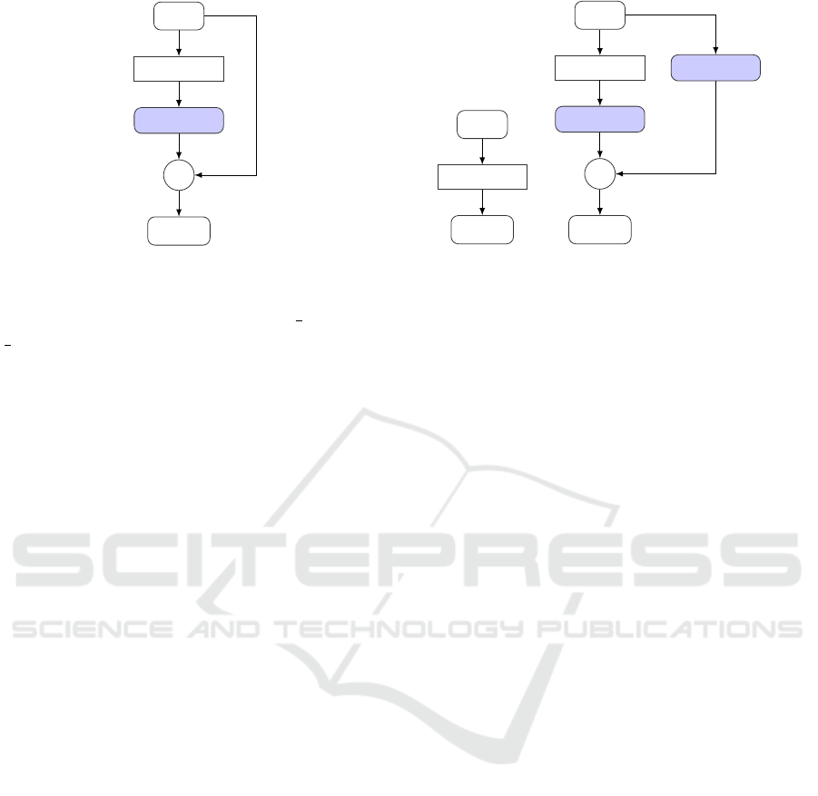
Input
GA
1 × 1
GA
w
+
Output
Figure 1: Computational graph of the global self-attention.
map for each input pixel, but rather on the local neigh-
borhood N
k
(i, j) = {a, b ∈ Z : |a − i| <
k
2
∧ |b − j| <
k
2
}, where k determines the neighborhood size. The
authors introduce this approach by replacing the fi-
nal convolutional layers of a CNN. The implementa-
tion is based on three 1 × 1 convolutions, which re-
quire fewer parameters compared to a k × k(k > 1)
convolution. Another advantage is its content-based
computation. However, it is not capable of imitating
the computation of the convolutional layer it replaces.
Therefore, adding it to a CNN destroys the pre-trained
behavior.
To overcome this issue, we propose an alternative
implementation, which we call embedded local self-
attention, henceforth “ELA”. The implementation is
inspired by the design of GA described above. Specif-
ically, we add a residual connection around the in-
troduced LA block, containing the original convolu-
tions and pre-trained weights. Similarly to GA, we
add a zero-initialized 1 × 1 × 1 convolution after the
LA block to conserve pre-trained behavior. Hence,
it is content-based and keeps pre-trained behavior at
initialization, at the cost of adding four 1 × 1 convo-
lutions. Both operations are visualized in Figure 2 as
computational graphs, showing the similarity of ELA
to GA.
2.3 Implementation Details
We base our analysis on two commonly used CNN
architectures, ResNet (He et al., 2016) and Efficient-
Net (Tan and Le, 2019). For all models, we rely on
commonly used ImageNet (Russakovsky et al., 2015)
pre-trained weights as initialization. In the following,
we describe our specific architecture choices.
2.3.1 ResNet (He et al., 2016)
Regarding ResNet architectures, we opt for
ResNet18, a well-established and frequently used
Input
LA
Output
(a) LA
Input
LA
k × k
CONV
1 × 1
LA
w
+
Output
(b) ELA
Figure 2: Computational graphs for both local approaches:
local self-attention (LA) and embedded local self-attention
(ELA).
variant. We insert the GA block after the first
and second blocks of ResNet18, as recommended
in (Wang et al., 2018). Regarding local attention,
for both LA and ELA, we replace all convolutional
layers of the last block. A similar setup is discussed
in (Ramachandran et al., 2019). Note that all ResNet
architectures feature four blocks. Hence, our inclu-
sion of attention mechanisms could be implemented
similarly for other variants.
2.3.2 EfficientNet (Tan and Le, 2019)
Again, we choose the smallest variant of this model
family, EfficientNet-B0, featuring seven blocks. To
insert the GA component, we again follow the rec-
ommendations of (Wang et al., 2018) and include it
after the second and third architecture blocks. Sim-
ilarly, we add LA and ELA by replacing convolu-
tions in the last block. Note that EfficientNet-B0 uti-
lizes so-called MBConv blocks (Sandler et al., 2018).
Hence, minimizing the number of parameters and in-
creasing efficiency. An MBConv block contains a
convolutional layer using groups, amongst other tech-
niques, to reduce parameters. Hence, naively replac-
ing this grouped convolution significantly increases
the parameter count. To avoid this issue, we replace
the whole MBConv block instead, leading to a sim-
ilar parameter reduction as observed for the ResNet
models described above. We argue that this parame-
ter reduction aligns better with the reasoning stated in
(Ramachandran et al., 2019) and increases compara-
bility between our selected CNN architectures.
2.3.3 Model Selection
Table 1 summarizes the different architectures we in-
vestigate in this study to analyze the influence of
self-attention on skin lesion and tissue classification.
When Medical Imaging Met Self-Attention: A Love Story That Didn’t Quite Work out
151
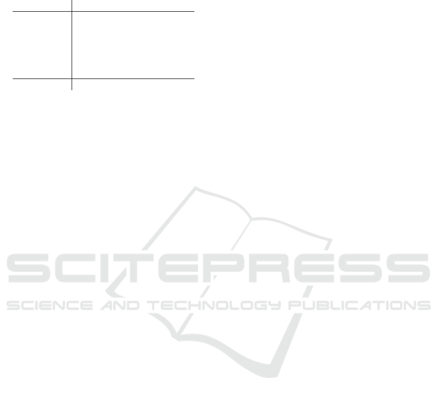
Table 1: Parameter counts of all architectures (in million)
and relative change compared to the corresponding base-
lines.
ResNet18 EfficientNet-B0
Base 11.18±0.00% 4.01±0.00%
+ GA 11.22+0.37% 4.02+0.12%
+ LA 5.67−49.25% 3.48−13.29%
+ GA + LA 5.71−48.87% 3.48−13.17%
+ ELA 14.98+34.02% 4.30+7.16%
+ GA + ELA 15.02+34.40% 4.30+7.27%
ViT 5.49±0.00%
Additionally, we report the parameter counts to en-
sure that none of our architecture changes widely
skew the model capacities. For comparison, we
also list our selected baselines: ResNet18 (He et al.,
2016), EfficientNet-B0 (Tan and Le, 2019), and two
attention-only ViT models (Kolesnikov et al., 2021).
Here, we select the ViT tiny variant with a parame-
ter count of 5.49 million parameters which is in be-
tween both selected CNN architectures. We investi-
gate a version pre-trained on the commonly used Ima-
geNet 1K challenge (Russakovsky et al., 2015) to stay
comparable to our other experiments and baselines,
as well as one ViT pre-trained on the larger ImageNet
21K dataset. We add the latter model, given empirical
observations that larger pre-training datasets increase
the downstream performance of transformer models
(Kolesnikov et al., 2021).
3 EXPERIMENTS
We start by describing the general experimental setup
we employed before discussing the achieved re-
sults. We then analyze the feature usage of the best-
performing skin lesion models. Finally, we visualize
the learned attention maps and compare them to lo-
cal Grad-CAM (Selvaraju et al., 2017) explanations
to gather additional insights.
3.1 Experimental Setup
To investigate the influence of additional self-
attention blocks, we analyze the performance of our
hybrid approaches in three distinct ways. First, we
trained the models on skin-lesion and tumor tissue
classification, comparing the balanced accuracies on
in-distribution and out-of-distribution test sets. Con-
cerning skin lesions, we use data from the ISIC
archive (ISIC, 2022) as training data and rely on the
PAD-UFES-20 dataset (Pacheco et al., 2020) as out-
of-distribution (OOD) test data. To align these tasks,
we simplified the classification problem to the three
classes benign nevi, melanoma, and others. Regard-
ing tumor tissue classification, we use the Came-
lyon17 dataset (Bandi et al., 2018), which already
provides an out-of-distribution split in addition to the
training data. The domain shift in Camelyon17 is in-
troduced by sampling the OOD set from a different
hospital.
To increase reliability, we performed a small hy-
perparameter search, considering the learning rate
and weight decay. To reduce the search space, we
fixed our batch size to 32. For data augmentation,
we followed the recommendations from (Perez et al.,
2018) and chose their best-performing augmentation
scheme. This scheme randomly applies cropping,
affine transformations, flipping, and changing the hue,
brightness, contrast, and saturation of the input. In
our experiments, we use SGD as an optimizer. To de-
termine the performance of a specific hyperparameter
combination, we perform the search with 2 random
data splits.
After determining the optimal learning rate and
weight decay for each model, which is shown in Ta-
ble 5, we train each architecture 10 times on different
data splits to determine the mean and standard devia-
tion of the achieved performances. Additionally, we
test for statistical significance by performing Welch’s
T-Test (Welch, 1947) on these samples. Here, we em-
ploy the widely used significance level of p < 0.05.
3.2 Performance Analysis
Table 2 summarizes the balanced accuracy for all ex-
amined models and corresponding baselines on both
datasets and the corresponding OOD sets.
On the ISIC archive data, the ResNet18 variants
with added attention mechanisms generally underper-
formed compared to the convolutional baseline. Fur-
ther, while the EfficientNet-B0 variants primarily ex-
hibit improvements, on average, over the baseline,
these improvements are not statistically significant.
On the Camelyon17 dataset, we observed similar per-
formances between the baselines and our hybrid mod-
els, with the only significant result being the Effi-
cientNet + ELA, which performs worse on average.
Hence, overall, we found no reliable upward trend
in performance when including self-attention mech-
anisms in a model.
We, however, noticed smaller differences between
the specific attention versions. Specifically, we found
that local approaches show, on average, increased per-
formance in combination with GA. Nevertheless, the
overall performance still decreases compared to the
baselines.
While the significant reduction of performance for
VISAPP 2024 - 19th International Conference on Computer Vision Theory and Applications
152
Table 2: Balanced Accuracies on each Dataset and each Model. Values inside the parentheses show the balanced accuracies
on the OOD-dataset. Bold values indicate statistically significant differences to the respective base model.
ISIC Dataset Camelyon17
ResNet EfficientNet ResNet EfficientNet
Base 73.9±1.74 (57.4±8.87) 75.4±1.75 (57.6±15.18) 98.4±0.18 (94.4±1.66) 98.5±0.16 (94.5±2.33)
+GA 72.1±1.31 (56.3±12.92) 76.6±1.83 (55.8±12.67) 98.4±0.07 (94.0±1.73) 98.6±0.17 (95.4±0.24)
+LA 70.8±2.96 (56.3±5.83) 75.5±1.58 (55.5±7.77) 98.2±0.36 (92.9±3.15) 98.4±0.21 (94.3±2.01)
+GA+LA 71.2±1.44 (55.5±4.64) 75.8±1.14 (55.4±12.48) 98.4±0.16 (92.2±5.90) 98.6±0.26 (94.3±0.63)
+ELA 72.3±1.45 (58.4±7.05) 73.8±2.29 (57.9±8.43) 98.2±0.23 (91.4±8.22) 98.3±0.07 (94.1±2.09)
+GA+ELA 73.5±1.35 (58.8±10.41) 75.9±1.77 (54.7±11.84) 98.4±0.14 (95.1±0.44) 98.5±0.40 (94.3±3.01)
1k 21k 1k 21 k
ViT 66.0±17.36 (52.6±113.45) 75.0±0.90 (56.7±6.94) 98.3±0.43 (94.8±0.91) 98.3±0.11 (94.5±0.96)
some models could be attributed to the reduced pa-
rameter count (LA models), we also observe similar
behavior in models with increased parameter count
(ELA). This is further corroborated by the observed
higher performance of the EfficientNet models in the
skin lesion task despite a lower parameter count com-
pared to the ResNet variants.
Regarding OOD performance, we see that there
are substantial differences between the datasets. For
the Camelyon17 dataset, the degradation of perfor-
mance is minimal for all models, and they still achieve
high balanced accuracies. We observe higher average
OOD performance for the EfficientNet + GA hybrid
model. However, this increase is not statistically sig-
nificant. Further, the only statistically reliable results
regarding the OOD performance are the observed de-
creases for some of the ResNet hybrid models. In
the skin lesion classification task, the drop in perfor-
mance is much more severe, also leading to higher
standard deviations. Hence, we did not observe any
significant improvements of the self-attention hybrid
models over the fully convolutional baselines in this
task.
The performance of the ViT heavily depends on
the pretraining data size in the skin lesion task. While
pretraining, the model with the 21k classes version of
ImageNet (Russakovsky et al., 2015) outperforms the
ResNet baseline in the skin lesion classification task,
pretraining with default ImageNet 1K does perform
significantly worse. This result confirms previous ob-
servations, e.g., (Kolesnikov et al., 2021), that larger
pretraining datasets can increase downstream perfor-
mance. In our experiment, this specifically meant that
two of our ten ViT 1K models diverged, resulting in
random guessing capabilities and explaining the huge
observed standard deviation.
3.3 Global Explanations: Feature Usage
To further investigate the model adaptations described
in Sec. 2.3.3, we investigated our hybrid models and
baselines on a feature level in the skin lesion classi-
fication task. Here we employ the feature attribution
method described in (Reimers et al., 2020). Previ-
ous work (Reimers et al., 2021; Penzel et al., 2022)
also applied this method to skin lesion classification
and investigated the usage of features related to the
ABCD rule (Nachbar et al., 1994) and known biases
in skin lesion data (Mishra and Celebi, 2016).
Here, we specifically follow (Reimers et al., 2021;
Penzel et al., 2022) and extract the same four fea-
tures related to the ABCD rule, namely Asymmetry,
Border irregularity, Color, and Dermoscopic struc-
tures, as well as the four bias features age of a pa-
tient, sex of the patient, skin color, and the occur-
rence of large colorful patches (Scope et al., 2016).
For a detailed description of these features, we re-
fer the reader to the original work (Reimers et al.,
2021). Furthermore, we follow the hyperparameter
settings described in (Penzel et al., 2022) and select
three conditional independence tests, namely cHSIC
(Fukumizu et al., 2007), RCoT (Strobl et al., 2019),
and CMIknn (Runge, 2018). We report the majority
decision and use a significance level of p < 0.01 (Pen-
zel et al., 2022).
Table 3 contains the results of our feature analy-
sis. Regarding the bias features, the age and skin color
of the patients are both learned by all analyzed mod-
els. The colorful patches and patient’s sex features
are also often learned, however, predominantly by the
ResNet variants. The EfficientNet models seem to be
more robust in that regard. Looking at the meaningful
ABCD rule features, we observe that the models often
learn the asymmetry or border irregularity while the
color feature is rarely, and the dermoscopic structures
feature is never learned. In contrast, both ViT models
learn to incorporate the color of the lesion into their
decision process. In general, GA seems to produce
slightly better results, given that all extended models
that utilize the color feature also include GA. How-
ever, this observation is inconsistent as other combi-
nations of GA do not result in this behavior. Some
When Medical Imaging Met Self-Attention: A Love Story That Didn’t Quite Work out
153

Table 3: Feature usage of our best-performing skin lesion models according to balanced accuracy. We abbreviate Asymmetry,
Border irregularity, Color, and Dermoscopic structures with the associated ABCD rule letter. We use significance level
p < 0.01 (Reimers et al., 2021).
A B C D Age Sex
Skin Colorful
Model color patches
ResNet
Base ✓ ✓ ✗ ✗ ✓ ✓ ✓ ✓
+GA ✓ ✓ ✓ ✗ ✓ ✓ ✓ ✓
+LA ✗ ✓ ✗ ✗ ✓ ✓ ✓ ✓
+GA+LA ✓ ✓ ✓ ✗ ✓ ✗ ✓ ✓
+ELA ✓ ✓ ✗ ✗ ✓ ✓ ✓ ✓
+GA+ELA ✓ ✓ ✗ ✗ ✓ ✓ ✓ ✓
EfficientNet
Base ✓ ✓ ✗ ✗ ✓ ✗ ✓ ✗
+GA ✗ ✓ ✗ ✗ ✓ ✗ ✓ ✓
+LA ✓ ✓ ✗ ✗ ✓ ✓ ✓ ✓
+GA+LA ✓ ✓ ✓ ✗ ✓ ✗ ✓ ✓
+ELA ✓ ✓ ✗ ✗ ✓ ✓ ✓ ✗
+GA+ELA ✗ ✓ ✗ ✗ ✓ ✓ ✓ ✓
ViT
1K ✓ ✓ ✓ ✗ ✓ ✗ ✓ ✓
21K ✓ ✓ ✓ ✗ ✓ ✓ ✓ ✓
models containing GA regress and do not learn to
utilize the skin lesion asymmetry, which is consis-
tently learned by the CNN baselines and which has
also been reported previously (Reimers et al., 2021).
To conclude, we do not find a systematic improve-
ment of the extended models incorporating attention
mechanisms over the fully convolutional baselines.
There is a small benefit in some attention-based mod-
els with respect to the color feature, especially for the
ViT architecture. Nevertheless, these results are simi-
lar to the empirical evaluation described above, where
some of the extended models achieved a performance
increase, which was not statistically significant.
3.4 Local Explanations: Visualizations
Introducing self-attention into CNNs allows for a di-
rect visualization of important image areas, which can
help to gain further insights about the model deci-
sion process. We leverage the method from (Wang
et al., 2018), which creates a map showing how a
certain pixel pays attention to all others and calcu-
lates the average over all of these maps. We com-
pare this attention map with Grad-CAM (Selvaraju
et al., 2017) and the visualization method of the de-
fault ViT (Kolesnikov et al., 2021). Table 4 contains
these visualizations generated using five images (all
classes for both datasets) and additionally displays the
mean of the attention maps over 500 images. To get
visually comparable results, we normalized the values
between 0 and 255. To stay comparable and only gen-
erate one visualization per image, we select the Grad-
CAM for the predicted class for all examples. We find
that while models focus on different parts of the im-
ages, we observe no visible improvements or biases.
Noteworthy, the highlighted area of Grad-CAM is
typically larger than the one for the GA visualization
due to the way Grad-CAM is implemented in (Sel-
varaju et al., 2017), which we directly follow. Grad-
CAM highlights mostly the whole object of interest.
This can be seen in the mean image for the ISIC task
because the center, where most of the skin lesions are
placed, gets highlighted the most. On the other hand,
the attention map visualization highlights more spe-
cific features like the border of the skin lesion, fram-
ing both interpretability techniques as uniquely dif-
ferent. Interestingly GA, as well as the ViT, put a lot
of attention on the corner of the image. While this is
initially quite unintuitive, we suggest that this is sim-
ilar behavior as reported in (Darcet et al., 2023; Xiao
et al., 2023). Here, transformers often produce atten-
tion spikes in the image background, as visible in our
mean images, to store information for further calcula-
tions. Unfortunately, this behavior cannot be further
unraveled by looking at the attention maps, therefore,
revealing a possible disadvantage of these visualiza-
tions in comparison to Grad-CAM.
Since we do not carry extensive medical knowl-
edge, we refrain from more interpretations into the
visualization here. We, however, note that the atten-
tion maps do not provide extensive visual explana-
tions that could not be provided by Grad-CAM ap-
proaches in a similar fashion. Additionally, we do not
observe specific structural biases since models seem
VISAPP 2024 - 19th International Conference on Computer Vision Theory and Applications
154
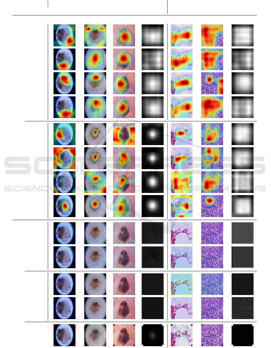
Table 4: Qualitative examples of explanations generated using either Grad-CAM (Selvaraju et al., 2017) or by visualizing the
learned attention maps. We choose one image per class per dataset and the best performing models of either the baselines or
adapted with GA or ELA (here abbreviated with G and E).
ISIC Camelyon17
Model Melanoma Nevus Other Mean Tumor No Tumor Mean
Grad-CAM
ResNet
-
+G+E+GE
EfficientNet
-
+G+E+GE
AttentionMaps
ResNet
+G+GE
EfficientNet
+G+GE
ViT
When Medical Imaging Met Self-Attention: A Love Story That Didn’t Quite Work out
155

to set a reasonable focus concerning the input data
typically. Therefore, we attribute no systematic ben-
efit to attention maps neither concerning local feature
usage nor interpretability.
4 CONCLUSIONS
This work investigated the usage of self-attention
mechanisms for medical imaging, specifically clas-
sification. Toward this goal, we extended two
widely used convolutional architectures with self-
attention mechanisms and empirically compared
these extended models against fully convolutional and
attention-only baselines. We conducted this compar-
ison on a dataset constructed from the well-known
ISIC archive (ISIC, 2022) and on the Camelyon17
dataset (Bandi et al., 2018). Additionally, we analyze
the OOD generalization on domain-shifted test sets.
While we did observe some minor improvements,
none of the attention-featuring architectures led to
a statistically significant increase in performance.
Sometimes, we even observe a significant decrease
in balanced accuracy. In some instances, we find
the reduction of performance on the OOD test data
is decreased when using self-attention. However, we
observe that this behavior is somewhat sensitive to
the used backbone model, dataset, and in-distribution
performance. Our investigation suggests that self-
attention alone does not result in a direct increase in
performance or generalization of medical image clas-
sification models.
To explain our findings further, we conducted two
related analyses, taking a closer look at explanations
and feature usage of our architectures. First, we
performed a feature analysis of the best-performing
skin lesion classification models using the attribution
method described in (Reimers et al., 2020). We ob-
served no systematic improvements in medically rel-
evant features or bias features over the fully convo-
lutional baselines, only slight deviations. Important
global features, such as dermoscopic structures, are
still not learned by employing self-attention.
Second, we performed a qualitative analysis of the
learned attention maps. While including attention in-
creases interpretability, e.g., revealing biased feature
usage, we find that out-of-the-box explanation meth-
ods, we used Grad-CAM (Selvaraju et al., 2017), per-
form similarly for that purpose. To conclude: our
work indicates that merely including self-attention
does not directly lead to benefits. Of course, more
work is required to ultimately conclude the suitability
of self-attention mechanisms for medical image anal-
ysis. In any case, we hope to inspire future work to in-
vestigate new architectural changes more thoroughly
than merely comparing performance scores since we
believe this can provide valuable insights.
REFERENCES
Bandi, P., Geessink, O., Manson, Q., Van Dijk, M., Balken-
hol, M., Hermsen, M., Bejnordi, B. E., Lee, B., Paeng,
K., Zhong, A., et al. (2018). From detection of in-
dividual metastases to classification of lymph node
status at the patient level: the camelyon17 challenge.
IEEE Transactions on Medical Imaging.
Bera, K., Schalper, K. A., Rimm, D. L., Velcheti, V., and
Madabhushi, A. (2019). Artificial intelligence in digi-
tal pathology — new tools for diagnosis and precision
oncology. Nature Reviews Clinical Oncology, pages
1–13.
Celebi, M. E., Codella, N., and Halpern, A. (2019). Der-
moscopy image analysis: overview and future direc-
tions. IEEE journal of biomedical and health infor-
matics, 23(2):474–478.
Codella, N., Rotemberg, V., Tschandl, P., Celebi,
M. E., Dusza, S., Gutman, D., Helba, B., Kalloo,
A., Liopyris, K., Marchetti, M., Kittler, H., and
Halpern, A. (2019). Skin Lesion Analysis Toward
Melanoma Detection 2018: A Challenge Hosted by
the International Skin Imaging Collaboration (ISIC).
arXiv:1902.03368 [cs]. arXiv: 1902.03368.
Darcet, T., Oquab, M., Mairal, J., and Bojanowski, P.
(2023). Vision transformers need registers. arXiv
preprint arXiv:2309.16588.
Fukumizu, K., Gretton, A., Sun, X., and Sch
¨
olkopf, B.
(2007). Kernel measures of conditional dependence.
Advances in neural information processing systems,
20.
He, K., Zhang, X., Ren, S., and Sun, J. (2016). Deep resid-
ual learning for image recognition. In Proceedings of
the IEEE conference on computer vision and pattern
recognition, pages 770–778.
He, X., Tan, E.-L., Bi, H., Zhang, X., Zhao, S., and Lei,
B. (2022). Fully transformer network for skin lesion
analysis. Medical Image Analysis, 77:102357.
ISIC (2022). Isic archive home page. Last accessed 23 July
2023.
Khened, M., Kori, A., Rajkumar, H., Krishnamurthi, G.,
and Srinivasan, B. (2021). A generalized deep learn-
ing framework for whole-slide image segmentation
and analysis. Scientific Reports, 11:11579.
Kolesnikov, A., Dosovitskiy, A., Weissenborn, D., Heigold,
G., Uszkoreit, J., Beyer, L., Minderer, M., Dehghani,
M., Houlsby, N., Gelly, S., Unterthiner, T., and Zhai,
X. (2021). An image is worth 16x16 words: Trans-
formers for image recognition at scale.
Krishna, G., Supriya, K., K, M., and Sorgile, M. (2023).
Lesionaid: Vision transformers-based skin lesion gen-
eration and classification.
Lee, B. and Paeng, K. (2018). A robust and effective ap-
proach towards accurate metastasis detection and pn-
stage classification in breast cancer. In Frangi, A. F.,
VISAPP 2024 - 19th International Conference on Computer Vision Theory and Applications
156

Schnabel, J. A., Davatzikos, C., Alberola-L
´
opez, C.,
and Fichtinger, G., editors, Medical Image Comput-
ing and Computer Assisted Intervention – MICCAI
2018, pages 841–850, Cham. Springer International
Publishing.
Mishra, N. K. and Celebi, M. E. (2016). An Overview
of Melanoma Detection in Dermoscopy Images
Using Image Processing and Machine Learning.
arXiv:1601.07843 [cs, stat]. arXiv: 1601.07843.
Nachbar, F., Stolz, W., Merkle, T., Cognetta, A. B., Vogt,
T., Landthaler, M., Bilek, P., Braun-Falco, O., and
Plewig, G. (1994). The ABCD rule of dermatoscopy.
High prospective value in the diagnosis of doubtful
melanocytic skin lesions. Journal of the American
Academy of Dermatology, 30(4):551–559.
Pacheco, A. G., Lima, G. R., Salom
˜
ao, A. S., Krohling,
B., Biral, I. P., de Angelo, G. G., Alves Jr, F. C., Es-
gario, J. G., Simora, A. C., Castro, P. B., Rodrigues,
F. B., Frasson, P. H., Krohling, R. A., Knidel, H., San-
tos, M. C., do Esp
´
ırito Santo, R. B., Macedo, T. L.,
Canuto, T. R., and de Barros, L. F. (2020). Pad-ufes-
20: A skin lesion dataset composed of patient data and
clinical images collected from smartphones. Data in
Brief, 32:106221.
Penzel, N., Reimers, C., Bodesheim, P., and Denzler, J.
(2022). Investigating neural network training on a fea-
ture level using conditional independence. In ECCV
Workshop on Causality in Vision (ECCV-WS), pages
383–399, Cham. Springer Nature Switzerland.
Perez, F., Vasconcelos, C., Avila, S., and Valle, E. (2018).
Data augmentation for skin lesion analysis.
Ramachandran, P., Parmar, N., Vaswani, A., Bello, I., Lev-
skaya, A., and Shlens, J. (2019). Stand-alone self-
attention in vision models. Advances in Neural Infor-
mation Processing Systems, 32.
Reimers, C., Penzel, N., Bodesheim, P., Runge, J., and Den-
zler, J. (2021). Conditional dependence tests reveal
the usage of abcd rule features and bias variables in
automatic skin lesion classification. In CVPR ISIC
Skin Image Analysis Workshop (CVPR-WS), pages
1810–1819.
Reimers, C., Runge, J., and Denzler, J. (2020). Determin-
ing the relevance of features for deep neural networks.
In European Conference on Computer Vision, pages
330–346. Springer.
Runge, J. (2018). Conditional independence testing based
on a nearest-neighbor estimator of conditional mutual
information. In International Conference on Artificial
Intelligence and Statistics. PMLR.
Russakovsky, O., Deng, J., Su, H., Krause, J., Satheesh, S.,
Ma, S., Huang, Z., Karpathy, A., Khosla, A., Bern-
stein, M., et al. (2015). Imagenet large scale visual
recognition challenge. International journal of com-
puter vision, 115:211–252.
Sandler, M., Howard, A., Zhu, M., Zhmoginov, A., and
Chen, L.-C. (2018). Mobilenetv2: Inverted residu-
als and linear bottlenecks. In Proceedings of the IEEE
conference on computer vision and pattern recogni-
tion, pages 4510–4520.
Scope, A., Marchetti, M. A., Marghoob, A. A., Dusza,
S. W., Geller, A. C., Satagopan, J. M., Weinstock,
M. A., Berwick, M., and Halpern, A. C. (2016). The
study of nevi in children: Principles learned and impli-
cations for melanoma diagnosis. Journal of the Amer-
ican Academy of Dermatology, 75(4):813–823.
Selvaraju, R. R., Cogswell, M., Das, A., Vedantam, R.,
Parikh, D., and Batra, D. (2017). Grad-cam: Visual
explanations from deep networks via gradient-based
localization. In Proceedings of the IEEE international
conference on computer vision, pages 618–626.
Society, A. C. (2022). Cancer facts & figures 2022. Last
accessed 02 August 2022.
Strobl, E. V., Zhang, K., and Visweswaran, S. (2019).
Approximate kernel-based conditional independence
tests for fast non-parametric causal discovery. Jour-
nal of Causal Inference.
Tan, M. and Le, Q. (2019). Efficientnet: Rethinking model
scaling for convolutional neural networks. In Interna-
tional conference on machine learning, pages 6105–
6114. PMLR.
Tolstikhin, I. O., Houlsby, N., Kolesnikov, A., Beyer, L.,
Zhai, X., Unterthiner, T., Yung, J., Steiner, A., Key-
sers, D., Uszkoreit, J., Lucic, M., and Dosovitskiy, A.
(2021). Mlp-mixer: An all-mlp architecture for vision.
CoRR, abs/2105.01601.
Trockman, A. and Kolter, J. Z. (2022). Patches are all you
need? CoRR, abs/2201.09792.
Tschandl, P., Rosendahl, C., and Kittler, H. (2018). The
HAM10000 dataset, a large collection of multi-source
dermatoscopic images of common pigmented skin le-
sions. Sci. Data, 5(1):180161.
Vaswani, A., Shazeer, N., Parmar, N., Uszkoreit, J., Jones,
L., Gomez, A. N., Kaiser, Ł., and Polosukhin, I.
(2017). Attention is all you need. Advances in neural
information processing systems, 30.
Wang, X., Girshick, R., Gupta, A., and He, K. (2018). Non-
local neural networks. In Proceedings of the IEEE
conference on computer vision and pattern recogni-
tion, pages 7794–7803.
Welch, B. L. (1947). The generalization of ‘student’s’ prob-
lem when several different population variances are
involved. Biometrika, 34(1/2):28–35.
Xiao, G., Tian, Y., Chen, B., Han, S., and Lewis, M. (2023).
Efficient streaming language models with attention
sinks. arXiv preprint arXiv:2309.17453.
Yang, G., Luo, S., and Greer, P. (2023). A novel vision
transformer model for skin cancer classification. Neu-
ral Processing Letters, pages 1–17.
When Medical Imaging Met Self-Attention: A Love Story That Didn’t Quite Work out
157

APPENDIX
Table 5 contains the best hyperparameters, we deter-
mined for our models.
Table 5: Best determined hyperparameters.
ISIC Camelyon17
Model LR WD LR WD
ResNet
Base 0.001 0.0001 0.001 0.001
+GA 0.001 0 0.001 0.001
+LA 0.001 0.0001 0.0001 0.01
+GA+LA 0.001 0 0.001 0.0001
+ELA 0.001 0 0.01 0
+GA+ELA 0.001 0 0.001 0.0001
EfficientNet
Base 0.01 0 0.001 0.0001
+GA 0.01 0 0.001 0.0001
+LA 0.01 0 0.001 0
+GA+LA 0.01 0 0.001 0.0001
+ELA 0.001 0 0.001 0.001
+GA+ELA 0.01 0 0.01 0
ViT
1K 0.001 0 0.001 0
21K 0.0001 0.0001 0.001 0.0001
VISAPP 2024 - 19th International Conference on Computer Vision Theory and Applications
158
