
Oral Diseases Recognition Based on Photographic Images
Mazin S. Mohammed
1
, Salah Zrigui
1,3
and Mounir Zrigui
2
1
University of Monastir, Research Laboratory in Algebra,
Numbers Theory and Intelligent sys-tem (RLANTIS), Monstir 5019, Tunisia
2
University of Al-Mosul, University of the Presidency, Nineveh, Iraq
3
Lig Laboratory, Grenoble, France
Keywords: Oral Disease Classification, Dental Caries Detection, Dental Images, Deep Learning.
Abstract: Recently, the automation diagnosis process of dental caries plays a critical role in medical applications. This
paper presents a new dataset of photo-graphic images for six different types of oral diseases. The dataset is
gathered and labelled by professional medical operators in the dentistry field. We use the collected dataset to
train a binary classifier to determine whether the region of interests (ROI) needs detection or not inside the
input image. Then, we train a detector to detect and localize the required ROI. Finally, we use the detected
regions to train a CNN network by adopting transfer learning technique to classify various kinds of teeth
diseases. With this model, we obtained an almost 93 % accuracy by modifying and re-training the pre-trained
model VGG19.
1 INTRODUCTION
This increasing global vulnerability to diseases has
left health care systems worldwide strained. To
protect against the spread of disease, hospitals, clinics
and nearly all types of medical facilities had to adhere
to several protective guidelines. This led to a
significant decrease in the number of patients that can
be treated at any given moment. In response,
researchers, more specifically researchers in the field
of artificial intelligence have been innovating and
proposing novel methods and technologies to ensure
safe diagnosis and treatment with minimal direct
contact. One of the most prominent fields for such
innovation is the automated diagnosis of dental
imagery (Araújo et al., 2023).
AI techniques have been used successfully in
various types of disciplines such as nature language
processing (Merhbene, Zouaghi and Zrigui, 2010;
Mahmoud and Zrigui, 2019), computer vision
(Mansouri, Charhad and Zrigui, 2017; Farhani,
Terbeh and Zrigui, 2019; Daood, AL-Saegh and
Mahmood, 2023), speech recognition (Bellagha and
Zrigui, 2020; Slimi et al., 2020; Amari et. Al., 2022),
biometrics, smart home applications (Alhafidh et al.,
2018), medical imaging, healthcare, robotics,
banking & finance, agriculture, military & defence,
marketing & advertising, and even oil discovery &
gas exploration. Lately, computer vision has been
used as an efficient tool in medical applications to
offer an accurate diagnosis and avoid errors in human
judgement. The use of artificial intelligence in
dentistry appears to has a great potential and it is
expected to play a vital role in the future of dental
health-care and oral diseases diagnosis.
Deep learning strategies have achieved remarkable
progress in understanding and analysing dental images.
Some neural network architectures such as
Convolutional Neural Networks (CNNs) lend
themselves naturally to exploit the availability of X-ray
and photographic images dataset to perform teeth
segmentation, classification, numbering, and lesions
detection. With the long waiting time to receive dental
care and the importance of an early diagnosis. We
decided to build a tool that helps the average person get
an early evaluation of his dental state. In this paper we
build a dental care detection and classification system
that can provide an early diagnosis from a simple
picture taken via any smartphone. The system takes as
an input a dataset that is comprised of photographic
images collected from some local clinics with the help
of medical team of specialist dentists. The remainder
of the paper is structured as follows. Section 2
examines prior research works. Section 3 presents the
material data and the proposed methodology. Section 4
showcases the experimental results. Lastly, we present
486
Mohammed, M., Zrigui, S. and Zrigui, M.
Oral Diseases Recognition Based on Photographic Images.
DOI: 10.5220/0012361500003636
Paper published under CC license (CC BY-NC-ND 4.0)
In Proceedings of the 16th International Conference on Agents and Artificial Intelligence (ICAART 2024) - Volume 3, pages 486-493
ISBN: 978-989-758-680-4; ISSN: 2184-433X
Proceedings Copyright © 2024 by SCITEPRESS – Science and Technology Publications, Lda.

our conclusions in Section 5.
2 LITERATURE REVIEW
In this section we will review some of the methods
that have been proposed to automate the diagnosis
process of dental diseases. As we mentioned before,
all the previous researches were either based on X-ray
images or photographic images. For example, the
authors in (Liu et al., 2019) proposed intelligent
dental Health-IoT system which was implemented on
smart hardware. Mobile phone was used as a terminal
to capture images in order to perform the diagnosis.
MASK R-CNN was used to perform the detection
process by applying the training on 12,6000 collected
images. The trained model achieved accuracy of 90%
to detect and recognize 7 different types of dental
diseases. The researchers in (Al Kheraif et al.,2019)
collected 800 of X-ray images and then used adaptive
histogram equalization which helped to divide the
images into back-ground bones and foreground teeth.
After that, they used hybrid graph cut to perform the
segmentation process to separate the oral cavity and
the tissues. Finally, deep learning networks were
trained using the segmented images to perform the
predication with accuracy of 97%.
Orthopantomogram (OPD) images were collected in
(Laishram and Thongam, 2020), and then pre-
processing techniques were applied to prepare for the
training process of faster-RCNN to perform the
detection and the classification on the same time. The
trained convolutional neural network achieved 90%
in the detection process and 99% in the classification
process. Mobile app (OralCam) was proposed in
(Liang et al., 2020) to offer an end to end complete
system with self- examination of five different
diseases. 3,182 oral images were taken from 500
participants to train a conventional neural network
which was tested to give on average detection
sensitivity of 78.7%. 620 photographic images were
captured of extracted molars using smartphone in
(Duong et al., 2021). The collected images were
labelled manually into three classes by four dentists.
After that, a series of image pre-processing
techniques were applied to enhance the gathered
pictures and perform the segmentation process.
Finally, the classification process was implemented
using SVM classifier which was trained using colour
intensity features of the collected dataset. 640
photographic images of different patients’ oral
cavities were captured using a smartphone in (Ding et
al., 2021). Images enchantment and data
augmentation were applied on the collected dataset.
Data augmentation was used to increase the number
of images to 3,990 to prepare the collected data for
the training process. Then, transfer learning technique
was used by retraining YOLOv3 CNN model to
detect and recognize two types of caries.
The authors in (Zhu et al., 2022) presented a deep
learning network as U-shape architecture to perform
the segmentation process of 3127 panoramic
radiograph images. The pro-posed network was
called CariesNet to determine three different degrees
of caries based on panoramic X-ray images.
Additionally, they used full- scale axial attention
module to enhance the segmentation process and
improve the results. The proposed method achieved
93.61% of accuracy. The researchers in (Rashid et al.,
2022) proposed A hybrid system to localize regions
of caries by combining photographic and X-ray
images. They used the collected dataset to train mask
R-CNN deep learning model to perform the
segmentation process to detect regions of cavities and
oral diseases. The proposed system achieved about
92% of accuracy. 1902 photographic images were
taken using a smartphone of 695 participants in
(Thanh et al., 2022) to detect three different classes of
caries. Four different deep learning architecture were
re-trained to detect the oral lesions from the collected
images. The trained models were Faster R-CNNs,
YOLOv3, RetinaNet, and SSD. A retrospective study
was presented in (Keser et al., 2023) by collecting
photographic pictures of 65 healthy and 72 oral
lesions. Inception V3 deep learning network was
trained using the collected dataset to create a binary
classifier. The trained architecture achieved accuracy
of 100% for healthy and Oral lichen planus lesions
cases. The re-searchers in (Gomes et al., 2023)
collected 5069 images for six different types of oral
mucosal lesions. The images were labelled and
cropped manually by specialists. They trained four
different convolutional neural networks using 70% of
the collected dataset, the rest of the data was used to
test the trained models. ResNet-50, VGG16,
InceptionV3 and Xception were used as base
classifiers to perform the learning process of the
proposed models. A dataset of 470 Panoramic X-ray
images was labelled and segmented in (Haghanifar et
al., 2023). A genetic algorithm was proposed to
perform the segmentation process with image
processing operations to slice each tooth individually.
Finally, capsule network was trained using the
extracted features from different deep learning
networks to achieve accuracy of 86.05%.
Oral Diseases Recognition Based on Photographic Images
487

3 DATA MATERIAL AND
METHODOLOGY
The first step of our project is data collecting and
images gathering. The data collection process is
carried out at some local clinics with the help of
medical team of specialist dentists. We collect 1600
photographic images representing six common dental
diseases. The collected dataset is assembled from
both genders (male/female) with ages between 7 to 65
years. Since, the capturing process of the
photographic images is an easy process, simply using
a smartphone, we were able to collect images even
from children. On the other hand, capturing X-ray
images for such age is complicated task and prone to
errors and mistakes. The collected dataset was
obtained in an anonymous manner all recordings of
any private information regarding the patients' names,
ages, medical history, or even their status have been
omitted. Figure 1 shows some samples of the
collected images for the six cases of the oral diseases.
In the second stage of our project, we perform the
annotation process by applying image labelling to
separate our dataset into six categories of dental
diseases. This process requires some manual effort to
ensure accurate labelling. Unfortunately, the manual
labouring of labelling process cannot be carried out
by ordinary labellers. Hence, the manual annotation
of the collected images is required to be performed by
professionals with expertise in the field of dentistry.
So, three dentists examined the collected dataset and
categorized the images into six cases of oral diseases.
Then we divide our dataset into two segments with
the ratio of 80%:20%. The first part of our data is used
for the training process to learn predication models to
perform the detection and the recognition process.
The second part of our data is used to test and evaluate
the trained models and measure their performance.
Convolutional Neural Networks (CNNs) have shown
great success in dealing with image related learning
tasks. This is due to their natural compatibility with
the grid like structure of an image. Therefore, we
adopt CNNs to implement the detection and the
recognition operations. In our project, we propose a
deep learning network to detect the region of teeth to
localize the region of interest. Then, we use the
detection model to crop the images of our dataset to
train a CNN network to perform the classification of
oral diseases. We use data augmentation to create
more images and increase the dataset size. Data
augmentation can be considered as a regularization
technique by manipulating the original data to create
more copies and synthesize a different version of the
images through applying various types of
transformation such as rotation, translation, scaling,
and even light (brightness) changes. This technique is
used to improve the performance and reduce
overfitting by exposing the trained models to
augmented versions of the original dataset which
helps the models to generalize better and become
more robust. The collected images are categorized
into two kinds, as shown in figure 1. The first type
contains only the teeth (which is our region of interest
ROI). While the second type contains the teeth and
some other parts of the face such as cheeks, nose, lips,
and jaws. Therefore, we need a mechanism to
separate the two types of images in our dataset.
Manual splitting is not an option as it requires time
and effort. More importantly, manual split will
interfere with the automation process of the diagnosis
because we need to detect our region of interest and
then send the localized part for the classification
process.First, we determine whether the detection
process of ROI is required or not. After that, we
localize our ROI to send the cropped parts of the
images to training process. Finally, we use the
training data to train deep learning models and apply
the assessment and evaluation using the testing data
to measure the accuracy of the trained models. We
select 50 images for each case from our dataset,
where the first 25 images require detection to localize
the region of interest and the other 25 images do not
require any detection. The purpose of this collection
of images is to train a binary classifier to determine
whether the tested image needs detection to find the
region of interest or not, so we can use the important
parts of the images and remove any unnecessary
segments. To achieve the training process of this
binary classifier, we need an efficient and accurate
model. Therefore, we utilize the concept of the
transfer learning by selecting a pre-trained network
and modifying the chosen model to perform the
binary classification.
Hence, we use MobileNetV2 network as our base
model for the training process. Applying the transfer
learning approach leverages the prior knowledge of
MobileNetV2 network.
Firstly, we eliminate the last layer of the
MobileNetV2 network and flatten the resulted
features from the final layer. Subsequently, a fully
connected layer with 64 nodes is attached to the
model. Finally, we add the output layer with two
nodes of SoftMax layer to classify the images into
two classes (requires detection and does not require).
After that, we perform the training process using the
selected images.
ICAART 2024 - 16th International Conference on Agents and Artificial Intelligence
488
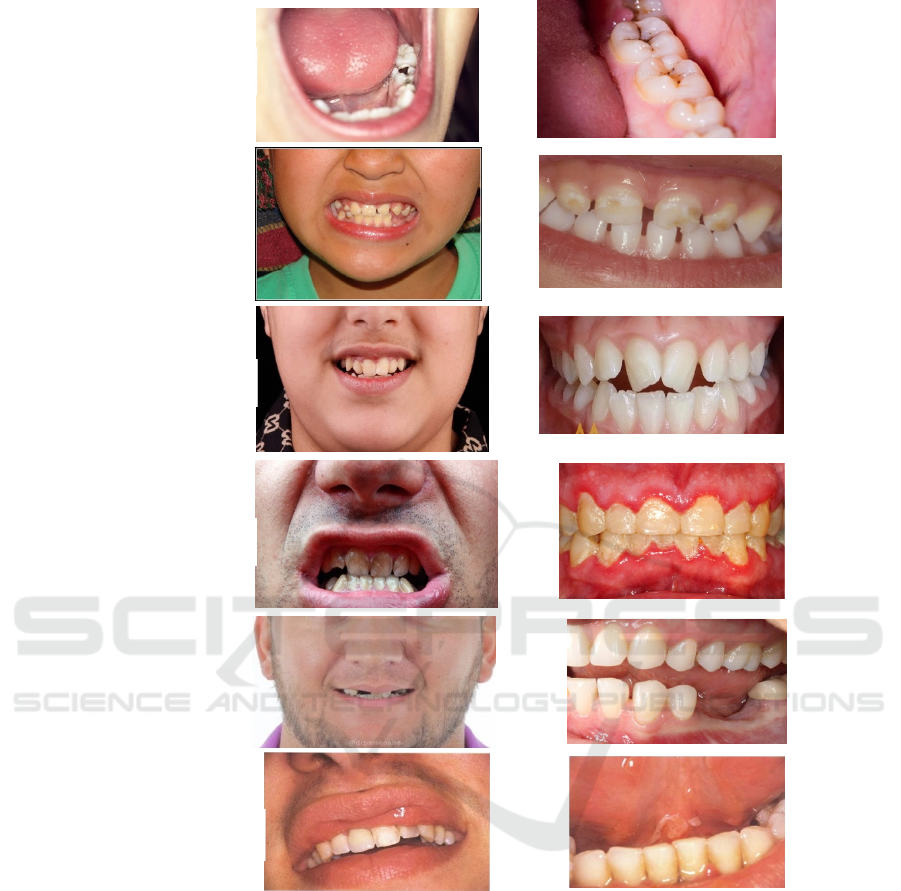
Figure 1: Photographic samples of our dataset.
During this stage, we freeze all the layers of
MobileNetV2 network (base layers) except for the
additional layers we added explicitly for binary
classification. This strategy allows us to optimize the
parameters for newly added layers while retaining the
original parameters of the MobileNetV2 network to
keep the previous knowledge of the model. In the
next phase, we need to train a fast and accurate
detector to determine the region of interest in the
contents of our images. For this particular task, we
use a light deep learning network to achieve the
training process of our detector model.
Adopting the transfer learning technique with
light pre-trained model can give us an efficient model
to implement the detection process. Hence, YOLO
V4-tiny model is used for this particular task. YOLO
V4-tiny is a single step object detector model which
means it can accomplish both the detection and
classification process in the same time in one step
instead of performing the two operations in separate
stages by applying initial detection in one step and
subsequent classification in the next step.
YOLO V4-tiny is a scaled down version of the
YOLO model has a smaller number of convolution
layers (smaller number of parameters) than the
ordinary YOLO. Therefore, adopting this model to
Case 1: Restorative
Dentistry
Case 2:
Pediatric
Case 3:
Orthodontic
Case 4:
Periodontics
Case 5:
Prosthodontics
Case 6: Oral medicine
Oral Diseases Recognition Based on Photographic Images
489

apply the training process reduces the cost of the
training time and the need of huge resources.
Additionally, by using transfer learning the pre-
trained YOLO V4-tiny returns the optimal values of
small number of parameters in the selected network.
Adopting this technique makes the learning process
possible despite the small size of the dataset.
Before the training process, we need to select
images from our data set to perform the detection
process to localize the region of interest inside the
images. Therefore, we selected 50 images, from each
case, which need detection. Then, we need to label
these images by providing a bounding box to
determine the coordinates of the region of interest for
each individual image. Since, we intend to train
YOLO V4-tiny model to perform the detection
process, the coordinates of bounding box for the
labelling operation should match the format of YOLO
network. Thus, we use Bbox-Label-Tool-Multi-Class
of the Darknet-library for the labelling process. It is
labelling tool that is completely compatible with the
YOLO format. This tool offers a programmable
configuration to initialize different modes of setting.
BBox-Label-Tool can create a simple GUI window to
input images and give the facility to label the region
of interest by applying a bounding box manually by a
user.
After the user localizes the region of interest,
BBOX tool will create the necessary files with the
required information for the training process. Figure
2 shows samples of the image labelling. Once the
labelling process of the selected images is done, we
can utilize these images to train our customized
detection model. As we mention before, we use for
this particular task YOLO V4-tiny network. We
retrain the pre-trained YOLO V4-tiny model with our
dataset to implement the learning process of the
detector. When the training process is completed, we
use the trained detector to localize the region of
interest to crop these regions. The cropped images are
used to prepare the training data to perform the
training of the disease classification. Figure 4 shows
the pipeline of the proposed method.
As shown in figure 3, after applying the detection
process, to determine the regions of interest, we
create database of the training images to learn
recognition models to classify 6 different types of
dental caries. Transfer learning approach is adopted
to achieve the training process of the classification
models. The analysis of the transfer learning achieves
the learning process by relying on the prior
knowledge from a pre-trained model. So, instead of
starting the training from scratch the learning process
starts with trained parameters of a base model which
has been trained using extensive amount of data.
Currently, numerous numbers of pre-trained models
are available to be utilized as base classifiers to
perform the training.
To implement the training of the disease’s
recognizer, we use VGG16 network as a foundational
classifier to exploit the prior knowledge of the
selected model. We modify the architecture of the
chosen network to achieve the training process with
our own images. We replace the last layer of VGG16
(which is responsible for classification of 1000
classes) with new classification layers for our 6
classes.
As shown in Figure 4, we include flatten layer to
make the size of vector features compatible with the
new attached layers. Then, we add a fully connected
layer of 512 nodes with a drop out layer of 0.5
dropping factor. The primary advantages of the drop
out layer is to decrease the effect of the overfitting
problem by skipping the update of the parameter’s
values during the training. Then, we add a second
fully connected layer of 256 nodes and followed by
another drop out layer. Finally, we wrap up the
designed network with a soft-max layer with 6 output
nodes to represent each individual disease.
After we complete the architecture of the
proposed network, we need to retrain the designed
model by applying fine-tune process to update the
weights and the parameters of our network to adjust
their values in a suitable configuration which allows
the trained model to capture and learn the most
relevant features from our dataset to achieve the
diseases classification task.
In order to improve the results, we expand our set
of experiments by testing different types of network
architectures as a base classifier. Hence, we use
additional pre-trained models to boost the accuracy of
our classifier. We exploit the prior knowledge
obtained from the following models: VGG16,
VGG19, GoogleNet, Xception, InceptionV3,
InceptionResNetV2, DenseNet201, MobileNetV2,
and NASNetLarge. Practically, we repeat the same
procedure of the fine-tune process to retrain these
models.
First, we remove the classification layer from the
selected network and we attach a fully connected
layers of 512 nodes with drop out layer, and followed
by another fully connected of 265 nodes with another
drop out layer. At the end of the network, we add a
soft max layer of 6 outputs nodes to classify 6
different diseases. All the results of these experiments
will be shown in the next section.
ICAART 2024 - 16th International Conference on Agents and Artificial Intelligence
490
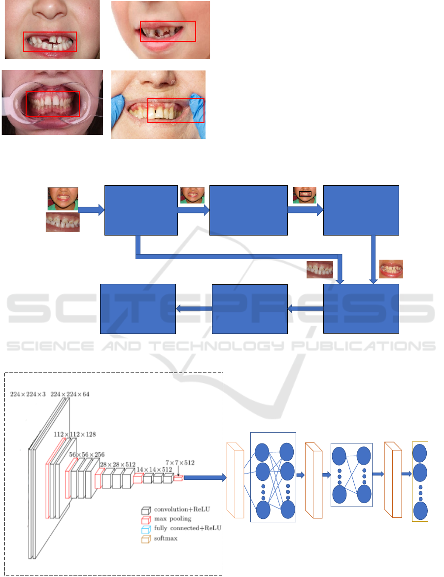
Figure 2: Data annotation to train the detector model.
4 THE RESULTS
In the current section, we provide the finding results
of our experiments. We use the training data set to
learn the optimal features of our images to create
diseases classification model. Transfer learning
method is used to perform the recognition process.
First, we modify VGG16 network to implement the
re-training process to reshape the weights and the
parameters of the selected network to capture the
optimal representation of the learnt features of our
dataset. We utilize the testing segment of our dataset
to assess the quality of the trained models. Therefore,
we compute the accuracy, precision, and recall as
metric measurements to conduct comprehensive
Figure 3: Flowchart of the proposed algorithm.
Figure 4: The architecture of the designed CNN network.
Binary classifier ( to
deter mine whether
the i mage needs
detection or not )
YOLO Tiny(to
localize the region of
interest )
Crop the the region
of i nter est
Input imag
e
s
Pr epar e the dat a f or
tr aining
Train deep learning
models
Evaluate the tr ained
models
Soft max
layer (5 nodes)
Base Network(
V
GG 16)
Flatten
layer
Fully connected
layer (512
nodes)
Fully connected
layer (256 nodes)
Dropout
layer
Dropout
layer
Oral Diseases Recognition Based on Photographic Images
491
assessments, validations, and comparative analyses
of the trained classifiers. By adopting VGG16, we
obtain almost 92% of accuracy. Clearly, the prior
knowledge obtained by training a model using
extensive amount of dataset achieve reasonable
performance by applying a fine-tuning process to
learn the optimal features representation of
recognizing 6 dental caries in our dataset.
Table 1: Accuracy of deep learning networks.
Base network
model
Accuracy Precision Recall
VGG16
92.11% 92.42% 92.99%
VGG19
93.55%
93.27
%
93.13%
AlexNet
89.19% 88.99% 89.41%
Resnet50
92.74% 92.02% 92.83%
GoogleNet
91.25% 91.14% 90.79%
NasNet-Mobile
85.86% 86.32% 85.95%
DenseNet201
92.76% 92.58% 92.39%
MobileNetV2
87.82 % 88.01% 88.91%
InceptionResNet
V2
90.68% 90.49% 90.18%
Xception
90.45% 90.61% 90.71%
InceptionV3
90.41% 90.97% 90.39%
It is important to highlight that all the experiment
and the obtained results are conducted by a personal
laptop type Lenovo where the processor is Core I7
with memory of 16 G RAM. The re-trained models
which are used in our experiments exhibit diversity in
their characteristics presenting variations in the
architecture design, connection mappings, layer
depth, and parameters quantities. Therefore, they
offer different performance and efficiency based on
their variations and properties. Basically, these
models may respond differently to the new given task
with unseen dataset to their previous knowledge.
Upon examining the results of Table 1, the
comparisons clearly demonstrate that VGG19
outshines as the most prominent base model which
offered an almost 93% of accuracy. The conducted
experiments show that the modified version of
VGG19 stands out by learning the best features of our
dataset to encapsulate the optimal representation of
different patterns for the oral diseases. Furthermore,
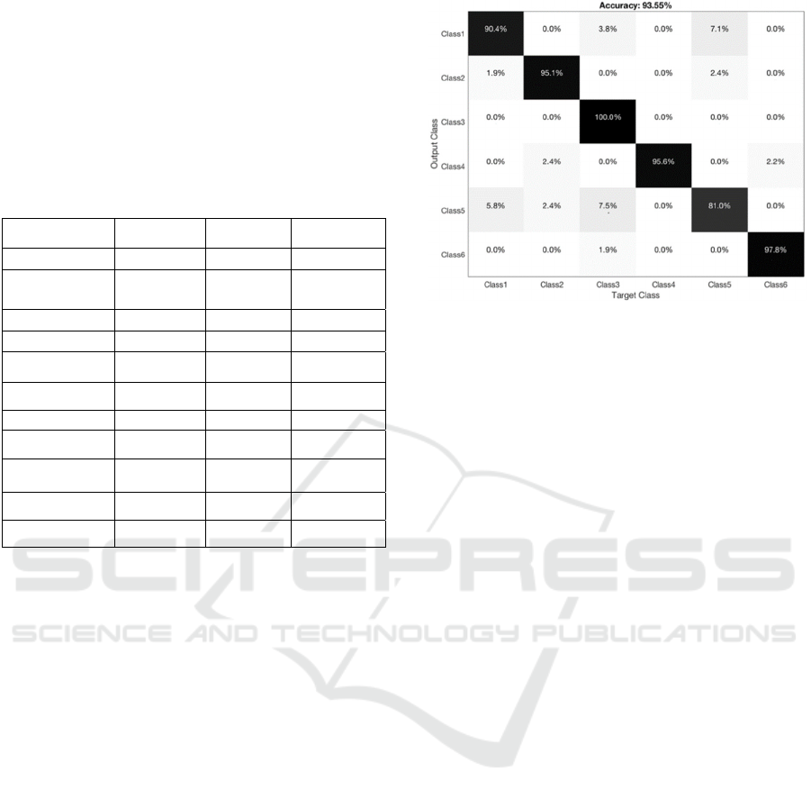
measured the confusion matrix of the modified
version of VGG19, these results are shown in figure
5.
Figure 5: The confusion matrix of the re-trained model
VGG19.
5 CONCLUSIONS
In this paper, we propose a new dataset of
photographic images to train a predication model to
diagnose 6 different kinds of oral diseases. The
gathered images are annotated by expert dentists. The
collected images are used to train a binary recognizer
to determine whether detection is necessary inside the
dental images to find the region of interest (ROI).
After that, we deploy a modified version of YOLO
V4-tiny network to perform the detection process of
ROI. The detected parts of ROI within our data are
cropped to prepare our dataset for the classification
process. Finally, we adopt the transfer learning
strategy to train multiple pre-trained models to
implement the recognition process. The modification
of these models allows us to exploit their previous
knowledge and achieve 93% accuracy to classify six
different types of oral diseases.
REFERENCES
Araújo, A. L. D., da Silva, V. M., Kudo, M. S., de Souza,
E. S. C., Saldivia‐Siracusa, C., Giraldo‐Roldán, D., ...
& Moraes, M. C. (2023). Machine learning concepts
applied to oral pathology and oral medicine: A
convolutional neural networks' approach. Journal of
Oral Pathology & Medicine, 52(2), 109-118.
Amari, R., Noubigh, Z., Zrigui, S., Berchech, D., Nicolas,
H., & Zrigui, M. (2022, September). Deep
Convolutional Neural Network for Arabic Speech
Recognition. In International Conference on
Computational Collective Intelligence (pp. 120-134).
Cham: Springer International Publishing.
ICAART 2024 - 16th International Conference on Agents and Artificial Intelligence
492

Alhafidh, B. M. H., Daood, A. I., Alawad, M. M., & Allen,
W. (2018). FPGA hardware implementation of smart
home autonomous system based on deep learning. In
Internet of Things–ICIOT 2018: Third International
Conference, Held as Part of the Services Conference
Federation, SCF 2018, Seattle, WA, USA, June 25-30,
2018, Proceedings 3 (pp. 121-133). Springer
International Publishing.
Al Kheraif, A. A., Wahba, A. A., & Fouad, H. (2019).
Detection of dental diseases from radiographic 2d
dental image using hybrid graph-cut technique and
convolutional neural network. Measurement, 146, 333-
342.
Bellagha, M. L., & Zrigui, M. (2020, November). Speaker
naming in tv programs based on speaker role
recognition. In 2020 IEEE/ACS 17th International
Conference on Computer Systems and Applications
(AICCSA) (pp. 1-8). IEEE.
DAOOD, A., AL-SAEGH, A. L. I., & MAHMOOD, A. F.
(2023). HANDWRITING DETECTION AND
RECOGNITION OF ARABIC NUMBERS AND
CHARACTERS USING DEEP LEARNING
METHODS. Journal of Engineering Science and
Technology, 18(3), 1581-1598.
Ding, B., Zhang, Z., Liang, Y., Wang, W., Hao, S., Meng,
Z., ... & Lv, Y. (2021). Detection of dental caries in oral
photographs taken by mobile phones based on the
YOLOv3 algorithm. Annals of Translational Medicine,
9(21).
Farhani, N., Terbeh, N., & Zrigui, M. (2019). Object
recognition approach based on generalized hough
transform and color distribution serving in generating
arabic sentences. International Journal of Computer
and Information Engineering, 13(6), 339-344.
Duong, D. L., Kabir, M. H., & Kuo, R. F. (2021).
Automated caries detection with smartphone color
photography using machine learning. Health
Informatics Journal, 27(2), 14604582211007530.
Gomes, R. F. T., Schmith, J., Figueiredo, R. M. D., Freitas,
S. A., Machado, G. N., Romanini, J., & Carrard, V. C.
(2023). Use of artificial intelligence in the classification
of elementary oral lesions from clinical images.
International Journal of Environmental Research and
Public Health, 20(5), 3894.
Haghanifar, A., Majdabadi, M. M., Haghanifar, S., Choi,
Y., & Ko, S. B. (2023). PaXNet: Tooth segmentation
and dental caries detection in panoramic X-ray using
ensemble transfer learning and capsule classifier.
Multimedia Tools and Applications, 1-21.
Karaoglu, A., Ozcan, C., Pekince, A., & Yasa, Y. (2023).
Numbering teeth in panoramic images: A novel method
based on deep learning and heuristic algorithm.
Engineering Science and Technology, an International
Journal, 37, 101316.
Keser, G., Bayrakdar, İ. Ş., Pekiner, F. N., Çelik, Ö., &
Orhan, K. (2023). A deep learning algorithm for
classification of oral lichen planus lesions from
photographic images: A retrospective study. Journal of
Stomatology, Oral and Maxillofacial Surgery, 124(1),
101264.
Kim, H. N., Kim, K., & Lee, Y. (2023). Intra-oral
photograph analysis for gingivitis screening in
orthodontic patients. International Journal of
Environmental Research and Public Health, 20(4),
3705.
Laishram, A., & Thongam, K. (2020, February). Detection
and classification of dental pathologies using faster-
RCNN in orthopantomogram radiography image.
In
2020 7th international conference on signal processing
and integrated networks (SPIN) (pp. 423-428). IEEE.
Liang, Y., Fan, H. W., Fang, Z., Miao, L., Li, W., Zhang,
X., ... & Chen, X. A. (2020, April). OralCam: enabling
self-examination and awareness of oral health using a
smartphone camera. In Proceedings of the 2020 CHI
conference on human factors in computing systems (pp.
1-13).
Liu, L., Xu, J., Huan, Y., Zou, Z., Yeh, S. C., & Zheng, L.
R. (2019). A smart dental health-IoT platform based on
intelligent hardware, deep learning, and mobile
terminal. IEEE journal of biomedical and health
informatics, 24(3), 898-906.
Mansouri, S., Charhad, M., & Zrigui, M. (2017). Arabic
Text Detection in News Video Based on Line Segment
Detector. Res. Comput. Sci., 132, 97-106.
Mahmoud, A., & Zrigui, M. (2019). Deep neural network
models for paraphrased text classification in the Arabic
language. In Natural Language Processing and
Information Systems: 24th International Conference on
Applications of Natural Language to Information
Systems, NLDB 2019, Salford, UK, June 26–28, 2019,
Proceedings 24 (pp. 3-16). Springer International
Publishing.
Merhbene, L., Zouaghi, A., & Zrigui, M. (2010, June).
Ambiguous Arabic words disambiguation. In 2010 11th
ACIS International Conference on Software
Engineering, Artificial Intelligence, Networking and
Parallel/Distributed Computing (pp. 157-164). IEEE.
Rashid, U., Javid, A., Khan, A. R., Liu, L., Ahmed, A.,
Khalid, O., ... & Nawaz, R. (2022). A hybrid mask
RCNN-based tool to localize dental cavities from real-
time mixed photographic images. PeerJ Computer
Science, 8, e888.
Slimi, A., Hamroun, M., Zrigui, M., & Nicolas, H. (2020,
November). Emotion recognition from speech using
spectrograms and shallow neural networks. In
Proceedings of the 18th International Conference on
Advances in Mobile Computing & Multimedia (pp. 35-
39).
Thanh, M. T. G., Van Toan, N., Ngoc, V. T. N., Tra, N. T.,
Giap, C. N., & Nguyen, D. M. (2022). Deep learning
application in dental caries detection using intraoral
photos taken by smartphones. Applied Sciences, 12(11),
5504.
Zhu, H., Cao, Z., Lian, L., Ye, G., Gao, H., & Wu, J. (2022).
CariesNet: a deep learning approach for segmentation
of multi-stage caries lesion from oral panoramic X-ray
image. Neural Computing and Applications, 1-9.
Oral Diseases Recognition Based on Photographic Images
493
