
3D Nuclei Segmentation by Combining GAN Based Image Synthesis and
Existing 3D Manual Annotations
Xareni Galindo
1,∗
, Thierno Barry
1,∗
, Pauline Guyot
2
, Charlotte Rivi
`
ere
2,3,4 a
, R
´
emi Galland
1 b
and Florian Levet
1,5 c
1
CNRS, Interdisciplinary Institute for Neuroscience, IINS, UMR 5297, University of Bordeaux, Bordeaux, France
2
Univ. Lyon, Universit
´
e Claude Bernard Lyon 1, CNRS, Institut Lumi
`
ere Mati
`
ere, UMR 5306, 69622, Villeurbanne, France
3
Institut Universitaire de France (IUF), France
4
Institut Convergence PLAsCAN, Centre de Canc
´
erologie de Lyon, INSERM U1052-CNRS, UMR5286, Univ. Lyon,
Universit
´
e Claude Bernard Lyon 1, Centre L
´
eon B
´
erard, Lyon, France
5
CNRS, INSERM, Bordeaux Imaging Center, BIC, UAR3420, US 4, University of Bordeaux, Bordeaux, France
fl
Keywords:
Bioimaging, Deep Learning, Miscrocopy, Nuclei, 3D, Image Processing, GAN.
Abstract:
Nuclei segmentation is an important task in cell biology analysis that requires accurate and reliable methods,
especially within complex low signal to noise ratio images with crowded cells populations. In this context,
deep learning-based methods such as Stardist have emerged as the best performing solutions for segmenting
nucleus. Unfortunately, the performances of such methods rely on the availability of vast libraries of ground
truth hand-annotated data-sets, which become especially tedious to create for 3D cell cultures in which nuclei
tend to overlap. In this work, we present a workflow to segment nuclei in 3D in such conditions when no
specific ground truth exists. It combines the use of a robust 2D segmentation method, Stardist 2D, which
have been trained on thousands of already available ground truth datasets, with the generation of pair of 3D
masks and synthetic fluorescence volumes through a conditional GAN. It allows to train a Stardist 3D model
with 3D ground truth masks and synthetic volumes that mimic our fluorescence ones. This strategy allows to
segment 3D data that have no available ground truth, alleviating the need to perform manual annotations, and
improving the results obtained by training Stardist with the original ground truth data.
1 INTRODUCTION
The popularity of 3D cell cultures, such as organoids
or spheroids, has recently exploded due to their abil-
ity to offer valuable models to study human biol-
ogy, far more physiologically relevant than 2D cul-
tures (Jensen and Teng, 2020; Kapalczynska et al.,
2018). Nowadays, automatically acquiring hundreds
of organoids in 3D has become a reality thanks to the
advances in microscopy systems (Beghin et al., 2022).
Life scientists have therefore access to distributions
of nuclei in 3D for a wide diversity of cell types and
growing conditions, a key feature forming the basis of
advanced quantitative analysis of important cell func-
tions. However, 3D cellular cultures inherently dis-
a
https://orcid.org/0000-0002-5048-5662
b
https://orcid.org/0000-0001-7117-3281
c
https://orcid.org/0000-0002-4009-6225
∗
These two authors contributed equally
play a large diversity of nuclei features, shapes and
arrangements. And 3D microscopy of such complex
samples most often leads to images with lower con-
trast and/or signal to noise ratio as compared to 2D
microscopy of single cellular layer, with overlapping
nuclei according to the achieved optical sectioning.
Hence, accurate and automated nuclei segmentation
in these conditions has turned out to be highly com-
plex. The bottleneck has therefore shifted from the
acquisition to the downstream analysis and quantifi-
cation steps.
Many computational solutions have been pro-
posed over the years that uses traditional image pro-
cessing methods to tackle this segmentation prob-
lem (Caicedo et al., 2019; Malpica et al., 1997; Li
et al., 2007), in 2D and 3D. However, they are usually
tailored for a specific application and do not general-
ize well, resulting in the necessity to adapt their pa-
rameters and ultimately preventing an automatic and
bias-free analysis.
Galindo, X., Barry, T., Guyot, P., Rivière, C., Galland, R. and Levet, F.
3D Nuclei Segmentation by Combining GAN Based Image Synthesis and Existing 3D Manual Annotations.
DOI: 10.5220/0012309200003657
Paper published under CC license (CC BY-NC-ND 4.0)
In Proceedings of the 17th International Joint Conference on Biomedical Engineering Systems and Technologies (BIOSTEC 2024) - Volume 1, pages 265-272
ISBN: 978-989-758-688-0; ISSN: 2184-4305
Proceedings Copyright © 2024 by SCITEPRESS – Science and Technology Publications, Lda.
265
In parallel, the last few years have witnessed the
rapid emergence of convolutional neural networks
(CNNs) as a method of choice for microscopy image
segmentation, achieving an accuracy unheard of for a
wide range of segmentation tasks. Among those, deep
learning-based nuclei segmentation has been particu-
larly active, with more than one hundred methods be-
ing developed since 2019 (Mougeot et al., 2022).
Supervised approaches, in which paired of im-
ages and masks are used for training, are the cur-
rent state of the art for nuclei segmentation, with
Stardist (Schmidt et al., 2018; Weigert et al., 2020)
and Cellpose (Stringer et al., 2021) having reached
a prominent position. Their adoption has been fa-
cilitated by their integration into several open-source
platforms, in which life scientists are able to directly
use several pre-trained models (Caicedo et al., 2019)
to segment their images. Unfortunately, these pre-
trained models are limited to the segmentation of nu-
clei in 2D, a fact directly related to the lack of avail-
able ground truth (GT) datasets in 3D. Determining
the organization of nuclei in 3D therefore requires life
scientists to annotate themselves their data, a daunting
and time-consuming task which turns out to be much
more challenging than in 2D due to the lower image
quality and more crowded cell populations.
Generating synthetic training datasets has there-
fore emerged as a potential solution to this problem
through the use of conditional Generative Adversar-
ial Networks (cGANs) (Goodfellow et al., 2014; Isola
et al., 2018; Zhu et al., 2017). cGANs learn a map-
ping from an observed source image x and a ran-
dom noise vector z, to a “transformed” target image
y, G : x, z → y. For nuclei segmentation, the use of
such generative networks aims to generate realistic
microscopy images (background, signal to noise ra-
tio, inhomogeneity, . . . ) of nuclei (target style) from
binary masks (source style) by training the cGAN
with paired or unpaired datasets (Baniukiewicz et al.,
2019; Fu et al., 2018; Wang et al., 2022). Never-
theless, GANs are notoriously known to be difficult
to train, with a large number of hyperparameters re-
quired to be tuned.
In this work, we present a workflow to segment
nuclei in 3D when no specific GT exists by leverag-
ing on cGANs to generate fluorescence synthetic vol-
umes of nuclei from 3D masks. While life scientists
start to be accustomed to using packaged deep learn-
ing methods for segmentation or classification, image
synthesis is still far from being accessible to regu-
lar users. We therefore made the choice to specif-
ically design our workflow to be accessible to non-
expert life-scientists, leveraging on already packaged
methods, both for the segmentation and the image
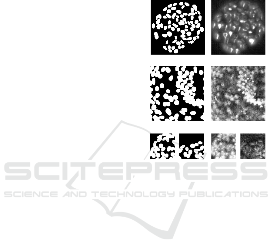
Binary mask
SpCycleGAN
s = 1024*1024
size = 512*512, crop = 128*128
size = 512*512, crop = 256*256
Figure 1: SpCycleGAN fails to generate realistic mi-
croscopy images even with different scaling and cropping.
generation, with the aim to avoid any tedious hand-
annotating steps. With that in mind, our workflow
first relies on the segmentation of the individualized
2D planes of our acquired volumes using the already
pre-trained, well established and now robust 2D seg-
mentation model Stardist 2D db2018 (Schmidt et al.,
2018). After modification of the instance masks by
applying a distance transform and a Gaussian filter,
we pair these transformed ‘GT’ masks with the flu-
orescent nuclei images to train a cGAN specifically
designed for biological images, that do not require
complex hyper-parameters tuning or specific crop-
ping data selection (Han et al., 2020). It allows to
generate 3D volumes of nuclei that resemble the flu-
orescent volumes we acquired, from existing 3D GT
masks that were typically created for a different cell
type or microscopy modality. Finally, those pairs of
GT mask volumes and synthetic fluorescent volumes
of nuclei are used to train a Stardist model in 3D ded-
icated to our 3D samples type and imaging modality.
Through this workflow, we demonstrate that us-
ing existing works to their full extent can (i) cir-
cumvent the requirement of tedious hand-annotating
steps for the segmentation of nuclei in 3D in com-
plex and crowded environment, and (ii) alleviate the
need to develop new deep learning architectures in an
BIOIMAGING 2024 - 11th International Conference on Bioimaging
266
already crowded field (Mougeot et al., 2022), while
also facilitating their usage by life scientists. We
applied this workflow on a couple of cell lines and
microscopy modalities and show that we managed
to have good qualitative segmentation results with-
out having to specifically hand annotate new volumes.
This approach greatly widens the scope of use of 3D
segmentation methods in the rapidly growing and di-
versifying field of 3D cell culture analysis, where
many imaging modalities are explored and cell types
with their own morphology characteristics used.
2 MOTIVATION
We recently acquired several oncospheres in 3D of
the colorectal cancer cell line HCT-116 expressing
the nucleus fluorescent label Fucci with a confocal
microscope, denoted as I
c
, that we aim to segment
while not having any GT mask for that cell culture
type and imaging modality. A couple of years ago
we trained a Stardist 3D model (Beghin et al., 2022),
Stardist
pre
3D
, by creating a GT dataset composed of 7
volumes that we manually annotated and that we will
denote as I
LS
GT
and M
LS
GT
for the images and masks, re-
spectively. These 7 volumes were composed of dif-
ferent 3D cell culture types (oncospheres and neu-
roectoderms), nuclear staining (DAPI and SOX) and
z-steps (500 nm and 1 µm). In addition, we acquired
them with the soSPIM imaging technology (Galland
et al., 2015), a single-objective light-sheet microscope
different from the confocal imaging modality used
to acquire the new oncospheres we aimed to seg-
ment. With this Stardist
pre
3D
model, we managed at
that time to automatically segment more than one
hundred oncospheres and neuroectoderms acquired
with the soSPIM imaging modality (Beghin et al.,
2022). However, applying Stardist
pre
3D
to I
c
gave poor
results, as it only managed to identify a portion of the
nuclei with an overall over-segmentation. This was
most probably due to the different cell morphology
and imaging modality of I
c
as compared to the GT
datasets used to train the model.
We therefore tried to generate synthetic volume
having the same style than I
c
with SpCycleGAN (Fu
et al., 2018), with the final objective of being able to
train a new Stardist 3D model more adapted to our
data. SpCycleGAN allows to generate synthetic vol-
umes from binary masks without GT by being built on
top of the unpaired image-to-image translation model
CycleGAN (Zhu et al., 2017). Being unpaired, the
network can use I
c
as target style without requir-
ing the corresponding nuclei segmentation as source.
Originally, the authors of (Fu et al., 2018; Wu et al.,
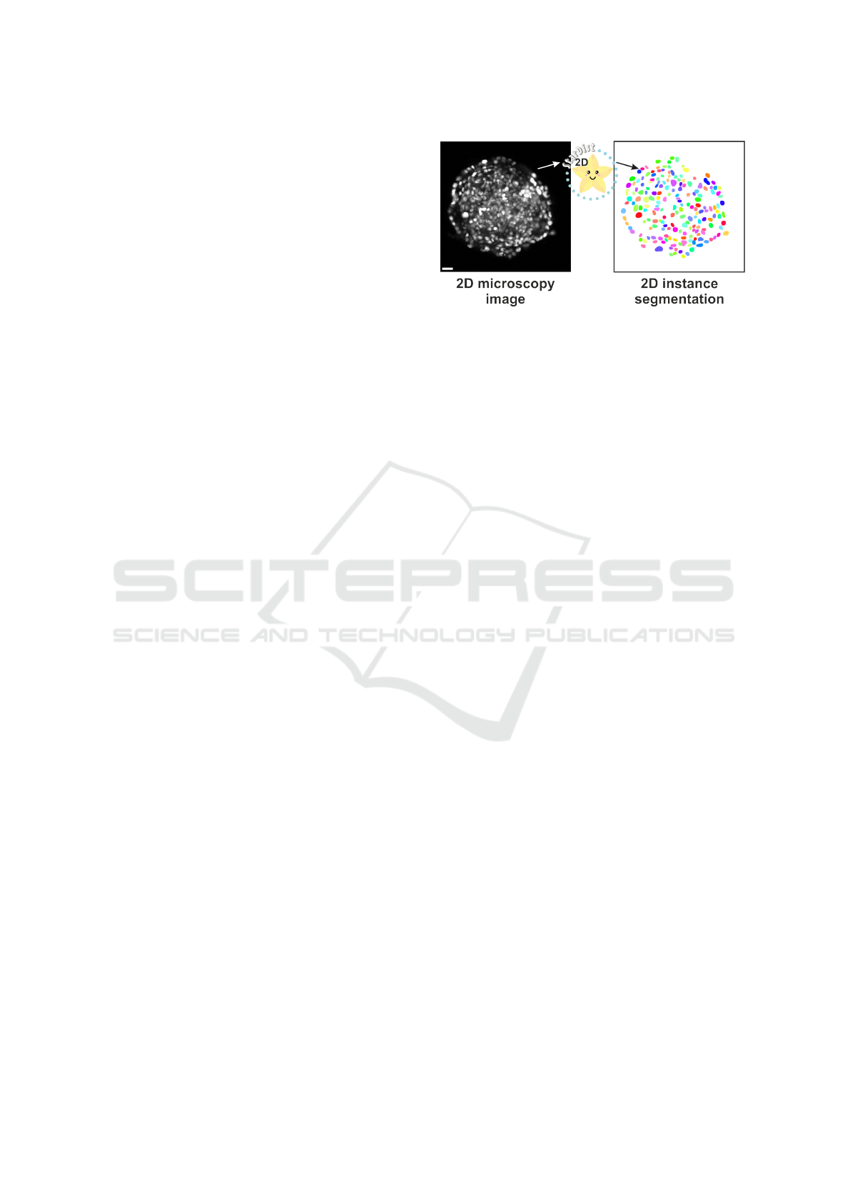
Figure 2: Nuclei segmentation with the 2D db2018 pre-
trained Stardist model results in sufficiently good results to
train a conditional GAN, scale bar = 20 µm.
2023) generated 3D nuclei masks by filling volumes
with binarized (deformed) ellipsoids even though it
could be limiting as it may not faithfully represent
the nuclei morphology. Having already a GT nuclei
masks, we therefore decided to train SpCycleGAN
with M
LS
GT
and I
c
to avoid this pitfall.
In our hand, SpCycleGAN failed to generate re-
alistic microscopy images (Fig. 1). Scrutinizing the
toy datasets available with the network, we tested dif-
ferent scaling and cropping of the data, as well as re-
moving any crops having void regions during train-
ing. Unfortunately, none of our tests were conclu-
sive. We hypothesize that this may be related to two
reasons. First, volumes composing I
c
exhibit a high
level of noise and an overall crowded and overlapping
nuclei population. SpCycleGAN may fail to transfer
all these features from binary masks. Second, SpCy-
cleGAN may require parameters fine-tuning to work
properly, but this goes against our objective of having
a more generic workflow that could be used by non-
experts.
3 METHOD
The proposed method is based upon the use of well es-
tablished and more robust deep-learning methods for
nuclei segmentation (Stardist (Schmidt et al., 2018;
Weigert et al., 2020)) and image synthesis (condi-
tional GANs in a slightly modified version (Han et al.,
2020)). Ours choices are motivated by the fact that
we wanted an as-simple-as-possible backbone. In this
manner, we intend to show that a simple approach fo-
cusing on data manipulation combined with existing
networks can perform qualitatively well. In addition,
we propose a workflow that could become generic
to enable the segmentation of a large variety of 3D
biological samples acquired upon different imaging
modalities.
3D Nuclei Segmentation by Combining GAN Based Image Synthesis and Existing 3D Manual Annotations
267
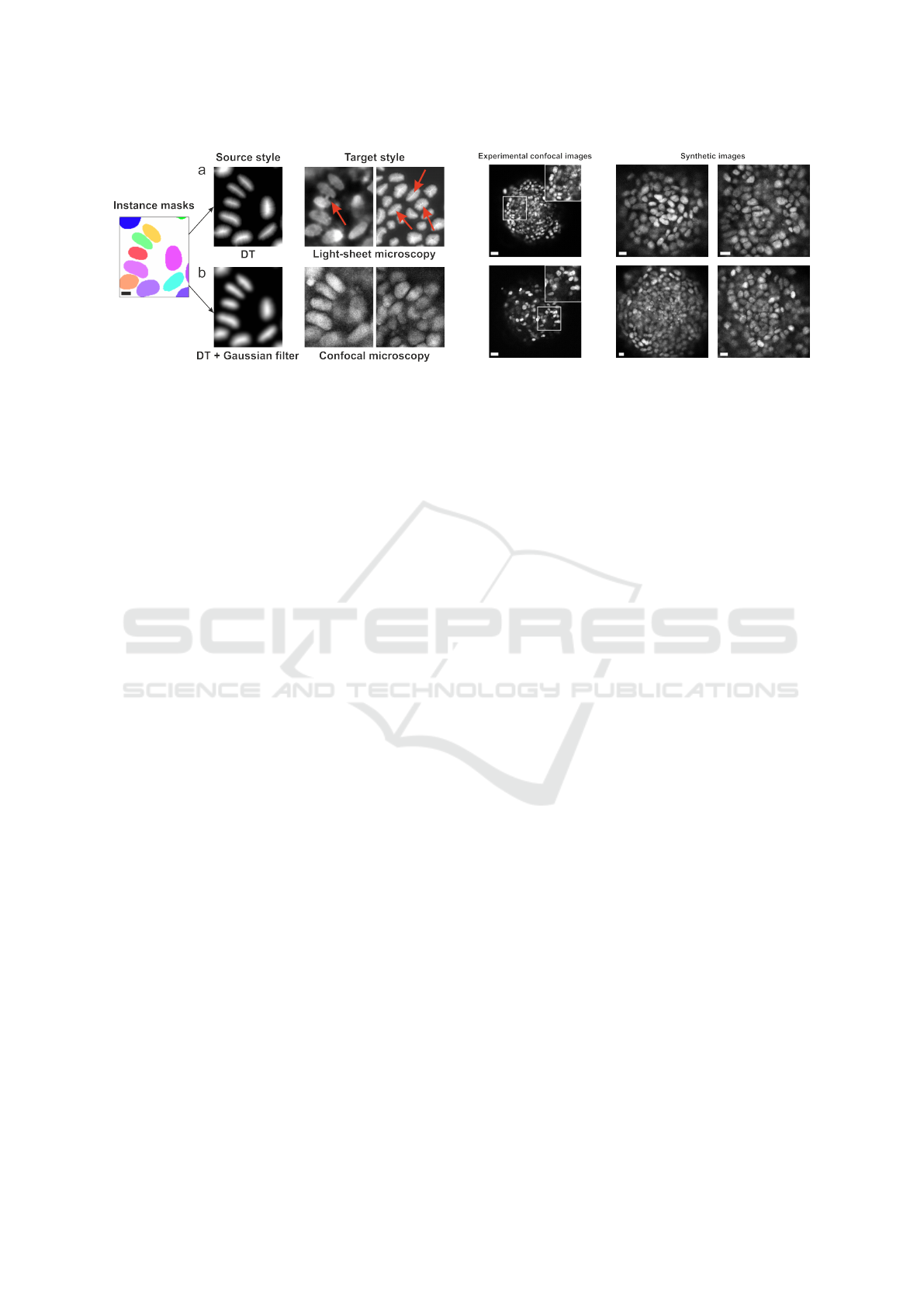
Figure 3: Manipulating instance masks to generate the FC-
GAN’s source style, scale bar = 5 µm. (a) After applying a
distance transform on the binarized instance masks, the FC-
GAN generates plausible images but with visible artefacts
related to the rough gradient of the transform (red arrows).
(b) Applying a Gaussian filter after the mask binarization
helps the FCGAN to learn a proper mapping between the
source and target style.
3.1 Fully-Conditional GAN
Contrary to natural images in which every local re-
gion contains relevant information, biological images
are composed of a mix of informative and void re-
gions. They are also inherently multiscale with both
the large-scale spatial organization of cells and their
individual morphology and texture being essential.
Since GANs have been originally developed for nat-
ural images, they tend to struggle to capture these in-
tertwined features and to fail to generate realistic void
regions.
We therefore based our synthesis method in the
fully-conditional GAN (FCGAN) architecture (Han
et al., 2020), an improvement of GAN focused on
modifications that allow to synthesize multi-scale bio-
logical images. This architecture consists of a genera-
tor built upon the cascaded refinement network (Chen
and Koltun, 2017) instead of the more traditional U-
Net (Ronneberger et al., 2015; Isola et al., 2018) ar-
chitecture as it is less prone to mode collapse. They
also added two modifications to the traditional GAN
architecture. First, they modified the input noise vec-
tor z to a noise ”image”, i.e. a 3D tensor with the
first two dimensions corresponding to the spatial po-
sitions. Instead of a noise vector that limits the size of
output images, modifying the noise image size allows
to output synthetized images of arbitrary sizes. Sec-
ond, they used a multi-scale discriminator. Having
a fixed size discriminator limits the quality and co-
herency of synthetized images to a micro (object) or
macro (global organization) level. Using a multi-scale
discriminator ensure the generator to produce images
both globally and locally accurate, a desired feature
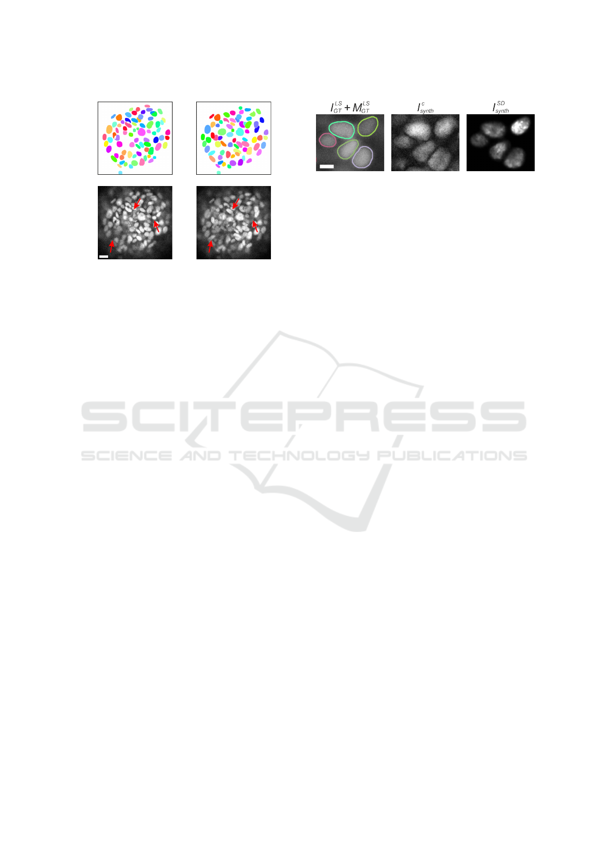
Figure 4: Comparison of experimental confocal images
(scale bar = 20 µm) and synthetic images generated with
the FCGAN style transfer (scale bar = 10 µm).
when dealing with biological images that exhibit sev-
eral levels of organization. All these modifications
allow to directly train the FCGAN with the acquired
images, alleviating the need to fine-tune the parame-
ters and resize or crop the void regions.
3.2 Image Synthesis Based 3D Stardist
Training
In spite of its numerous advantages, FCGAN requires
however paired images for training, therefore com-
pelling us to provide corresponding mask and im-
age pairs. Fortunately, 2D pre-trained models of
well-established nuclei segmentation methods such as
Stardist (Schmidt et al., 2018) or Cellpose (Stringer
et al., 2021) have been trained with thousands of 2D
GT masks and achieve now a high degree of robust-
ness over a large variety of sample types. Conse-
quently, we can directly determine the nuclei spa-
tial distribution by segmenting each of the 2D frame
(1024 ∗ 1024) of the volumes of I
c
with the 2D pre-
trained db2018 Stardist
db
2D
model (Fig. 2) instead of
simulating them. These 2D segmentation will be de-
noted M
c
2D
.
For image generation through the FCGAN model,
binary and instance masks can lead to fuzzy results
because their gradient can be too rough during back-
propagation (Long et al., 2021) (Fig. 3(a)). We
therefore applied a distance transform to our instance
masks (Long et al., 2021) followed by an intensity
normalization and a Gaussian filter, which allows to
remove intensity stiff jumps and dependency to the
nuclei length, ultimately facilitating the style trans-
fer performed by the FCGAN model (Fig. 3(b)). Ap-
plying this process to each mask image of M
c
2D
, we
obtained a set of transformed mask images dt(M
c
2D
).
Consequently, pairing dt(M
c
2D
) (source style) with the
corresponding images from I
c
(target style), allowed
BIOIMAGING 2024 - 11th International Conference on Bioimaging
268
x
y
x
y
x
z
x
z
yy
(z = 50)
(y = 302)
(x = 512)
x
y
x
y
x
z
x
z
yy
(z = 50)
(y = 302)
(x = 512)
x
y
x
y
x
z
x
z
yy
(z = 50)
(y = 302)
(x = 512)
x
y
x
y
x
z
x
z
yy
(z = 50)
(y = 302)
(x = 512)
I
c
Stardist
3D
pre
Stardist
3D
c
Stardist
3D
bin
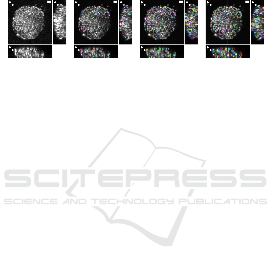
Figure 5: Comparison of segmentation obtained with Stardist
pre
3D
(Beghin et al., 2022), Stardist
bin
3D
and our Stardist
c
3D
model
trained with synthetic volumes.
us to train the FCGAN
c
model.
Finally, we generated I
c
synth
, synthetic confocal-
styled volumes (Fig 4) obtained by applying FCGAN
c
to the transformed masks dt(M
LS
GT
) of the 3D GT
dataset M
LS
GT
. This was carried out after ensuring that
the sizes of the masks composing dt(M
LS
GT
) were sim-
ilar to the sizes of the nuclei present in I
c
. We then
trained a 3D Stardist model called Stardist
c
3D
with the
paired I
c
synth
volumes and the corresponding 3D masks
M
LS
GT
.
4 RESULTS
To assess both qualitatively and quantitatively the seg-
mentation accuracy of our workflow, we compared it
to two baselines and used two different cell cultures
acquired with different microscopy modalities. In ad-
dition to I
c
, we thus acquired with a spinning-disc
confocal microscope 4 suspended S180-E-cad–GFP
cells (Fu et al., 2022) spheroids which nuclei were
stained with DAPI (denoted as I
SD
). Indeed, the vol-
umes composing I
c
are highly challenging because of
their crowded overlapping nuclei population and the
overall level of noise. This makes them unsuitable to
evaluate the segmentation accuracy of our workflow
other than qualitatively, since it is even difficult by
eye to determine the limits of each nucleus. On the
contrary, I
SD
were composed of a lower number of
cells over a shorter thickness (5-10 µm) ensuring to
have a sparser distribution of nuclei per volume (90
nuclei for the 4 volumes) easier to asses. On the side
of the segmentation, and in addition to Stardist
pre
3D
,
the second baseline was obtained by training a 3D
Stardist model Stardist
bin
3D
to learn how to reconstruct
3D instance masks from binarized masks, i.e. by pair-
ing M
LS
GT
with its binarization. 3D segmentation with
Stardist
bin
3D
were therefore done by applying the model
directly on the binarized 2D masks obtained with the
pre-trained Stardist
db
2D
model on I
c
and I
SD
.
4.1 Qualitative Assessment with I
c
Applying Stardist
pre
3D
(Beghin et al., 2022) gave poor
results with the volumes composing I
c
for two rea-
sons (Fig. 5). First, this model was trained with masks
smaller than the nuclei present in I
c
, resulting in over-
segmentation. Second, due to the different imaging
modality, I
c
exhibited more noise with its nuclei hav-
ing a different texture than the ones from I
LS
GT
. It re-
sulted with Stardist
pre
3D
missing a lot of nuclei when
applied to I
c
. On the other hand, Stardist
bin
3D
man-
aged to segment more nuclei than Stardist
pre
3D
since the
2D segmentation provided by Stardist
db
2D
managed to
identify more nuclei on a per frame basis. Neverthe-
less, results shows that Stardist
bin
3D
struggles to handle
isolated 2D masks, resulting in over-segmented small
nuclei (Fig. 6 and Fig. 5). On the contrary, the seg-
mentation provided by Stardist
c
3D
gave better qualita-
tive results than Stardist
pre
3D
and Stardist
bin
3D
(Fig. 5).
In particular, Stardist
c
3D
managed to segment nuclei
exhibiting a wide range of intensity values, from dim
to intense, while preventing over-segmentation at the
same time.
4.2 Quantitative Assessment with I
SD
Contrary to I
LS
GT
and I
c
that exhibited cells having a
bright and homogeneous texture, I
SD
is composed of
cells having a bright region surrounded by a dimer re-
gion which corresponds to the cell cytoplasm (Fig. 7-
left). Consequently, even if Stardist
pre
3D
managed to
identify more than 90% of the nuclei (Table 1), the
overall segmentation is imperfect. While the model
identified almost all the brightest nuclei (Fig. 7), ex-
plaining the high number of True Positives (TP), False
Positives (FP) results from the model separating the
bright and dim regions of a cell as 2 objects. False
Negatives (FN), on the other hand, mostly originates
from some nuclei exhibiting a different texture, for
which the model only identified a small part of the
membrane. Stardist
bin
3D
identified almost 99% of the
3D Nuclei Segmentation by Combining GAN Based Image Synthesis and Existing 3D Manual Annotations
269
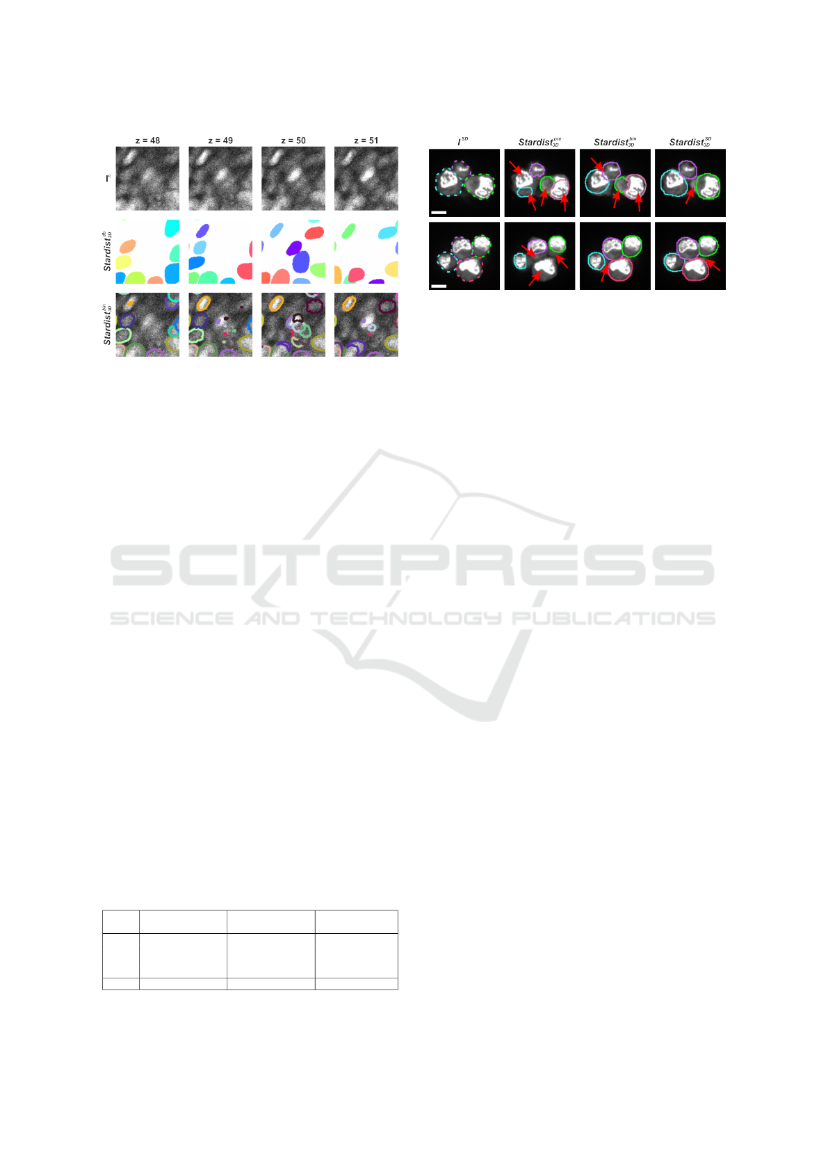
Figure 6: Stardist
bin
3D
fails to properly handle isolated 2D
masks by over-segmenting them.
nuclei present in I
SD
(Table 1), thanks to Stardist
db
2D
performing well in segmenting the nuclei in this low
density nuclei distribution. Nevertheless, the seg-
mentation being done in a per-frame basis, it led to
the wrong identification of some cytoplasm as nu-
cleus, explaining the large number of FP. Finally,
we used our workflow to train a new Stardist 3D
model Stardist
SD
3D
with paired of synthetic volumes
I
SD
synth
(generated by transferring the style of I
SD
to the
transformed masks dt(M
LS
GT
)) and masks M
LS
GT
. Sim-
ilarly to Stardist
bin
3D
, Stardist
SD
3D
successfully detected
99% of the nuclei (Table 1). Nevertheless, directly ac-
counting the axial direction for segmentation, in com-
parison to merging 2D masks to 3D as in Stardist
bin
3D
,
ended up with a lower number of errors with only 3
FP resulting from over-segmented cells.
4.3 Discussion
Our workflow relies heavily on the FCGAN image
synthesis capabilities which are, in our context, much
more reliable than a CycleGAN thanks to the FC-
GAN’s multi-scale feature and paired training dataset.
We have shown that using the pre-trained Stardist
db
2D
model directly on the acquired microscopy images
and modifying the obtained instance masks was suffi-
cient to properly train a FCGAN model. On the con-
Table 1: Comparison of the segmentation accuracy between
Stardist
pre
3D
, Stardist
bin
3D
and Stardist
SD
3D
(Data 1, n = 33; Data
2, n = 23; Data 3, n = 11; Data 4, n = 23.
I
SD
Stardist
pre
3D
Stardist
bin
3D
Stardist
SD
3D
TP FP FN TP FP FN TP FP FN
Data 1 97% 15% 3% 100% 0% 0% 97% 9% 3%
Data 2 91% 13% 8% 100% 30% 0% 100% 0% 0%
Data 3 91% 27% 9% 100% 36% 0% 100% 0% 0%
Data 4 87% 9% 13% 96% 17% 4% 100% 0% 0%
Total 92% 14% 8% 99% 17% 1% 99% 3% 1%
Figure 7: Comparison of the segmentation provided by
Stardist
pre
3D
, Stardist
bin
3D
and Stardist
SD
3D
, scale bar = 5 µm.
Red arrows pinpoint regions with segmentation errors.
trary, the segmentation provided by Stardist
db
2D
was
not accurate enough to directly train a Stardist model
in 3D such as Stardist
bin
3D
. This discrepancy is related
to the fact that Stardist
db
2D
does not account for the ax-
ial direction, with several nuclei being therefore only
segmented over one or two frames. It leads to a nu-
clei over-segmentation by Stardist
bin
3D
, and therefore
a possibly larger number of FP and FN. In our work-
flow, Stardist
db
2D
is only used to learn the style transfer.
Creation of the synthetic volumes is done by apply-
ing the new style to dt(M
LS
GT
), a dataset composed of
hand annotated masks in which all the nucleus are ac-
curately identified. Since our models Stardist
c
3D
and
Stardist
SD
3D
are trained with these synthetic volumes,
they are mostly insensitive to the segmentation ac-
curacy of Stardist
db
2D
and are tailored to segment nu-
clei having the same texture as the acquisitions they
have been trained on, therefore outperforming both
Stardist
bin
3D
and Stardist
pre
3D
.
Importantly, our synthetic volumes I
c
synth
and I
SD
synth
are composed of stacked generated 2D frames that
does not guarantee intensity coherency in the axial di-
rection (Fig. 8). They also lack smooth transitions be-
tween some appearing and disappearing nuclei as one
would expect from a real acquisition. Nevertheless,
our results confirm precedent findings (Baniukiewicz
et al., 2019; Wu et al., 2023) for which training seg-
mentation networks with synthetic volumes allows to
achieve good performance.
As already explained by the authors of
Stardist (Schmidt et al., 2018; Weigert et al.,
2020) and Cellpose (Stringer et al., 2021), we want
to reinforce the fact that it is critical to train these
models with data that have objects of similar sizes
and features than the images we want to segment.
Training Stardist with synthetic volumes as proposed
in our workflow facilitates this process. It becomes
indeed sufficient to resize M
LS
GT
to generate images
with nuclei exhibiting the same sizes and textures
than the ones composing the acquired volumes we
BIOIMAGING 2024 - 11th International Conference on Bioimaging
270
Frame n
Frame n + 1
Instance masks
Synthetic images
Figure 8: As synthetic volumes are composed of stack gen-
erated frames, a lack of smooth transition between appear-
ing and disappearing nuclei is visible (red arrows), scale bar
= 10 µm.
want to segment.
Finally, the border detection of the cells compos-
ing I
SD
by Stardist
SD
3D
was not perfect and can be ex-
plained by two reasons (Fig. 7). First, the 3D seg-
mentation provided by Stardist is highly convex while
I
SD
exhibits cells with irregular and possibly concave
borders. Second, I
SD
is composed of volumes with
saturated intensity, resulting in a compressed dynam-
ics. This combined with the small number of volumes
prevented FCGAN to learn a perfect style mapping.
5 CONCLUSION
In this work, our objective was not to develop a
method achieving state of the art accuracy for seg-
menting nuclei in 3D. In the past years (Mougeot
et al., 2022), nuclei segmentation has seen the de-
velopment of hundreds of methods competing for
the leading position in term of segmentation accu-
racy. Unfortunately, most of these techniques are out
of reach for life science labs and imaging facilities
because they can be limited to one operating sys-
tem or do not provide source code, tutorial or toy
datasets (Mougeot et al., 2022). We therefore wanted
to show that it is also possible to achieve good qualita-
tive segmentation by using already established meth-
ods such as Stardist (Schmidt et al., 2018; Weigert
et al., 2020) that are widely used in labs and facilities.
Image synthesis with GANs is still, however, far from
being easily accessible to life scientists. We there-
fore focused on a GAN architecture designed for the
multi-scale organization of biological images. The
FCGAN of Han (Han et al., 2020) was therefore an
ideal choice as it allowed us to directly use our ac-
Figure 9: From the same mask image, two synthetic images
were generated with a different style.
quired microscopy images without cropping or fine-
tuning parameters of the model.
All things considered, our workflow resulted in
qualitatively good segmentation of microscopy vol-
umes for which no GT existed. We were able to
generate synthetic volumes having the style of dif-
ferent cell types and microscopy modalities from the
same set of 3D GT masks (Fig. 9). Pairing them to-
gether, we trained several Stardist models tailored for
each acquisition, managing to segment datasets with-
out spending weeks in annotating volumes. We also
quantitatively demonstrated that our workflow per-
formed better than a pre-trained Stardist 3D model on
a limited set of 4 volumes. In the future, we plan to
push further this quantification as wells as further test
its generalization capability by acquiring new datasets
having a higher complexity than the I
SD
dataset. We
also plan to test Omnipose (Cutler et al., 2022) in our
workflow, with the aim to better identify irregular cell
borders.
Another point to consider is that the image syn-
thesis provided by FCGAN will always be heavily
dependent on the quality of the nuclei 2D segmen-
tation. In our case, the pre-trained Stardist
db
2D
model
gave satisfactory segmentation for feeding the FC-
GAN model. Otherwise, it will be necessary to gener-
ate new 2D manual annotations, a task still a lot easier
than 3D annotations that can be accelerated by tech-
niques such as SAM (Kirillov et al., 2023).
Finally, we think that this workflow could be
improved by further manipulating the existing GT
masks. GANs require similar distributions of the ob-
jects morphology and spatial organization between
the source and target styles to generate realistic im-
ages (Liu et al., 2020). Similarly, Stardist would also
benefit from training with pairs having the same dis-
tributions that the volumes to segment. This requires
the ability to quantify the differences between these
distributions, as it would allow to modify the GT
masks to make them similar to the objects one would
want to segment.
3D Nuclei Segmentation by Combining GAN Based Image Synthesis and Existing 3D Manual Annotations
271

REFERENCES
Baniukiewicz, P., Lutton, E. J., Collier, S., and Bretschnei-
der, T. (2019). Generative adversarial networks for
augmenting training data of microscopic cell images.
Frontiers in Computer Science, 1.
Beghin, A., Grenci, G., Sahni, G., Guo, S., Rajendiran, H.,
Delaire, T., Mohamad Raffi, S. B., Blanc, D., de Mets,
R., Ong, H. T., Galindo, X., Monet, A., Acharya, V.,
Racine, V., Levet, F., Galland, R., Sibarita, J.-B., and
Viasnoff, V. (2022). Automated high-speed 3d imag-
ing of organoid cultures with multi-scale phenotypic
quantification. Nature Methods, 19(7):881–892.
Caicedo, J. C., Goodman, A., Karhohs, K. W., Cimini,
B. A., Ackerman, J., Haghighi, M., Heng, C., Becker,
T., Doan, M., McQuin, C., Rohban, M., Singh, S., and
Carpenter, A. E. (2019). Nucleus segmentation across
imaging experiments: the 2018 data science bowl. Na-
ture Methods, 16(12):1247–1253.
Chen, Q. and Koltun, V. (2017). Photographic image syn-
thesis with cascaded refinement networks.
Cutler, K. J., Stringer, C., Lo, T. W., Rappez, L., Strous-
trup, N., Brook Peterson, S., Wiggins, P. A., and
Mougous, J. D. (2022). Omnipose: a high-precision
morphology-independent solution for bacterial cell
segmentation. Nature Methods, 19(11):1438–1448.
Fu, C., Arora, A., Engl, W., Sheetz, M., and Viasnoff, V.
(2022). Cooperative regulation of adherens junction
expansion through epidermal growth factor receptor
activation. Journal of Cell Science, 135(4):jcs258929.
Fu, C., Lee, S., Ho, D. J., Han, S., Salama, P., Dunn, K. W.,
and Delp, E. J. (2018). Three dimensional fluores-
cence microscopy image synthesis and segmentation.
In 2018 IEEE/CVF Conference on Computer Vision
and Pattern Recognition Workshops (CVPRW), pages
2302–23028.
Galland, R., Grenci, G., Aravind, A., Viasnoff, V., Studer,
V., and Sibarita, J.-B. (2015). 3d high- and super-
resolution imaging using single-objective spim. Na-
ture Methods, 12(7):641–644.
Goodfellow, I. J., Pouget-Abadie, J., Mirza, M., Xu, B.,
Warde-Farley, D., Ozair, S., Courville, A., and Ben-
gio, Y. (2014). Generative adversarial networks.
Han, L., Murphy, R. F., and Ramanan, D. (2020). Learning
generative models of tissue organization with super-
vised gans.
Isola, P., Zhu, J.-Y., Zhou, T., and Efros, A. A. (2018).
Image-to-image translation with conditional adversar-
ial networks.
Jensen, C. and Teng, Y. (2020). Is it time to start transition-
ing from 2d to 3d cell culture? Frontiers in Molecular
Biosciences, 7.
Kapalczynska, M., Kolenda, T., Przybyla, W., Za-
jaczkowska, M., Teresiak, A., Filas, V., Ibbs, M., Bliz-
niak, R., Luczewski, L., and Lamperska, K. (2018). 2d
and 3d cell cultures – a comparison of different types
of cancer cell cultures. Archives of Medical Science,
14(4):910–919.
Kirillov, A., Mintun, E., Ravi, N., Mao, H., Rolland, C.,
Gustafson, L., Xiao, T., Whitehead, S., Berg, A. C.,
Lo, W.-Y., Doll
´
ar, P., and Girshick, R. (2023). Seg-
ment anything.
Li, G., Liu, T., Tarokh, A., Nie, J., Guo, L., Mara, A., Hol-
ley, S., and Wong, S. T. (2007). 3d cell nuclei seg-
mentation based on gradient flow tracking. BMC Cell
Biology, 8(1):40.
Liu, Q., Gaeta, I. M., Millis, B. A., Tyska, M. J., and
Huo, Y. (2020). Gan based unsupervised segmenta-
tion: Should we match the exact number of objects.
Long, J., Yan, Z., Peng, L., and Li, T. (2021). The geometric
attention-aware network for lane detection in complex
road scenes. PLOS ONE, 16(7):1–15.
Malpica, N., de Sol
´
orzano, C. O., Vaquero, J. J., Santos,
A., Vallcorba, I., Garc
´
ıa-Sagredo, J. M., and del Pozo,
F. (1997). Applying watershed algorithms to the seg-
mentation of clustered nuclei. Cytometry, 28(4):289–
297.
Mougeot, G., Dubos, T., Chausse, F., P
´
ery, E., Grau-
mann, K., Tatout, C., Evans, D. E., and Desset, S.
(2022). Deep learning – promises for 3D nuclear
imaging: a guide for biologists. Journal of Cell Sci-
ence, 135(7):jcs258986.
Ronneberger, O., Fischer, P., and Brox, T. (2015). U-
net: Convolutional networks for biomedical image
segmentation. In Navab, N., Hornegger, J., Wells,
W. M., and Frangi, A. F., editors, Medical Image Com-
puting and Computer-Assisted Intervention – MICCAI
2015, pages 234–241, Cham. Springer International
Publishing.
Schmidt, U., Weigert, M., Broaddus, C., and Myers, G.
(2018). Cell detection with star-convex polygons.
In Frangi, A. F., Schnabel, J. A., Davatzikos, C.,
Alberola-L
´
opez, C., and Fichtinger, G., editors, Med-
ical Image Computing and Computer Assisted In-
tervention – MICCAI 2018, pages 265–273, Cham.
Springer International Publishing.
Stringer, C., Wang, T., Michaelos, M., and Pachitariu, M.
(2021). Cellpose: a generalist algorithm for cellular
segmentation. Nature Methods, 18(1):100–106.
Wang, J., Tabassum, N., Toma, T. T., Wang, Y., Gahlmann,
A., and Acton, S. T. (2022). 3D GAN image synthesis
and dataset quality assessment for bacterial biofilm.
Bioinformatics, 38(19):4598–4604.
Weigert, M., Schmidt, U., Haase, R., Sugawara, K., and
Myers, G. (2020). Star-convex polyhedra for 3d object
detection and segmentation in microscopy. In 2020
IEEE Winter Conference on Applications of Computer
Vision (WACV), pages 3655–3662, Los Alamitos, CA,
USA. IEEE Computer Society.
Wu, L., Chen, A., Salama, P., Winfree, S., Dunn, K. W.,
and Delp, E. J. (2023). Nisnet3d: three-dimensional
nuclear synthesis and instance segmentation for flu-
orescence microscopy images. Scientific Reports,
13(1):9533.
Zhu, J.-Y., Park, T., Isola, P., and Efros, A. A. (2017).
Unpaired image-to-image translation using cycle-
consistent adversarial networks. In Computer Vision
(ICCV), 2017 IEEE International Conference on.
BIOIMAGING 2024 - 11th International Conference on Bioimaging
272
