
Spatial Analysis of Blastocystosis Patients in Lampung Province:
Study on Malignancy Patients who Received Chemotherapy at
RSUDAM Lampung Province in 2022
Jhons Fatriyadi Suwandi
1,* a
, Noviany
2b
, Juspeni Kartika
3
and Agnes Kurniawan
4c
1
Department of Microbiology and Parasitologi, Faculty of Medicine, University of Lampung, Bandar Lampung, Indonesia
2
Department of Chemistry, Faculty of Mathematics and Natural Sciences, University of Lampung,
Bandar Lampung, Indonesia
3
Department of Internal Medicine, Faculty of Medicine, University of Lampung / Dr. H Abdul Moeloek General Hospital of
Lampung Province, Bandar Lampung, Indonesia
4
Department of Parasitology, Faculty of Medicine, University of Indonesia, Jakarta, Indonesia
Keywords: Blastocystis sp, Immunosuppressive, Spatial Analysis, Malignancy.
Abstract: Blastocystosis is an intestinal protozoan infection that can cause diarrhea. In the immunocompetent group, it
can be asymptomatic. However, in immunosuppressed patients, it can be severe. The disease is transmitted
by the oro-faecal route through food that has been contaminated with the faeces of patients. Spatial analysis
can be used to map people to determine the risk of the parasite spreading. The aim of this study was to
investigate the mapping of blastocystosis patients, studies in patients with malignancies at Lampung
Provincial Hospital. We diagnosed Blastocystis infection in 62 fecal samples from study subjects through
fecal microscopic examination. To determine the location of the patients' residences, we used Google Maps
and Google Art applications based on the addresses listed on the questionnaire. The coordinate data were
analyzed, and mapped using the ArcGIS 10.8.2. The results of the microscopic examination showed that
32.26% of the samples were positive for Blastocystis infection. Patients are distributed within the coordinates
of 104.6380950 to 109.1035280 East and 3.9533070 to 7.0776440 South. The majority of patients are located
in Bandar Lampung City, with the farthest origin of residence being Purbalingga Regency in Central Java
Province. The buffering pattern was obtained using a radius of 10 kilometers.
1
INTRODUCTION
Several species of intestinal protozoa have been
reported to be one of the causes of intestinal disorders
such as diarrhea, which can be persistent and chronic
diarrhea or acute diarrhea. Intestinal protozoal
infections in immunocompetent groups are usually
asymptomatic or mildly symptomatic.
In immunosuppressive groups such as patients
with HIV/AIDS, malignancies, patients on
immunosuppressive therapy and several other
conditions of impaired immunity can be severe and
can cause death (Esteghamati et al., 2019; Fletcher et
a
https://orcid.org/0000-0002-6938-1628
b
https://orcid.org/0000-0002-4046-6134
c
https://orcid.org/0000-0003-1969-5503
al., 2014). Intestinal protozoa that have been reported
as causes of infection are Giardia lamblia,
Cryptosporidium sp, Amoeba, Isospora sp,
Cyclospora sp and Blastocystis sp (Boughattas et al.,
2017; Esteghamati et al., 2019; Giannakopoulos et al.,
2019; Laksemi et al., 2020).
Intestinal protozoal infections are often overlooked
and underdiagnosed. This is because the symptoms are
mild and even asymptomatic, especially in
immunocompetent groups. However, intestinal
protozoal infection is one of the infections that can
cause increased morbidity and mortality in the
community. Impaired nutrient absorption, resulting in
impaired growth and development, especially in
Suwandi, J. F., Noviany, , Kartika, J. and Kurniawan, A.
Spatial Analysis of Blastocystosis Patients in Lampung Province: Study on Malignancy Patients who Received Chemotherapy at RSUDAM Lampung Province in 2022.
DOI: 10.5220/0013672300003873
Paper published under CC license (CC BY-NC-ND 4.0)
In Proceedings of the 1st International Conference on Medical Science and Health (ICOMESH 2023), pages 353-358
ISBN: 978-989-758-740-5
Proceedings Copyright © 2025 by SCITEPRESS – Science and Technology Publications, Lda.
353

children under five years of age, may lead to cognitive
impairment in the future. It is therefore important to
overcome this infection (Faria et al., 2017).
Blastocystis sp is a cosmopolitan protozoan
parasite. Symptoms are generally atypical and include
abdominal pain, constipation, diarrhea, flatulence, and
irritable bowel syndrome (IBS). Several species of this
parasite are found in the gastrointestinal tract of
animals and humans. Several subtypes have also been
identified. Not all subtypes infect humans, with only 9
of the 17 reported subtypes found in humans (Asfaram
et al., 2019; Dacal et al., 2018; Ramírez et al., 2016;
Villamizar et al., 2019; Wawrzyniak et al., 2013) This
protozoan is thought to be transmitted by the fecal-oral
route. Transmission can occur from person to person,
but the possibility of transmission from person to
animal and vice versa is unclear (Ramírez et al., 2016).
Geographic Information Systems (GIS) are an
important tool for studying the distribution of disease
within a location or region. Disease mapping is useful
for understanding the distribution of disease incidence
and identifying underlying geographic risk factors. In
addition, disease mapping facilitates decision making
and rapid response for disease management. There are
not many studies that discuss the spatial analysis of
intestinal protozoan infections, especially
blastocystosis in Indonesia. In fact, one of the factors
in the spread of diseases, especially orofaecally
transmitted diseases, is closely related to
geographical, environmental and socio-cultural
conditions (Fletcher et al., 2014). The use of spatial
analysis on the spread of parasitic diseases has been
done in Lampung Province (Dharmamedula et al.,
2017; Suwandi et al., 2014; Wardani et al., 2018), but
blastocystosis infection has never been done. This
paper discusses the spatial analysis of blastocystosis
patients in the group of malignancy patients who
received chemotherapy in Lampung Province.
2
MATERIALS AND METHODS
The design of this study was cross-sectional
analytical observational conducted at RSUDAM
Lampung Province. The population of this study were
patients with malignancies who received
chemotherapy with or without symptoms of diarrhea
at RSUDAM Lampung Province in the period
October to November 2022. The sample of this study
is part of the population determined based on
inclusion and exclusion criteria. Inclusion criteria for
determining research subjects, namely patients with
malignancies who receive chemotherapy with or
without diarrhea symptoms; age> 1 year; have
received at least 1 chemotherapy with a minimum
rarity of at least 1 week since the first chemotherapy;
willing to participate in the study by signing an
informed consent. Exclusion criteria are suffering
from severe complications, not willing to provide
feces.
To determine blastocystosis infection, fecal
parasitologic examination was performed on all fecal
samples of the study subjects. Fecal examination was
performed using the formalin ether concentration
technique. This technique is used to increase the
sensitivity, especially when the number of parasites is
low. Approximately 1 ml of fecal suspension in 5%
formalin was added to 5 ml of 10% formalin and
stirred until homogeneous. After filtering the feces to
separate the fiber, 2 ml of diethyl ether was added.
The solution was shaken vigorously and centrifuged
at 2500 rpm for 2 minutes. The precipitate was
removed and the supernatant was collected. The
sediment from this concentration test is used for
direct testing with 1% Lugol's solvent, which is then
read under a microscope at 400X magnification. The
result of the test is positive if Blastocystis sp is found
or negative if not found.
In order to determine the coordinates of the
location of the research subject's residence, it was
carried out with the help of google map and google
earth devices. The home address contained in the
questionnaire was entered into google map or google
earth to find the location of the research subject's
home. The coordinate points that were successfully
found were then analyzed to see the mapping of
patients and the buffering pattern formed using
ArcGIS 10.8.2 software. The patients were mapped
based on the results of the microscopic examination.
The buffering pattern refers to the gate distance of
houseflies as one of the mechanical vectors for the
spread of intestinal protozoa. This research has
received ethical approval from the Health Ethics
Committee of the Faculty of Medicine, University of
Lampung No. 2059/UN26.18/PP/05.02.00/2022.
3
RESULTS AND DISCUSSION
Geographically, the residence of the research
subjects is located at the coordinates 104.6380950
east longitude - 109.1035280 east longitude and
3.9533070 south latitude - 7.0776440 south latitude.
Administratively, Bandar Lampung City is the place
where most of the research subjects live.
Thesesubjects generally live in densely populated
neighborhoods. Figure 1 shows the distribution of
research subjects.
ICOMESH 2023 - INTERNATIONAL CONFERENCE ON MEDICAL SCIENCE AND HEALTH
354
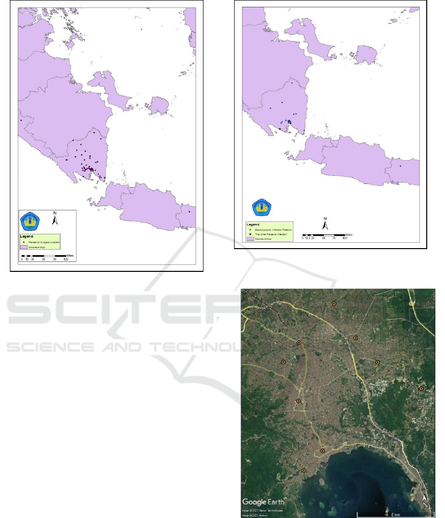
Figure 1: Distribution of All Research Subjects
Based on the results of microscopic examination
of fecal parasitology, 20 subjects (32.26%) were
positive for Blastocystis sp, 1 subject (1.61%) was
positive for Entamoeba histolytica, and 1 subject
(1.62%) was positive for hookworm. Almost all
subjects found to have parasites resided in Lampung
province, only 1 person came from Purbalingga,
Central Java. The distribution of patients found
positive for parasites by microscopic examination is
shown in Figure 2. Administratively, the location of
the most positive patients was found in Bandar
Lampung City.
Based on satellite imagery, it appears that the
research subjects who were positive for
blastocystosis in Bandar Lampung were located in
densely populated areas. This will certainly have an
impact on the risk area for the spread of this infection.
The satellite image is shown in Figure 3.
can result in the parasite entering the human body
(CDC, 2019). The presence of mechanical vectors
also increases the transmission process by these
vectors. Houseflies (Musca domestica) are one of the
mechanical vectors that are suspected to be one of the
factors that facilitate the spread of this parasite. The
ability to fly and move far from the housefly, as well
as the resistance of the parasite attached to the fly's
body, are important in the transmission process of
this parasite (Hastutiek & Fitri, 2013; Szostakowska
Figure 2: Distribution of Research Subjects Positive for
Blastocystis sp and Other Intestinal Parasitic Infections
Figure 3: Satellite images of blastocystosis in Bandar
Lampung
et al., 2004). In addition to houseflies as potential
mechanical vectors, cockroaches have been reported
as potential mechanical vectors in the transmission of
Blastocystis sp (Dokmaikaw & Suntaravitun, 2019;
Ma et al., 2020).
Spatial Analysis of Blastocystosis Patients in Lampung Province: Study on Malignancy Patients who Received Chemotherapy at RSUDAM
Lampung Province in 2022
355
The mechanism of transmission of Blastocystis sp
remains unclear. The infective stage that can be
transmitted to humans has not been identified. In
general, this transmission is fecal-oral. Parasite
contamination from feces on food or eating utensils
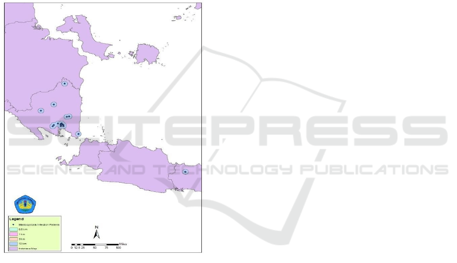
Buffer zones are areas of potential or risk for
transmission. The radius of this buffer area refers to
the area of contact and transmission of parasites by
mechanical vectors of houseflies, which are abundant
in the environment. The flight distance of houseflies
can reach 32 km (20 miles) (Hastutiek & Fitri, 2013;
Puspitarani et al., 2017; Szostakowska et al., 2004;
Zurek et al., 2001). In this study, the radius of the
buffer area is up to 10 km, and an overview of the
buffer area is shown in Figure 4.
Figure 4: Buffering patterns in patients with blastocystosis
Based on the prevalence obtained, this study has
a higher prevalence (32.26%) compared to research
in Sydney (5%) (Fletcher et al., 2014); in Rio de
Janeiro (12.7%) (Faria et al., 2017); in Iran (5.2%)
(Asfaram et al., 2019) and in Padang by 21.3%
(Nofita et al., 2015). Other studies in Brazil showed
a similar prevalence (31%) (Bertozzo et al., 2022)
and in Manado (36,4%) (Muflihatun et al., 2015).
This could be due to the different study populations.
In this study, the population was a high-risk group,
namely patients suspected of having a compromised
immune system. In the immunocompromised
population, the incidence of protozoal infections
tends to be high and can cause opportunistic
infections, including Blatocystosis (Laksemi et al.,
2020; Xu et al., 2021).
The incidence of intestinal protozoan infections
such as Blastocystis sp is inextricably linked to
favorable environmental conditions such as poor
sanitation, population density, availability of
mechanical vectors, and community behavior. Open
defecation practices may result in environmental
contamination by human feces containing protozoa.
This is consistent with the results of other spatial
analysis studies, such as those in Rio de Janeiro
(Faria et al., 2017) and Iran (Asfaram et al., 2019).
4
CONCLUSIONS
The prevalence of blastocystosis in risk groups such
as patients with malignancies was 32.26%. The
buffering pattern formed is quite extensive, especially
in Bandar Lampung City, although in general the
subtype of this parasite is not yet known.
ACKNOWLEDGEMENTS
We would like to thank all those who helped this
study, especially the patients (volunteers) who were
willing to be the subject of this study. As well as to
the University of Lampung HETI Project which has
funded this research.
REFERENCES
Asfaram, S., Daryani, A., Sarvi, S., Pagheh, A. S., Hosseini,
S. A., Saberi, R., Hoseiny, S. M., Soosaraei, M., &
Sharif, M. (2019). Geospatial analysis and
epidemiological aspects of human infections with
Blastocystis hominis in Mazandaran Province, northern
Iran. Epidemiology and Health, 41, e2019009.
https://doi.org/10.4178/epih.e2019009
Bertozzo, T. V., David, É. B., Oliveira-Arbex, A. P.,
Victória, C., & Guimarães, S. (2022). Frequency,
spatial distribution, and genetic diversity of
Blastocystis among referred individuals to a clinical
laboratory: First report of subtype 9 in Brazil. Acta
Tropica, 234(June), 1–8.
https://doi.org/10.1016/j.actatropica.2022.106608
Boughattas, S., Behnke, J. M., Al-Ansari, K., Sharma, A.,
Abu-Alainin, W., Al-Thani, A., & Abu-Madi, M. A.
(2017). Molecular analysis of the enteric protozoa
associated with acute diarrhea in hospitalized children.
Frontiers in Cellular and Infection Microbiology,
7(AUG), 1–10.
https://doi.org/10.3389/fcimb.2017.00343
ICOMESH 2023 - INTERNATIONAL CONFERENCE ON MEDICAL SCIENCE AND HEALTH
356

CDC. (2019). Blastocystis sp.
https://www.cdc.gov/dpdx/blastocystis/index.html
Dacal, E., Saugar, J. M., De Lucio, A., Hernández-De-
Mingo, M., Robinson, E., Köster, P. C., Aznar-Ruiz-
De-Alegría, M. L., Espasa, M., Ninda, A., Gandasegui,
J., Sulleiro, E., Moreno, M., Salvador, F., Molina, I.,
Rodríguez, E., & Carmena, D. (2018). Prevalence and
molecular characterization of Strongyloides stercoralis,
Giardia duodenalis, Cryptosporidium spp., and
Blastocystis spp. isolates in school children in Cubal,
Western Angola. Parasites and Vectors, 11(1), 1–18.
https://doi.org/10.1186/s13071-018-2640-z
Dharmamedula, Y. P., Mutiara, H., Suwandi, J. F., &
Setyaningrum, E. (2017). Mapping of infected students
and association of socioeconomic factors and parents'
knowledge level with incidence of soil-transmitted
helminth infections among primary school students in
Natar subdistrict, South Lampung District. Medula,
7(5).
https://juke.kedokteran.unila.ac.id/index.php/medula/a
rticle/view/1921
Dokmaikaw, A., & Suntaravitun, P. (2019). Prevalence of
parasitic contamination of cockroaches collected from
fresh markets in Chachoengsao province, Thailand.
Kobe Journal of Medical Sciences, 65(4), E118–E123.
Esteghamati, A., Khanaliha, K., Bokharaei-Salim, F.,
Sayyahfar, S., & Ghaderipour, M. (2019). Prevalence
of intestinal parasitic infection in cancer, organ
transplant and primary immunodeficiency patients in
Tehran, Iran. Asian Pacific Journal of Cancer
Prevention, 20(2), 495–501.
https://doi.org/10.31557/APJCP.2019.20.2.495
Faria, C. P., Zanini, G. M., Dias, G. S., da Silva, S., de
Freitas, M. B., Almendra, R., Santana, P., & Sousa, M.
do C. (2017). Geospatial distribution of intestinal
parasitic infections in Rio de Janeiro (Brazil) and its
association with social determinants. PLoS Neglected
Tropical Diseases, 11(3), 1–21.
https://doi.org/10.1371/journal.pntd.0005445
Fletcher, S., Caprarelli, G., Merif, J., Andresen, D., Van
Hal, S., Stark, D., & Ellis, J. (2014). Epidemiology and
geographical distribution of enteric protozoan
infections in Sydney, Australia. Journal of Public
Health Research, 3(2), 83–91.
https://doi.org/10.4081/jphr.2014.298
Giannakopoulos, X., Sakkas, H., Ragos, V., Tsiambas, E.,
Bozidis, P., Evangelou, A. M., Papadopoulou, C.,
Petrogian-Nopoulos, L., & Sofikitis, N. (2019). Impact
of enterococcal urinary tract infections in
immunocompromised – neoplastic patients. Journal of
B.U.ON., 24(5), 1768–1775.
Hastutiek, P., & Fitri, L. E. (2013). Potency of M. domestica
Linn. as a Vector for Several Diseases Jurnal
Kedokteran Brawijaya, 23(3), 125–136.
https://doi.org/10.21776/ub.jkb.2007.023.03.4
Laksemi, D. A., Suwanti, L. T., Suwanti, L. T., Mufasirin,
M., Mufasirin, M., Suastika, K., & Sudarmaja, M.
(2020). Opportunistic parasitic infections in patients
with human immunodeficiency virus/acquired
immunodeficiency syndrome: A review. Veterinary
World, 13(4), 716–725.
ttps://doi.org/10.14202/vetworld.2020.716-725
Ma, L., Zhang, Y., Qiao, H., Li, S., Wang, H., Zhang, N., &
Zhang, X. (2020). Cockroach as a vector of blastocystis
sp. Is risk for golden monkeys in zoo. Korean Journal
of Parasitology, 58(5), 583–587.
https://doi.org/10.3347/kjp.2020.58.5.583
Muflihatun, T., Bernadus, J. B. B., & Wahongan, G. J. P.
(2015). Comparison of Blastocystis hominis Detection
by Microscopic Examination and Copro Elisa
Examination. Jurnal E-Biomedik, 3(1), 1–4.
https://doi.org/10.35790/ebm.3.1.2015.7476
Nofita, E., Harminarti, N., & Rusjdi, S. R. (2015).
Microscopic and PCR Identification of Blastocystis
hominis in Stool Samples at the Laboratory of Dr. M.
Djamil Hospital Padang. Majalah Kedokteran Andalas,
37(1), 26. https://doi.org/10.22338/mka.v37.i1.p26-
31.2014
Puspitarani, F., Sukendra, D. M., & Siwiendrayanti, A.
(2017). Application of Ultraviolet Light on Fly Trap
Equipment on the Number of Houseflies Trapped.
Higeia Journal of Public Health Research and
Development, 1(3), 84–94.
Ramírez, J. D., Sánchez, A., Hernández, C., Flórez, C.,
Bernal, M. C., Giraldo, J. C., Reyes, P., López, M. C.,
García, L., Cooper, P. J., Vicuña, Y., Mongi, F., &
Casero, R. D. (2016). Geographic distribution of human
Blastocystis subtypes in South America. Infection,
Genetics and Evolution, 41, 32–35.
https://doi.org/10.1016/j.meegid.2016.03.017
Suwandi, J. F., Supargiyono, S., Asmara, W., & Kusnanto,
H. (2014). Mapping and Prevalence of Malaria
Falciparum Patients with ACT Failed Therapy, in
Hanura Public Health Center, Pesawaran, Lampung,
Indonesia. Open Journal of Epidemiology, 04(03), 169–
177. https://doi.org/10.4236/ojepi.2014.43023
Szostakowska, B., Kruminis-Lozowska, W., Racewicz, M.,
Knight, R., Tamang, L., Myjak, P., & Graczyk, T.
(2004). Cryptosporidium parvum and Giardia lamblia
recovered from flies on a cattle farm and in a landfill.
Applied and Environmental Microbiology, 70(6),
3742–3744. https://doi.org/10.1128/AEM.70.6.3742-
3744.2004
Villamizar, X., Higuera, A., Herrera, G., Vasquez-A, L. R.,
Buitron, L., Muñoz, L. M., Gonzalez-C, F. E., Lopez,
M. C., Giraldo, J. C., & Ramírez, J. D. (2019).
Molecular and descriptive epidemiology of intestinal
protozoan parasites of children and their pets in Cauca,
Colombia: A cross-sectional study. BMC Infectious
Diseases, 19(1), 1–11. https://doi.org/10.1186/s12879-
019-3810-0
Wardani, A. B., Suwandi, J. F., & Sari, R. D. P. (2018).
Mapping of Potential Mosquito Breeding Sites in
Hanura Health Center Area. Medula, 8(1).
https://juke.kedokteran.unila.ac.id/index.php/medula/a
rticle/view/2105
Wawrzyniak, I., Poirier, P., Texier, C., Delbac, F.,
Viscogliosi, E., Dionigia, M., & Alaoui, H. E. (2013).
Blastocystis, an unrecognized parasite: An overview of
pathogenesis and diagnosis. Therapeutic Advances in
Spatial Analysis of Blastocystosis Patients in Lampung Province: Study on Malignancy Patients who Received Chemotherapy at RSUDAM
Lampung Province in 2022
357

Infectious Disease, 1(5), 167–178.
https://doi.org/10.1177/2049936113504754
Xu, N., Jiang, Z., Liu, H., Jiang, Y., Wang, Z., Zhou, D.,
Shen, Y., & Cao, J. (2021). Prevalence and genetic
characteristics of Blastocystis hominis and
Cystoisospora belli in HIV/AIDS patients in Guangxi
Zhuang Autonomous Region, China. Scientific Reports,
11(1), 1–10. https://doi.org/10.1038/s41598-021-
94962-3
Zurek, L., Denning, S. S., Schal, C., & Watson, D. W.
(2001). Vector competence of Musca domestica
(Diptera: Muscidae) for Yersinia pseudotuberculosis.
Journal of Medical Entomology, 38(2), 333–335.
https://doi.org/10.1603/0022-2585-38.2.333
ICOMESH 2023 - INTERNATIONAL CONFERENCE ON MEDICAL SCIENCE AND HEALTH
358
