
Automatic Coronary Angiogram Keyframe Extraction
Hounaida Moalla
1,2 a
, Aiman Ghrab
3 b
,Bassem Ben Hamed
2,4 c
, Amine Bahloul
3 d
,
Rania Hammami
3 e
and Leila Abid
3 f
1
Higher Institute of Technological Studies, University of Sfax, Sfax, Tunisia
fi
3
Hedi Chaker University Hospital of Sfax, University of Sfax, Sfax, Tunisia
4
National School of Electronics and Telecommunications of Sfax, University of Sfax, Sfax, Tunisia
Keywords:
Coronary Angiograms, Keyframes, Filters.
Abstract:
Coronary artery disease is one of the most feared atherosclerosis complications. Doctors use coronary angiog-
raphy as a diagnostic tool to diagnose a patient with obstructive coronary artery disease and treat it efficiently.
The effectiveness of the doctor’s intervention strongly depends on the quality of the diagnosis. Therefore,
good extraction of keyframes from coronary angiography will certainly improve the accuracy of the decision.
Hence the importance is given to this step. To determine the best way to extract keyframes from coronary
angiograms, we tested several methods for keyframe extraction. Our keyframe extraction method that we pro-
pose is based on the use of filters and the calculation of frame intensities of a given coronary angiogram. The
pilot frame is the brightest one, and the keyframes will be its six neighboring frames. Our method Contrast
Enhanced Sato filter, succeeded in extracting the right keyframes with an accuracy of around 85.74%.
1 INTRODUCTION
Coronary artery disease is the first cause of mortal-
ity and morbidity in developed countries and world-
wide (Ojha and Dhamoon, 2021). This recent in-
crease in this disease is secondary mainly to one’s
health style and to the development of diagnostic tools
(Lee et al., 2022). Coronary angiography remains
the best diagnostic method for obstructive coronary
artery disease (CAD). However, it has some limita-
tions: clinicians often use visual assessment to assess
the severity of a coronary plaque obstruction. This
method is the source of interobserver variability. Ac-
curate estimation is of great importance because it
will lead to treatment strategies such as Percutaneous
Coronary Intervention (PCI), Coronary Artery By-
pass Graft (CABG) surgery, or simply medical treat-
ment (Neumann et al., 2019). To mitigate these lim-
a
https://orcid.org/0000-0003-3180-9446
b
https://orcid.org/0000-0002-1974-9551
c
https://orcid.org/0000-0003-1586-9537
d
https://orcid.org/0000-0003-1632-5646
e
https://orcid.org/0000-0003-1168-6450
f
https://orcid.org/0000-0001-7793-5240
itations, the constructors of Catheterization labora-
tory (Cath lab) equipment provide many solutions:
a simple method is Quantitative Coronary Angiogra-
phy analysis (QCA) (Collet et al., 2017). QCA is a
great tool because it estimates obstruction percentage
and the artery reference diameter by semi-automatic
keyframe analysis. It requires third-party manual in-
put, the clinician, or the Cath lab technician. Even
though this method decreases interobserver variabil-
ity, it still has poor reproducibility (Avram et al.,
2021). Fully automated analysis solutions are still
in the development stage and have not yet been im-
plemented in Cath labs. To extract information from
an angiogram, a sequence of X-ray captured images,
determining the keyframe is the first and most impor-
tant step because it will affect the analysis. In this
work, we try to determine the most efficient method
of keyframe extraction.
For technical reasons, X-Ray Angiogram (XRA)
has low contrast between vessels and background,
many artifacts (such as bony structures, pacemaker
leads, . . . ), image noise, and non-uniform illumina-
tion (Kerkeni et al., 2016). All these characteristics
make automatic recognition of vascular structures a
hard task. It is, therefore, necessary to improve the
582
Moalla, H., Ghrab, A., Ben Hamed, B., Bahloul, A., Hammami, R. and Abid, L.
Automatic Coronary Angiogram Keyframe Extraction.
DOI: 10.5220/0011850700003411
In Proceedings of the 12th International Conference on Pattern Recognition Applications and Methods (ICPRAM 2023), pages 582-589
ISBN: 978-989-758-626-2; ISSN: 2184-4313
Copyright
c
2023 by SCITEPRESS – Science and Technology Publications, Lda. Under CC license (CC BY-NC-ND 4.0)

quality of these images by applying enhancing algo-
rithms (increasing contrast, noise reduction. . . ) as a
first step.
The last decade has been marked by the evolution
of computing techniques in imaging thanks to new
hardware (memory capacity, processing speed,...),
software (new languages, specific applications,...),
and architectural technologies that are efficient and
easy to use. All these advantages have been widely
exploited in the medical field, particularly in the de-
tection of stenoses by image processing.
In the literature, several approaches to pre-
processing have been proposed. They consist of trac-
ing the borders of the vessels more clearly to be as
precise as possible when determining the vascular
volume (Danilov et al., 2021; Lamy et al., 2021).
These methods are mainly based on the application
of filters. The choice of filters highly depends on the
dataset and the quality of the images to be processed.
This paper demonstrates the results of differ-
ent filter-based algorithms applied to a coronary an-
giogram dataset and assesses them to determine the
most efficient algorithm.
2 RELATED WORKS
Video processing can consist of finding the keyframe
using different techniques such as motion-based in-
formation gathering, video frame aggregation, and
shot detection. On the other hand, the clas-
sic keyframe identification algorithms (in their raw
states) do not perform well in the processing of coro-
nary angiography videos. that’s why adaptations to
the field of application are useful (Kavipriya and Hire-
math, 2022). In fact, in recent research about auto-
matic coronary angiogram analysis, few acceptable
solutions were found. This may be due to the charac-
teristics of X-ray acquisitions (Kerkeni et al., 2016).
Since a keyframe is defined as a frame that con-
tains a vessel full of dye, some authors used vessel ex-
traction to identify it. Moon et al. (Moon et al., 2021)
automated keyframe detection method used this def-
inition: first, they applied a contrast enhancing treat-
ment using a multi-scale top-hat transform-based al-
gorithm, then, vessel structure was extracted using
a Frangi filter: the keyframe was defined as the one
the highest number of surviving pixels. Avram et al.
(Avram et al., 2021) used a similarity index, a param-
eter that translates the difference between each frame
and the first frame (empty vessel). The keyframe is
the one with the lowest index.
On the other hand, Zhou et al. (Zhou et al., 2021)
used a two-phase algorithm to train a deep learning
keyframe classification model using a manually la-
beled database. They used a ResNet 18 architecture
and a combination of neural network optimizers.
Keyframe extraction is based on image processing
techniques to detect the vessels. This process is pre-
ceded by applying filters on the images to increase the
contrasts (Zhou et al., 2021). The choice of the ade-
quate filter is not based on standard rules but rather
empirical, depending on the images’ quality and the
dataset used (Lamy et al., 2021). The first compar-
ative study of several filters was proposed by (Lamy
et al., 2021) such as Frangi, Sato, and Canny. Another
filter research was conducted by (Sazak et al., 2019)
to compare Hessian filters, Phase Congruency Tensor
(PCT), and mathematical morphology-based method.
(Qin et al., 2022) do not filter images but suggest
a new method for extracting vessels from coronary
angiography images. Their architecture is based on
a PCA unrolling network containing a pooling layer
and a long-term convolutional memory network.
On the other hand, the choice of keyframes has
been discussed in several previous works (Kerkeni
et al., 2016; Lamy et al., 2021; Gawande et al., 2020)
based on the calculations of intensity, similarity, dis-
tance, histograms, clustering,... Other means that are
also useful consist of applying deep learning models
to extract keyframes by eliminating those with great
similarities (Gawande et al., 2020). A combination of
the two strategies has also been proposed in (Jo et al.,
2018).
Closer to the domain, the process of extracting
keyframes depends on the context: it can mean ex-
tracting a summary of a sequence of images. There-
fore it chooses frames that sum up the whole se-
quence, just like film processing (Thakre et al., 2016;
Gawande et al., 2020; Jiang and Shi, 2021). Extrac-
tion can also focus on a sample of frames contain-
ing the maximum amount of data; we can cite the
example of medical angiograms (Zhou et al., 2021;
Gawande et al., 2020). These two have been the sub-
ject of several studies. The algorithms that have been
proposed eventually depend on the subject.
3 LITERATURE OF USED
METHODS
3.1 Hessian Filter
Most filtering methods are based on the calculation of
image intensity. The calculation of the Hessian matrix
then turns out to be the best method to give more per-
formance. For a 3D input image, the Hessian matrix
is a 3 × 3 matrix composed of second-order partial
Automatic Coronary Angiogram Keyframe Extraction
583

derivatives of the input image. At each point of the
image, Hessian matrix H is a function f(x1, x2, x3)
defined as in (1):
H( f ) =
h
11
h
12
h
13
h
21
h
22
h
23
h
31
h
32
h
33
=
∂
2
f
∂x
1
2
∂
2
f
∂x
1
∂x
2
∂
2
f
∂x
1
∂x
3
∂
2
f
∂x
2
∂x
1
∂
2
f
∂x
2
2
∂
2
f
∂x
2
∂x
3
∂
2
f
∂x
3
∂x
1
∂
2
f
∂x
3
∂x
2
∂
2
f
∂x
3
2
(1)
Hessian then applies a Gaussian kernel of standard
deviation σ to convolve the initial images. A blood
vessel will then be seen as a clear tube against a dark
background. Several filters are derived from Hessian
in order to improve the visibility of vessels such as
Sato, Canny, and Meijering.
3.2 Sato Filter
Sato is a method to improve the visibility of curvilin-
ear structures such as vessels and bronchi in 2D and
3D medical images (Sato et al., 1998). It is a ves-
sel enhancement approach based on the eigenvectors
of the Hessian matrix aimed at both the discrimina-
tion of linear structures from other structures and the
recovery of the original linear structures from the cor-
rupted structures. (2) expresses the Sato filter:
F =
λ
c
exp
−λ
2
1
2(α
1
λ
c
)
2
: λ
1
≤ 0,λ
c
̸= 0
λ
c
exp
−λ
2
1
2(α
2
λ
c
)
2
: λ
1
≥ 0,λ
c
̸= 0
0 : λ
c
= 0
(2)
with
λ
c
= min(−λ
2
,−λ
3
) (3)
3.3 Meijering Filter
The Meijering filter is a vessel function developed to
detect vascular structures. This approach was initially
proposed for 2D images (Meijering et al., 2004) and
then extended to 3D (Obara et al., 2012). It is also
based on the modified Hessian matrix defined by (4):
H(f ) =
h
11
+
α
2
(h
22
+ h
33
) (1 −
α
2
)h
12
(1 −
α
2
)h
13
(1 −
α
2
)h
21
h
22
+
α
2
(h
11
+ h
33
) (1 −
α
2
)h
23
(1 −
α
2
)h
31
(1 −
α
2
)h
32
h
33
+
α
2
(h
11
+ h
22
)
(4)
The Meijering filter is then defined as in (5):
F =
λ
max
/λ
min
: λ
max
≤ 0
0 : λ
max
≥ 0
(5)
where
λ
max
= max(λ
′
1
,λ
′
2
,λ
′
3
) (6)
is computed at each voxel.
3.4 Frangi Filter
This filter can also detect continuous ridges, such as
rivers, ripples, and ships in 2D and 3D images (Frangi
et al., 1998). It computes Hessian eigenvectors to
calculate the similarity of an image region to ves-
sels. The method relies on the use of three vectors to
be more discriminating. Three measures are derived
from these eigenvectors as shown in (7):
R
b
= |λ
1
|/
p
|λ
2
λ
3
|
R
a
= |λ
2
|/|λ
3
|
S =
q
λ
2
1
+ λ
2
2
+ λ
2
3
(7)
Where Rb is the blob-like structure measure and S
is the Frobenius norm of the Hessian matrix. These
measures are combined in a vesselness function as
given by (8):
V
σ
(p) = |x| =
(
0 i f λ
2
≤ 0
exp(−
R
2
b
2β
2
)(1 − exp(−
S
2
2c
2
)) otherwise
(8)
where β and c are thresholding parameters to con-
trol the sensitivity of the filter to Rb and S respec-
tively.
3.5 Canny Filter
It is a multi-step algorithm (Canny, 1986) :
• Noise reduction: this step is based on the appli-
cation of the Gaussian filter to remove the noise.
The equation for a Gaussian filter kernel of size
(2k+1)×(2k+1) is given as in (9):
H
i j
=
1
2πσ
2
exp(−
(i − (k + 1))
2
+ ( j − (k + 1))
2
2σ
2
)
(9)
with 1 ≤ i , j ≤ (2k+1).
The selection of the Gaussian kernel size will in-
fluence the performance of the detector. A small
size corresponds to a powerful sensitivity to noise.
A 5x5 or 3x3 can be good choices depending on
the input.
• Gradient calculation: computing intensity and di-
rection of edges by calculating the gradient of
the image applying edge detection operators. The
change in intensity of the pixels can mean the ex-
istence of edges whose detection can be done by
applying Sobel filters to show this change in inten-
sity in both directions: horizontal (x) and vertical
(y) as (10):
K
x
=
−1 0 1
−2 0 2
−1 0 1
,K
y
=
1 2 1
0 0 0
−1 −2 −1
(10)
ICPRAM 2023 - 12th International Conference on Pattern Recognition Applications and Methods
584

Then, the gradient is applied according to (11):
|G| =
q
I
2
x
+ I
2
y
θ(x,y) = arctan(
I
y
I
x
)
(11)
• Non-maximal suppression: A full image analysis
is performed to remove any unwanted pixels that
may not constitute the edge. The result will be a
binary image with ”thin edges”.
• And hysteresis threshold by setting two thresh-
old values for the intensity gradient (minVal and
maxVal) to detect the edges to keep and those to
delete. Thresholds must simply be well chosen.
Fig. 1(a) illustrates the effect of these filters on a
coronary angiogram.
3.6 Stacked Filters
The result of the filters mentioned above are modest.
For better output, we try the stack of filters. Adding
a gaussian filter to a frame before or after the applica-
tion of a Sato or a Frangi filter may lead to improved
results thanks to its ability to remove the noise. Also,
an after-treatment with a filter that improves the con-
trast could have much more outstanding outcomes.
This is highlighted in Fig. 1(b).
4 PROPOSED METHOD
To develop a fully automated keyframe extraction
method, we focused on vessel extraction techniques.
We transformed angiogram videos into a sequence of
frames. We then applied different vessel-enhancing
algorithms to determine the frame that contains a ves-
sel full of contract agents. Our contribution is sum-
marized in the extraction of keyframes from an an-
giographic video based on the use of filters in order to
improve the recognition of the heart vessels.
The filter technique in itself is not new, but the ex-
traction algorithm we propose has given good results.
In the first step, the set of filters we used was Canny,
Meijering and Sato. Each time, we apply a filter from
the list as shown in Algorithm 1.
#Algorithm 1: pre-treatment of frames
Algorithm Keyframes ( )
Begin
Input : set of frames
Output : list of keyframes
L=[ ]
for f in frames :
im=read(f)
gray=grayscale(im)
res=filter(gray)
int=intensity(res)
L_int.append(int)
M=max(L_int)
ind=L_int .index(M)
nb_k=7
keys=extract(ind, L_int,nb_k)
End.
#Algorithm 2: extraction of keyframes
Algorithm extract (ind, L_int, nb_k)
Begin
L_kf=[ ]
for i in range (ind-nb_k//2,ind+nb_k//2) :
if (i>0 and i < nb) :
frame=read(L_int[i])
L_kf.append(frame)
else :
pass
save ( L_kf )
End.
The Keyframes algorithm is run for each frame set
of a separate angiographic video. At each frame, we
apply the chosen filter, then we calculate the intensity
of the filtered image. All the intensities are saved in a
list. The pilot frame will be the one having the maxi-
mal intensity. From its position in the list, Algorithm
2 selects the 6 neighboring frames of the pilot frame.
Thereafter, to improve the results, we rounded the
percentages of conformity compared to the manual
annotation according to Algorithm 3.
#Algorithm 3: improvement of keyframes extraction
Algorithm improvement ( )
Begin
L_frames=liste of frames manually annotated
nbF=len(L_frames)
# iterate through the list of patients in the
datasets for p in patients :
pourcentagePatient=0
# iterate through the list of coros in the
dataset for c in coros :
Lf=liste of 6 keyFrames of the coro c
nb_fp=6
pourcentageCoro =0
n=0
for f in Lf :
if c in L_frames :
n+=1
if n > 4 :
pourcentageCoro=100
else :
pourcentageCoro = (nb_fp / nbF ) * 100
pourcentagePatient+=pourcentageCoro
pourcentagePatient = pourcentagePatient/nbCoro
pourcentageTotal+=pourcentagePatient
pourcentageTotal = pourcentageTotal / nbPatients
return pourcentageTotal
End.
Automatic Coronary Angiogram Keyframe Extraction
585
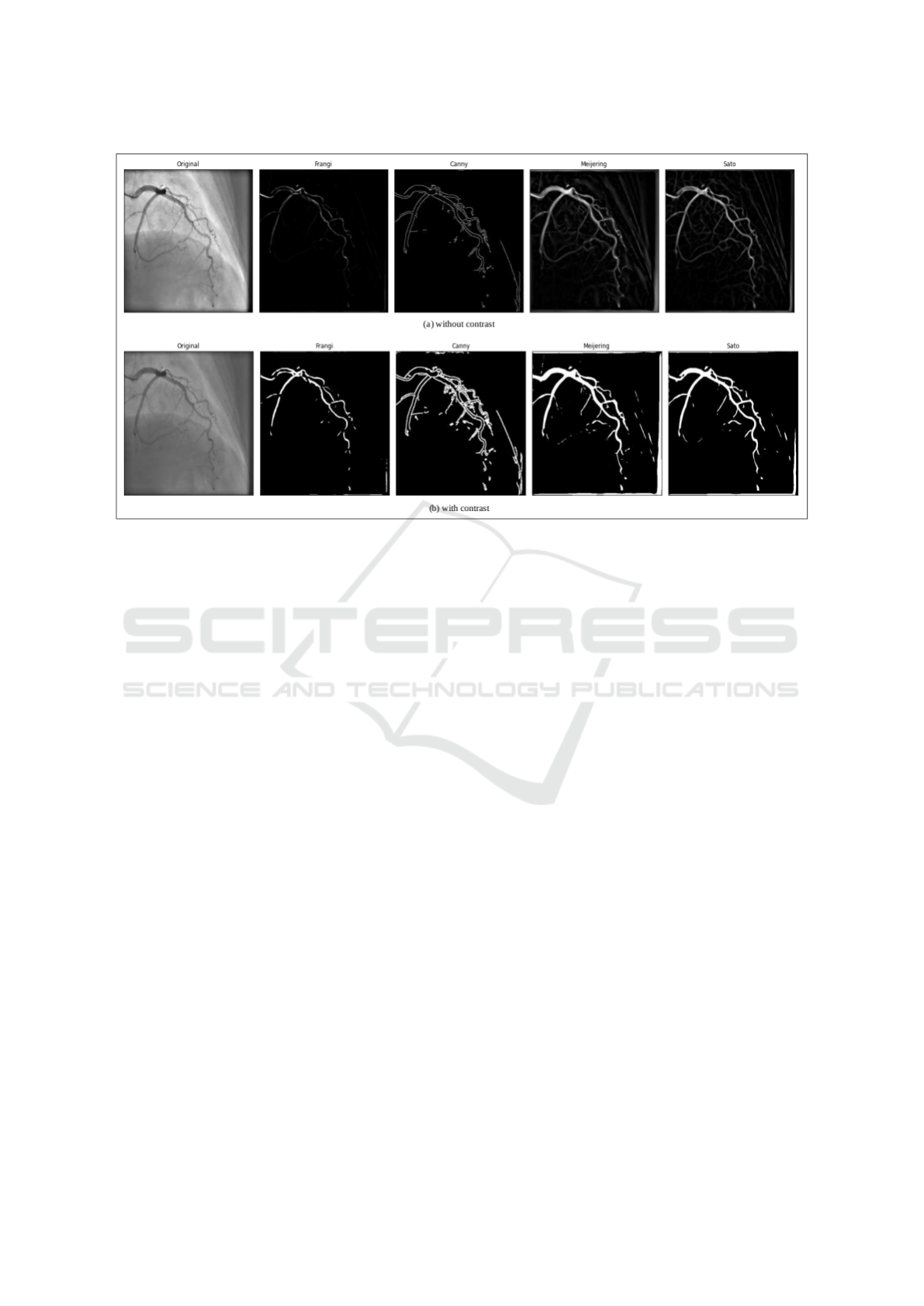
Figure 1: Original frame with results of filters.
5 EXPERIMENTAL RESULTS
5.1 Dataset
The full dataset was collected from exams performed
by a single catheterization laboratory during the pe-
riod between January 2018 and December 2021.
Dataset consisted of 3159 angiographic study: a to-
tal of 37209 coronary angiograms was extracted. We
used a sample of 45 angiograms to extract a total of
1434 frames of size 512 x 512 pixels. We developed a
web application to help two experienced cardiologists
to annotate a sample from our dataset. The manual an-
notation found 474 keyframes and 960 non-keyframe.
The frames were randomly split into 80% training and
20% test datasets.
5.2 Results of Vessel Extraction
In the first step, algorithms 1 and 2 were applied once
on the original dataset without filters, then we tested
them with filters. The results showed that the calcu-
lation with a fixed 6 keyframes gave the best result
provided by the Sato filter: 77,58%. Using an im-
provement algorithm, we improved our results from
77.58% to 85.74%.
In the second step, we applied a pre-processing com-
posed of two pipelined filters. We proposed to pre-
execute the Gaussian filter on the frames before or af-
ter applying the filters mentioned above. The choice
of the Gaussian filter is justified by his ability to elim-
inate the noise of the images. The two-filters pipeline
also gave acceptable results. Since angiograms usu-
ally have a thick black frame secondary to the acqui-
sition parameters, a third optimization technique is to
use a crop function. Cropping was applied once with
a number of 50 pixels on all four sides, then again
with a variable number of pixels.
Table 1 shows the keyframe extraction results using
the proposed algorithm. Meijering gave the worst re-
sults even when combined with a gaussian filter. On
the other hand, a Contrast-Sato had the best outcomes
with an overall accuracy of 85.74%. Fig. 2 shows an
example of the results obtained with each algorithm.
From these results, we conclude that cropping did
not have a major effect on the results and the com-
bination of filters seems to have an unpredictable out-
come except for the contrast filter. Fig. 3 shows the
intensity curve of the frames of the same example.
Our Contrast-Sato algorithm produced the keyframes
from 12 to 17. This set coincides with the manual an-
notation in 4 frames out of 6, therefore presenting a
satisfactory result.
6 CONCLUSION
We have developed an algorithm for extracting
keyframes from angiographic video sequences. The
proposed method detected keyframes with an accu-
ICPRAM 2023 - 12th International Conference on Pattern Recognition Applications and Methods
586
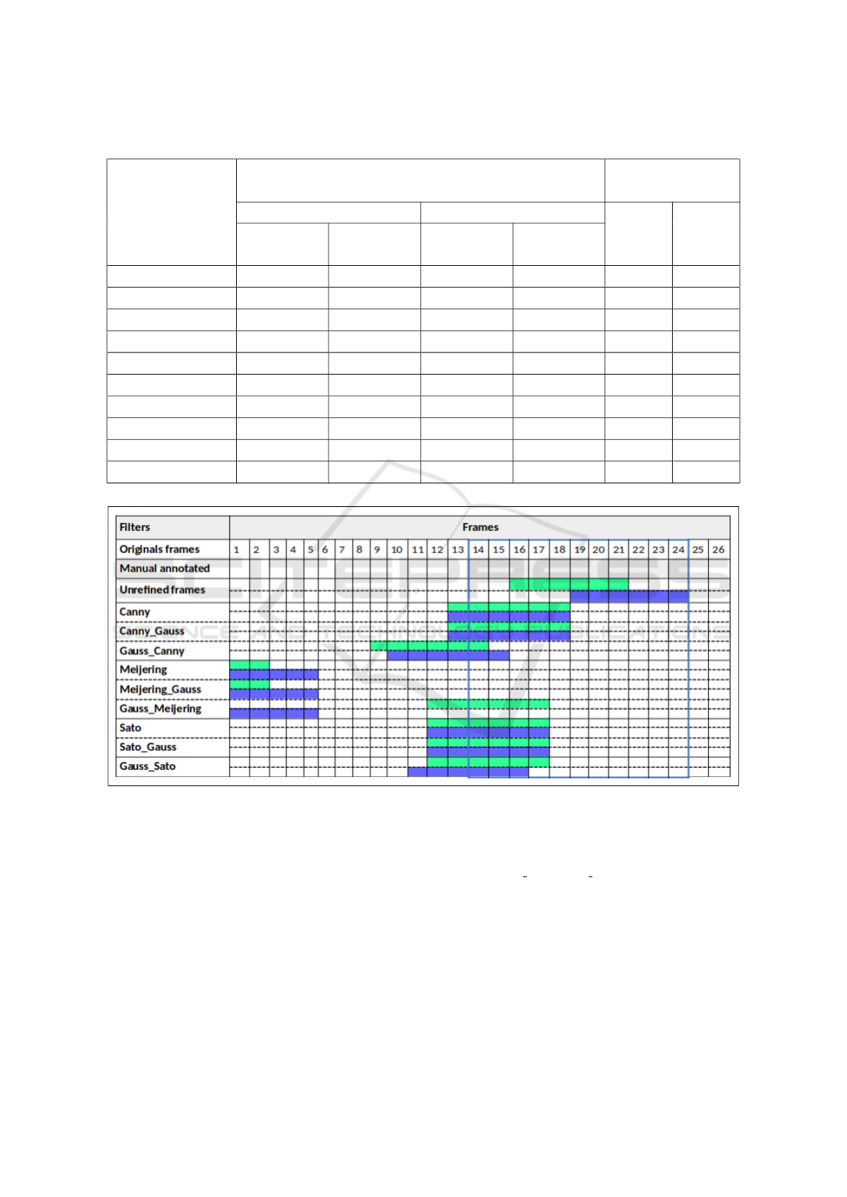
Table 1: Extraction percentages of keyframes using filters.
Filters
With fixed number of frames
With a variable
number of frames
Without cropping With cropping Without
cropping
With
croppingWithout
improvement
With
improvement
Without
improvement
With
improvement
Original images 19.01 20.08 38.39 40.80 16.68 37.89
Meijering 22.58 36.91 11.79 13.45 35.77 10.17
Canny 72.85 81.74 70.07 78.16 73.49 70.79
Sato 77.58 85.74 73.39 80.92 73.34 74.52
Canny-Gaussian 72.85 81.74 70.07 78.16 73.49 70.79
Gaussian-Canny 55.97 60.86 57.38 60.37 59.00 56.01
Meijering-Gaussian 33.58 36.91 11.79 13.45 35.77 10.17
Gaussian-Meijering 61.25 67.93 18.70 22.77 60.21 18.96
Sato-Gaussian 77.70 85.18 76.35 82.77 72.29 73.39
Gaussian-Sato 76.34 82.96 69.62 75.06 70.25 64.51
Figure 2: Keyframes extracted from a randomly drawn coro : (green) without cropping (blue) with cropping.
racy of 85.74% compared to manual annotation.
Greater attention will be devoted to this prelimi-
nary stage of processing the angiographic frames. Fu-
ture improvements include the use of deep learning
methods seeking closer performance.
REFERENCES
Avram, R., Olgin, J., Wan, A., Ahmed, Z., Verreault-
Julien, L., Abreau, S., Wan, D., Gonzalez, J. E.,
So, D., Soni, K., et al. (2021). Cathai: Fully auto-
mated coronary angiography interpretation and steno-
sis detection using a deep learning-based algorithmic
pipeline. Journal of the American College of Cardiol-
ogy, 77(18 Supplement 1):3244–3244.
Collet, C., Grundeken, M. J., Asano, T., Onuma, Y., Wi-
jns, W., and Serruys, P. W. (2017). State of the art:
coronary angiography. EuroIntervention: journal of
EuroPCR in collaboration with the Working Group on
Interventional Cardiology of the European Society of
Cardiology, 13(6):634–643.
Danilov, V. V., Klyshnikov, K. Y., Gerget, O. M., Kutikhin,
A. G., Ganyukov, V. I., Frangi, A. F., and Ovcharenko,
E. A. (2021). Real-time coronary artery stenosis de-
tection based on modern neural networks. Scientific
Automatic Coronary Angiogram Keyframe Extraction
587
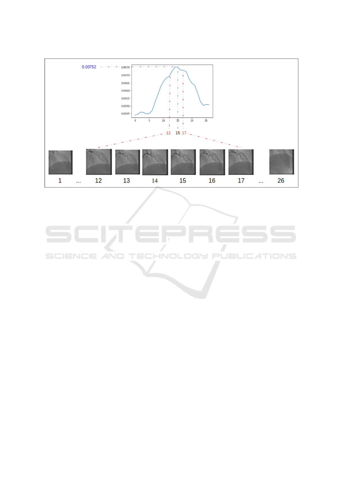
Figure 3: Temporal intensity score variation of coronary angiogram frames.
reports, 11(1):1–13.
Frangi, A. F., Niessen, W. J., Vincken, K. L., and Viergever,
M. A. (1998). Multiscale vessel enhancement filter-
ing. In International conference on medical image
computing and computer-assisted intervention, pages
130–137. Springer.
Gawande, U., Hajari, K., and Golhar, Y. (2020). Deep learn-
ing approach to key frame detection in human action
videos. Recent Trends in Computational Intelligence,
1:1–17.
Jiang, Z.-g. and Shi, X.-t. (2021). Application research of
key frames extraction technology combined with opti-
mized faster r-cnn algorithm in traffic video analysis.
Complexity, 2021.
Jo, K., Kweon, J., Kim, Y.-H., and Choi, J. (2018). Segmen-
tation of the main vessel of the left anterior descend-
ing artery using selective feature mapping in coronary
angiography. IEEE Access, 7:919–930.
Kavipriya, K. and Hiremath, M. (2022). Computational
method to extract the keyframe from angiogram
video. JOURNAL OF ALGEBRAIC STATISTICS,
13(3):3088–3097.
Kerkeni, A., Benabdallah, A., Manzanera, A., and Bedoui,
M. H. (2016). A coronary artery segmentation method
based on multiscale analysis and region growing.
Computerized Medical Imaging and Graphics, 48:49–
61.
Lamy, J., Merveille, O., Kerautret, B., Passat, N., and
Vacavant, A. (2021). Vesselness filters: A survey
with benchmarks applied to liver imaging. In 2020
25th International Conference on Pattern Recognition
(ICPR), pages 3528–3535. IEEE.
Lee, Y.-T. H., Fang, J., Schieb, L., Park, S., Casper, M., and
Gillespie, C. (2022). Prevalence and trends of coro-
nary heart disease in the united states, 2011 to 2018.
JAMA cardiology, 7(4):459–462.
Meijering, E., Jacob, M., Sarria, J.-C., Steiner, P., Hirling,
H., and Unser, e. M. (2004). Design and validation of
a tool for neurite tracing and analysis in fluorescence
microscopy images. Cytometry Part A: the journal
of the International Society for Analytical Cytology,
58(2):167–176.
Moon, J. H., Cha, W. C., Chung, M. J., Lee, K.-S., Cho,
B. H., Choi, J. H., et al. (2021). Automatic stenosis
recognition from coronary angiography using convo-
lutional neural networks. Computer methods and pro-
grams in biomedicine, 198:105819.
Neumann, F.-J., Sousa-Uva, M., Ahlsson, A., Alfonso, F.,
Banning, A. P., Benedetto, U., Byrne, R. A., Collet,
J.-P., Falk, V., Head, S. J., et al. (2019). 2018 esc/eacts
guidelines on myocardial revascularization. European
heart journal, 40(2):87–165.
Obara, B., Fricker, M., Gavaghan, D., and Grau, V. (2012).
Contrast-independent curvilinear structure detection
in biomedical images. IEEE Transactions on Image
Processing, 21(5):2572–2581.
Ojha, N. and Dhamoon, A. S. (2021). Myocardial infarc-
tion. In StatPearls [Internet]. StatPearls Publishing.
Qin, B., Mao, H., Liu, Y., Zhao, J., Lv, Y., Zhu, Y., Ding,
S., and Chen, X. (2022). Robust pca unrolling net-
work for super-resolution vessel extraction in x-ray
coronary angiography. IEEE Transactions on Medi-
cal Imaging.
Sato, Y., Nakajima, S., Shiraga, N., Atsumi, H., Yoshida, S.,
Koller, T., Gerig, G., and Kikinis, R. (1998). Three-
dimensional multi-scale line filter for segmentation
and visualization of curvilinear structures in medical
images. Medical image analysis, 2(2):143–168.
Sazak, C¸ ., Nelson, C. J., and Obara, B. (2019). The multi-
scale bowler-hat transform for blood vessel enhance-
ment in retinal images. Pattern Recognition, 88:739–
750.
ICPRAM 2023 - 12th International Conference on Pattern Recognition Applications and Methods
588

Thakre, K., Rajurkar, A., and Manthalkar, R. (2016). Video
partitioning and secured keyframe extraction of mpeg
video. Procedia Computer Science, 78:790–798.
Zhou, C., Dinh, T. V., Kong, H., Yap, J., Yeo, K. K., Lee,
H. K., and Liang, K. (2021). Automated deep learning
analysis of angiography video sequences for coronary
artery disease. arXiv preprint arXiv:2101.12505.
Automatic Coronary Angiogram Keyframe Extraction
589
