
EFL-Net: An Efficient Lightweight Neural Network Architecture for
Retinal Vessel Segmentation
Nasrin Akbari and Amirali Baniasadi
Department of Electrical and Computer Engineering, University of Victoria, Victoria, Canada
Keywords:
Blood Vessel Segmentation, Deep Learning, Image Processing.
Abstract:
Accurate segmentation of retinal vessels is crucial for the timely diagnosis and treatment of conditions like
diabetes and hypertension, which can prevent blindness. Deep learning algorithms have been successful in
segmenting retinal vessels, but they often require a large number of parameters and computations. To address
this, we propose an efficient and fast lightweight network (EFL-Net) for retinal blood vessel segmentation.
EFL-Net includes the ResNet branches shuffle block (RBS block) and the Dilated Separable Down block
(DSD block) to extract features at various granularities and enhance the network receptive field, respectively.
These blocks are lightweight and can be easily integrated into existing CNN models. The model also uses
PixelShuffle as an upsampling layer in the decoder, which has a higher capacity for learning features than
deconvolution and interpolation approaches. The model was tested on the Drive and CHASEDB1 datasets and
achieved excellent results with fewer parameters compared to other networks such as ladder net and DCU-Net.
EFL-Net achieved F1 measures of 0.8351 and 0.8242 on the CHASEDB1 and DRIVE datasets, respectively,
with 0.340 million parameters, compared to 1.5 million for ladder net and 1 million for DCU-Net.
1 INTRODUCTION
The retina is a layer of light-sensitive nerve tissue
located at the back of the eye that receives images
and transmits them to the brain as electric signals
through the optic nerve (Kolb, 2012). Changes in the
retina and optic nerve may indicate certain diseases
such as glaucoma (Salmon, 2019) or hypertensive
retinopathy (HR) (Irshad and Akram, 2014), which
can cause blurring of vision. As we age, the oxida-
tive load increases, leading to higher levels of oxida-
tive stress which can cause pathologies such as age-
related macular degeneration or neuropathic compli-
cations of diabetes in the eye (Payne et al., 2014).
Diabetic retinopathy (DR) is a condition that affects
individuals with diabetes, causing gradual damage to
the retina and potentially leading to vision loss. It is
a major complication of diabetes that can threaten vi-
sion.
Primary eye care (PEC) can use a funduscopy ex-
amination to give an early screening for drug-induced
retinal toxicity (Alberta et al., 2022). In the procedure
of funduscopy examination, an ophthalmologist looks
at the structures of the retina, retinal blood vessels,
and optic nerve head (disk) of the eye (Walker et al.,
1990). There are several ways to analyze retinal im-
ages and find diseases, one of which is retinal image
segmentation, which can be divided into manual and
automatic methods. Manual segmentation takes time
and expertise, while automated algorithms are useful
for early detection and treatment of eye diseases due
to their increased accuracy, reduced cost, and faster
speed compared to manual segmentation.
U-Net (Ronneberger et al., 2015) is an automatic
model used to segment vessels in retina images and
is one of the successful medical and biomedical im-
age segmentation methods based on deep neural net-
works. Humans often struggle to distinguish blood
vessel images from their distorted backgrounds, mak-
ing it more difficult to detect diseases. As a result,
developing practical algorithms to identify vessel im-
ages and their surroundings would be useful (Yang
et al., 2022).
Deep neural networks (DNNs) have been shown
to be effective in automatically learning reliable and
complex features from raw data without the need for
manual feature engineering (Ronneberger et al., 2015;
Zhou et al., 2021; Gu et al., 2019; Li et al., 2020).
These techniques have achieved significant success in
the fields of computer vision and medical health. Re-
search on retinal vessel segmentation using DNNs has
proposed various architectures for this task, however,
920
Akbari, N. and Baniasadi, A.
EFL-Net: An Efficient Lightweight Neural Network Architecture for Retinal Vessel Segmentation.
DOI: 10.5220/0011754700003417
In Proceedings of the 18th International Joint Conference on Computer Vision, Imaging and Computer Graphics Theory and Applications (VISIGRAPP 2023) - Volume 4: VISAPP, pages
920-927
ISBN: 978-989-758-634-7; ISSN: 2184-4321
Copyright
c
2023 by SCITEPRESS – Science and Technology Publications, Lda. Under CC license (CC BY-NC-ND 4.0)
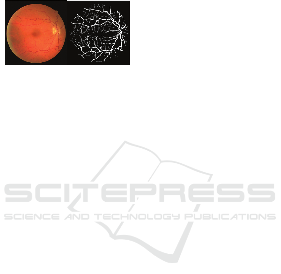
Figure 1: A retinal image from DRIVE dataset (left) and
retinal vessel segmentation (right) (Staal et al., 2004).
our observations indicate that many of these models
are not optimal in terms of architecture and num-
ber of parameters. Figure 2 illustrates the F1 mea-
sure and complexity of well-known DNN-based mod-
els (Azad et al., 2019; Zhou et al., 2021; Gu et al.,
2019; Mou et al., 2021; Li et al., 2020; Zhuang, 2018;
Zahangir Alom et al., 2018; Guo et al., 2021; Yang
et al., 2022). More research is needed to improve the
number of parameters and accuracy of current mod-
els. Our research aims to design a lightweight CNN
architecture with fewer parameters that can achieve
similar or better results in retinal vessel segmentation
compared to state-of-the-art networks.
The remainder of the paper is structured as fol-
lows: Section 2 describes the proposed network
architecture, Section 3 compares experimental re-
sults to state-of-the-art neural networks using the
CHASEDB1 and DRIVE datasets, and Section 4 pro-
vides concluding remarks and discussion of future re-
search directions.
2 METHODOLOGY
U-Net is a convolutional neural network that was
designed for image segmentation in the field of
biomedicine (Ronneberger et al., 2015). It is an im-
provement on the previously developed FCN - ”Fully
convolutional networks for semantic segmentation”
(Long et al., 2015). Its ability to perform well with
small training datasets has made it the most reliable
architecture for the semantic segmentation of biolog-
ical images.
The U-Net architecture consists of four encoder
blocks on the left side, known as the contracting path,
and four decoder blocks on the right side referred
to as the expansive path. The encoder captures fea-
tures from the input image and reduces its resolution
through pooling layers, while the decoder part recon-
structs the image and restores object details through
skip connections between the encoder and decoder
layers. While the U-Net model has been successful
in various tasks, it has several drawbacks including a
large number of parameters (31.031 million) and poor
performance on retinal vessel segmentation (F1 score
of 0.7783 on the CHASEDB1 dataset). In order to ad-
dress these issues, we analyzed additional papers and
their blocks and developed a solution that introduces
two new blocks for enhanced feature extraction: the
Resnet Branches Shuffle Block (RBSB) and the Di-
lated Separated Down Block (DSDB). We also em-
ployed efficient layers such as pixel shuffle decon-
volution and interpolation techniques in the decoder
path of the U-Net model (Shi and Caballero, 1874).
Our modified U-Net model aims to improve perfor-
mance on retinal vessel segmentation tasks
2.1 Our Proposed Architecture
Inspired by the U-Net (Ronneberger et al., 2015) and
ShuffleNetV2 (Ma et al., 2018) models, we propose
the Efficient and Fast Lightweight Neural Network
(EFL-Net) for retinal vessel segmentation. Our goal
is to create a lightweight and accurate deep learn-
ing model for this task. To increase the receptive
field of the U-Net model, we introduce the Resnet
Branches Shuffle Block (RBS block) and the Dilated
Separable Down Block (DSD block) to our architec-
ture. The encoder path of the U-Net model consists
of four stages, each comprising EFL-Net, RBS, and
DSD blocks for feature extraction and downsampling.
In the decoder path, we use the PixelShuffle layer
(Shi and Caballero, 1874) for upsampling instead of
the deconvolution layer and add the encoder and de-
coder features rather than concatenating them to re-
duce computation. A diagram of our architecture is
shown in Figure 3. A Dropout Block layer with a
batch normalization layer after each convolution layer
is included after each RBS block. In the following
section, we will provide a more in-depth discussion
of the core concepts that form the basis of our archi-
tecture and how they contribute to the overall design.
2.2 Resnet Branches Shuffle Block
In this paper, we present an improved feature extrac-
tion method for image classification tasks. Our ap-
proach is based on the Res2Net (Gao et al., 2019)
and ShuffleNetV2 (Ma et al., 2018) architectures, and
aims to enhance the feature extraction capabilities of
the ShuffleNetV2 basic unit.
To this end, we propose the RBS block (Figure 4),
which modifies the number of split channels in the in-
put feature maps of the ShuffleNetV2 basic unit. The
input, which has n channels, is divided into n/4 groups
with the same number of channels each, denoted as
EFL-Net: An Efficient Lightweight Neural Network Architecture for Retinal Vessel Segmentation
921
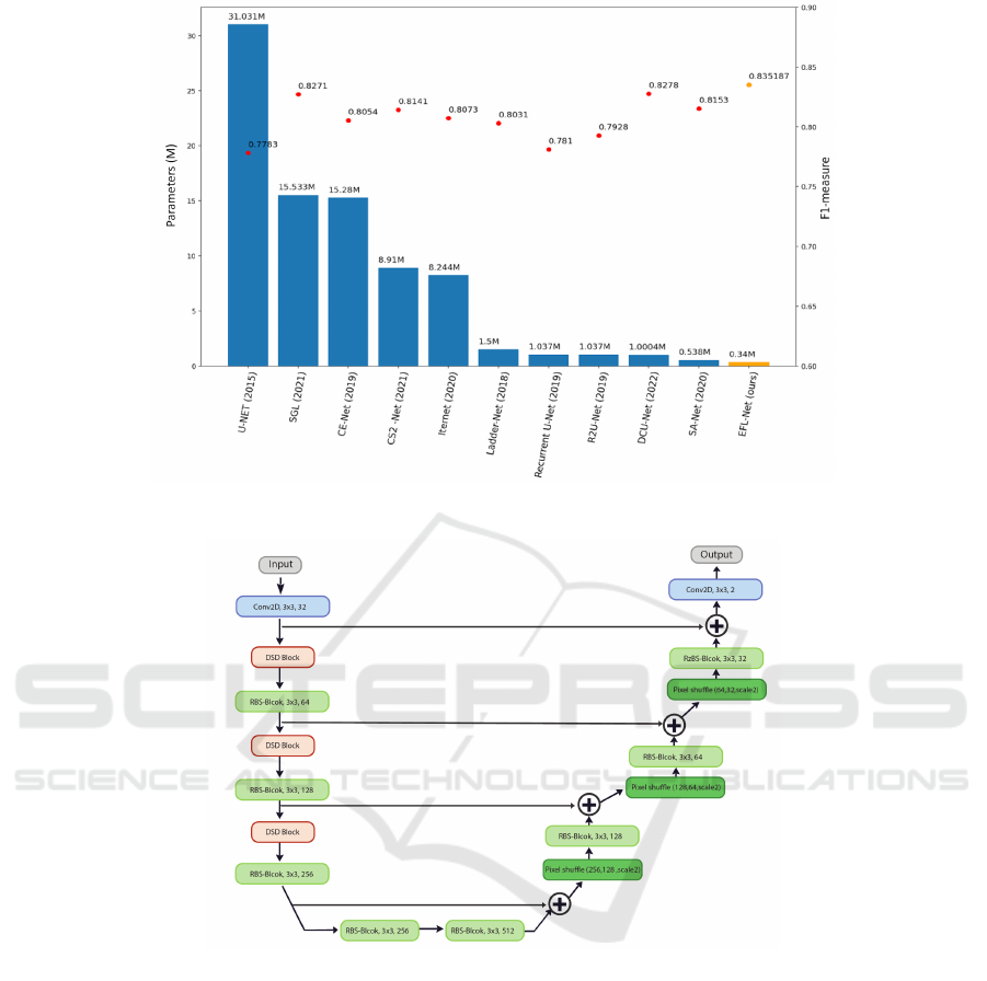
Figure 2: Accuracy and number of parameters of several retinal vessel segmentation papers in the past five years (CHASEDB1
dataset (Owen et al., 2009)).
Figure 3: EFL-Net Architecture.
xi, where i ∈
{
1, 2, 3, 4
}
. Each xi has the same spa-
tial size and is passed through a bottleneck unit Ci(),
resulting in the output yi. The output of the previous
bottleneck unit, Ci − 1(), is then added to the current
group xi and passed through the bottleneck unit Ci().
This results in an output with a larger receptive field
than xi. In addition, the channels xi for i > 1 are ag-
gregated with x1 to reuse features. The remainder of
the RBS block is identical to the ShuffleNetV2 con-
volution block.
RBS block can be easily incorporated into any
network as a lightweight feature extractor. Our ex-
perimental results demonstrate the superiority of the
RBS block over the original ShuffleNetV2 basic unit
in terms of multi-scale feature extraction and the num-
ber of parameters. As shown in Figure 4, the architec-
ture consists of several blocks, which we will describe
in detail in the following section.
2.3 ShuffleNetV2 Basic Unit
The basic unit in ShuffleNetV2 (Ma et al., 2018) is
a block of layers that includes a depthwise separa-
ble convolution (Chollet, 2017), a pointwise convolu-
tion (Chollet, 2017), and a shuffle operation (Zhang
et al., 2018). This unit is used to construct larger
network architectures in a way that reduces computa-
tional complexity while maintaining representational
capacity. The use of depthwise separable convolu-
tions and the shuffle operation also make the network
VISAPP 2023 - 18th International Conference on Computer Vision Theory and Applications
922
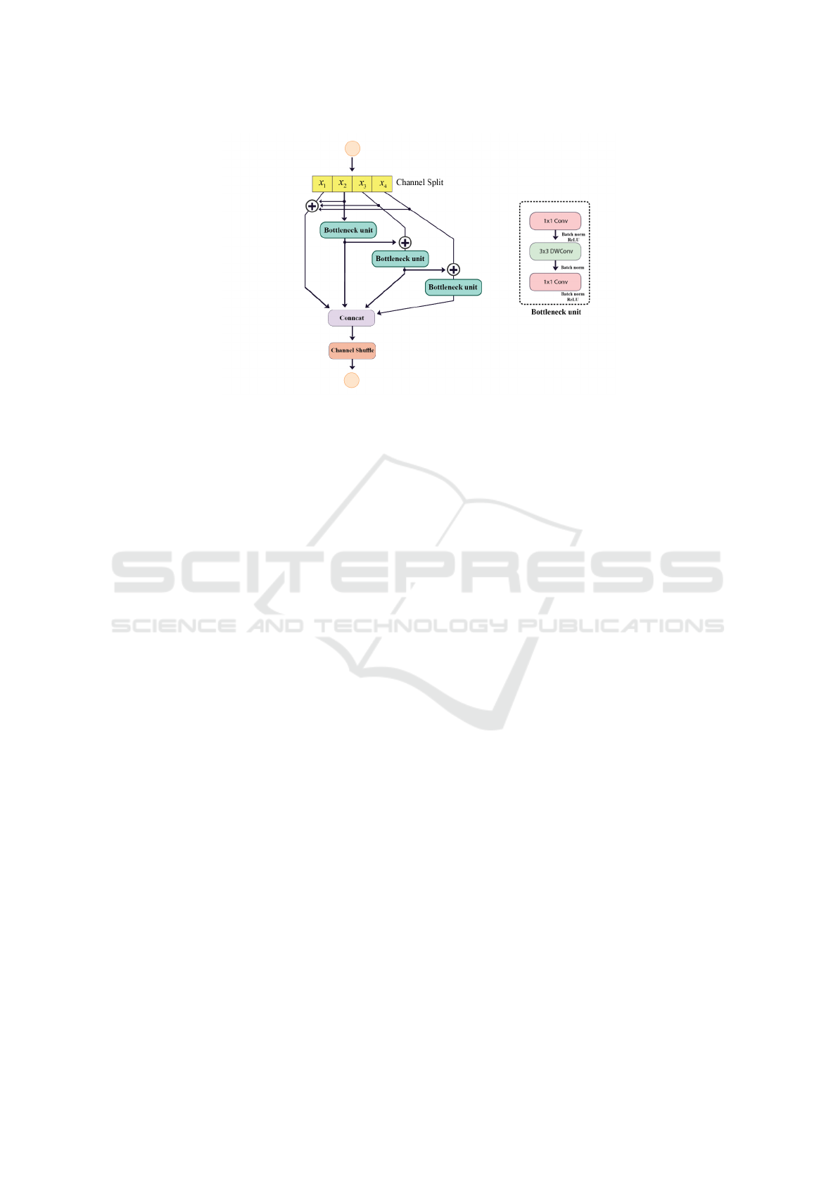
Figure 4: a) ShuffleNetV2 basic unit (Ma et al., 2018), b) Resnet Branches Shuffle Block (RBS block), DWConv stands for
depth-wise convolution.
more efficient by reducing the number of parameters
and computations required.
2.4 Depthwise Separable Convolution
A depthwise separable convolution (Chollet, 2017) is
a way of decomposing a standard convolution in a
convolutional neural network into two smaller con-
volutions: a depthwise convolution that applies a sep-
arate kernel to each input channel, and a pointwise
convolution that mixes the output channels as shown
in Figure 5) . This can reduce the number of param-
eters and computations in a model and make it more
efficient, particularly for mobile and embedded appli-
cations.
2.5 Channel Shuffle
A channel shuffle (Zhang et al., 2018) operation is a
way of rearranging the channels of a feature map by
interleaving them into groups. It is used to allow a
convolutional neural network to mix and combine the
information in different channels more flexibly and is
often used with depthwise separable convolutions to
increase representational capacity while maintaining
efficiency.
2.6 Dilated Separable down Block
(DSD)
The receptive field (RF) is a crucial concept in the
design of convolutional neural networks (CNNs). As
described in (Luo et al., 2016), the RF at each layer
is the size of the region in the input that contributes
to generating a particular feature in the output. In or-
der to accurately predict the boundaries of objects in
the input image, such as organs, tumors, or vessels, it
is necessary to provide the model with access to all
relevant parts of the image. In a CNN, each neuron
controls a specific part of the data and is exposed to
different parts of the input data during the convolu-
tion process, filling a segmented area known as the
local receptive field. In this paper, we propose the use
of the Dilated Separable Down block (DSD block) as
a method for increasing the RF of the network and
improving its ability to predict object boundaries.
The Dilated Separable Down block (DSD) block,
as shown in Figure 6, consists of two branches of 3x3
group convolutional layers with a stride size of 2 and
a pointwise convolutional layer. Note that different
dilation rates are applied to the different groups to
extract multi-scale features. The results of the two
branches are concatenated to improve the ability of
the network to represent features. This block can be
used in place of pooling layers to increase the expres-
sive power of the model. The use of dilated convo-
lution allows the model to increase its field of view
without increasing the number of parameters.
2.7 PixelShuffle
PixelShuffle (Shi and Caballero, 1874) is a type of
upsampling layer used in convolutional neural net-
works (CNNs) to increase the resolution of the out-
put feature maps. It has the advantage of being able
to achieve a higher resolution output than other up-
sampling methods and being more efficient, as it does
not require the use of additional convolutional kernels
or the insertion of zeros into the feature maps. These
EFL-Net: An Efficient Lightweight Neural Network Architecture for Retinal Vessel Segmentation
923
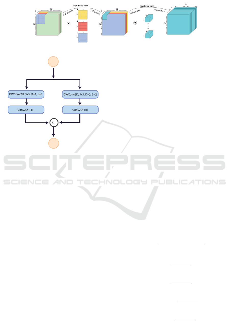
Figure 5: Depthwise separable convolution (Pandey, 2018).
Figure 6: Dilated Separable Down Block.
properties make PixelShuffle a useful tool for tasks
that require a high degree of spatial resolution and for
use in mobile and embedded applications where com-
putational resources are limited.
3 EXPERIMENTAL
ENVIRONMENT AND RESULT
3.1 DATASETS and Data Preprocessing
The DRIVE (Owen et al., 2009), and CHASEDB1
(Owen et al., 2009) are both publicly available
datasets for retinal segmentation. The DRIVE dataset
consists of 40 2D RGB images with a resolution of
565 x 584 pixels, with 20 images in both the train-
ing and test sets. The CHASEDB1 dataset includes
28 images, and has a resolution of 999 x 960 pixels.
There are 20 training images and 8 test images in the
CHASEDB1 dataset. The model’s performance was
evaluated on both datasets using the ground truth la-
bels provided by the first expert.
We enhanced the size of the dataset by imple-
menting data augmentation techniques. To focus on
the relevant information and eliminate unnecessary
processing, we employed a mask or field of view
(FOV) to extract patches from the input image that
only contained vessels. While the DRIVE dataset in-
cludes a binary mask, the CHASEDB1 dataset does
not. Therefore we manually created a mask for the
CHASEDB1 dataset.
Before training, the data was pre-processed to re-
move noise and uneven lighting in fundus images.
The green channel of the RGB image was chosen as
it allows for better visualization of blood vessels. The
data was then normalized and scaled, and contrast
limited adaptive histogram equalization (Zuiderveld,
1994) and gamma adjustment were applied to im-
prove the contrast between the foreground and back-
ground
3.2 Evaluation Approaches
The performance of a segmentation model can be
evaluated by comparing its results to the ground truth
(GT) and considering four scenarios: true positive
(TP), false positive (FP), false negative (FN), and true
negative (TN). TP is the number of correctly classi-
fied blood vessel pixels, FP is the number of incor-
rectly classified background pixels as vessels, FN is
the number of incorrectly classified vessel pixels as
background, and TN is the number of correctly clas-
sified background pixels. In addition to these four in-
dicators, the model’s performance can also be eval-
uated using the following criteria: sensitivity (SE),
specificity (SP), accuracy (ACC), precision (Pr), and
F-Measure (F1).
AC =
T P + T N
T P + T N + FP + FN
SE =
T P
T P + FN
SP =
T N
T N + FP
Precision =
T P
T P + FP
Recall =
T P
T P + FN
VISAPP 2023 - 18th International Conference on Computer Vision Theory and Applications
924
Table 1: Performance comparison between the EFL-Net and some state-of-the-art models on DRIVE.
Methods Year F1 SE SP Acc AUC Parameters (M)
U-NET (Ronneberger et al., 2015) 2015 0.7783 0.8288 0.9701 0.9578 0.9772 31.031
SGL (Zhou et al., 2021) 2021 0.8271 0.869 0.9843 0.9771 0.992 15.533
CE-Net (Gu et al., 2019) 2019 0.8054 0.8093 0.9797 0.9641 0.9834 15.28
CS2-Net (Mou et al., 2021) 2021 0.8141 0.8329 0.9784 0.9651 0.9851 8.91
Iternet (Li et al., 2020) 2020 0.8073 0.797 0.9823 0.9655 0.9851 8.244
Ladder-Net (Zhuang, 2018) 2018 0.8031 0.7978 0.9818 0.9656 0.9839 1.5
Recurrent U-Net (Zahangir Alom et al., 2018) 2019 0.781 0.7459 0.9836 0.9622 0.798 1.037
R2U-Net (Zahangir Alom et al., 2018) 2019 0.7928 0.7756 0.982 0.9634 0.9815 1.037
DCU-Net (Yang et al., 2022) 2022 0.8278 0.8075 0.9841 0.9664 0.9872 1.0004
SA-Net (Hu et al., 2021) 2020 0.8153 0.8573 0.9835 0.9755 0.9905 0.538707
EFL-Net (ours) 2022 0.8242 0.7957 0.9802 0.9567 0.9803 0.340
Table 2: Performance comparison between our EFL-Net and some state-of-the-art models on CHASEDB1.
Methods Year F1 SE SP Acc AUC Parameters (M)
U-NET (Ronneberger et al., 2015) 2015 0.8174 0.7537 0.982 0.9531 0.9755 31.031
BCDU-Net (d=3) (Azad et al., 2019) 2019 0.8224 0.8007 0.9786 0.956 0.9789 20.659
SGL (Zhou et al., 2021) 2021 0.8316 0.838 0.9834 0.9705 0.9886 15.533
CE-Net (Gu et al., 2019) 2019 0.8243 0.8276 0.9735 0.9545 0.9794 15.28
CS2-Net (Mou et al., 2021) 2021 0.8228 0.8154 0.9757 0.9553 0.9784 8.91
Iternet (Li et al., 2020) 2020 0.8205 0.7735 0.9838 0.9573 0.9816 8.244
Ladder-Net (Zhuang, 2018) 2018 0.8202 0.7856 0.981 0.9561 0.9793 1.5
Recurrent U-Net (Zahangir Alom et al., 2018) 2019 0.8155 0.7751 0.9816 0.9556 0.9782 1.037
R2U-Net (Zahangir Alom et al., 2018) 2019 0.8171 0.7792 0.9813 0.9556 0.9784 1.037
DCU-Net (Yang et al., 2022) 2022 0.8272 0.8115 0.978 0.9568 0.981 1.0004
SA-Net (Hu et al., 2021) 2020 0.8263 0.8212 0.984 0.9698 0.9864 0.538707
EFL-Net (ours) 2022 0.8351 0.7977 0.9860 0.9651 0.9868 0.340
F1 = 2 ×
Precision × Recall
Precision + Recall
Specificity (SP) is the ratio of correctly segmented
background pixels to the total number of actual back-
ground pixels, while sensitivity (SE) is the ratio of
correctly segmented blood vessel pixels to the total
number of actual blood vessel pixels. Accuracy (Acc)
shows the percentage of total image pixels that were
correctly segmented. Precision measures the quality
of the model’s positive predictions, while recall mea-
sures the quality of negative predictions. A higher
precision value indicates that the model’s architecture
is better trained on the given data. F1 is the weighted
harmonic mean of precision and recall.
3.3 Loss Function
The focal loss (Lin et al., 2017) is a method for ad-
dressing the class imbalance between foreground and
background pixels in a dataset, and is defined as fol-
lows:
FL(p
t
) = −α
t
∗ (1 −p
t
)
γ
∗ log(p
t
) where
(
p i f y = 1
1 − p else
(1)
In the focal loss, the predicted probability of the
network output is denoted by p and the focusing pa-
rameter, which can be adjusted, is denoted by γ. Sam-
ples that are easy to classify contribute less to the loss
values, while samples that are difficult to classify con-
tribute more, causing the model to focus more on the
latter.
3.4 Experimental Environment and
Parameters
In this work, we trained our network from scratch for
200 epochs using a batch size of 256. We initialized
the weights with random values and used the Adam
optimizer with default parameters and an initial learn-
ing rate of 0.001. The learning rate was updated at
each epoch using a cosine function attenuation strat-
egy. Our experiments were conducted on a server
with a Linux operating system, 2.30 GHz processor,
128 GB RAM, an NVIDIA TESLA P100 GPU, and
the Pytorch 1.7.0 framework.
3.5 Experimental Result
Tables 1 and 2 present a comparison of the per-
formance of our proposed architecture with existing
methods on the DRIVE and CHASEDB1 datasets.
The results show that our model outperforms other
methods on the CHASEDB1 dataset and produces
EFL-Net: An Efficient Lightweight Neural Network Architecture for Retinal Vessel Segmentation
925
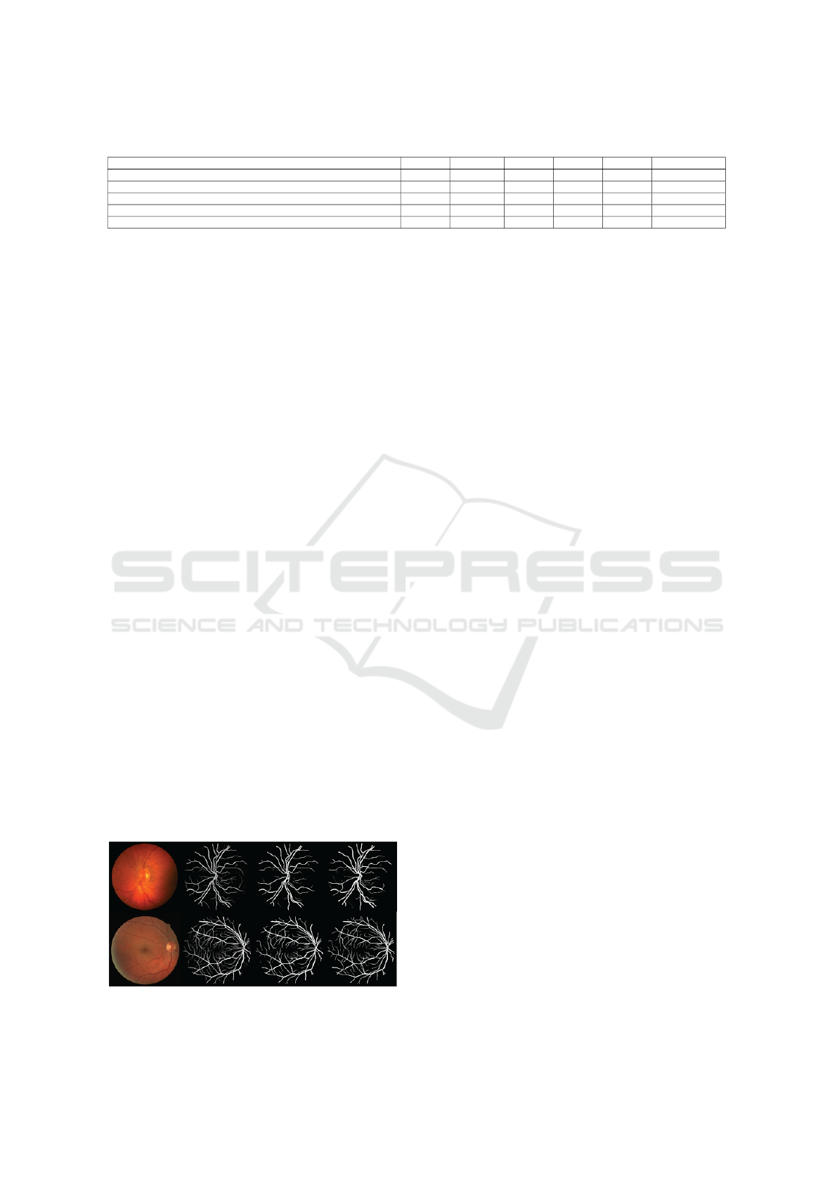
Table 3: Ablation experiment on RBS and DSD blocks. The EFL-Net is trained and evaluated on CHASEDB1.
Method F1 SE SP Acc AUC Parameters (M)
EFL-Net (Standard convolution + maxpooling2D + cross entropy loss) 0.747399 0.6839 0.9815 0.9489 0.82892 1.936738
EFL-Net (Standard convolution + DSD block + cross entropy loss) 0.770642 0.7031103 0.986 0.957 0.979272 1.788498
EFL-Net (ShuffleNetV2 basic unit + DSD block + cross entropy loss) 0.815429 0.739147 0.990829 0.962996 0.985666 0.410922
EFL-Net (RBS block + DSD block + cross entropy loss) 0.801086 0.950963 0.947362 0.9648 0.988168 0.340950
EFL-Net (RBS block + DSD block+ Focal loss) 0.8351 0.7693 0.9891 0.9648 0.9871 0.340950
satisfactory results on the DRIVE dataset. In par-
ticular, our model achieves the highest F1 score of
0.8351 and the highest specificity of 0.9860 on the
CHASEDB1 dataset, demonstrating its superiority
in retinal vessel segmentation compared to previous
works. When comparing the results of our model
(EFL-Net) with other networks, it is clear that our net-
work achieves equivalent or better results compared to
the best performing networks with a smaller number
of parameters.
We conducted ablation experiments on the
CHASEDB1 dataset to study the impact of different
components of our proposed architecture. In the first
experiment, we compared the performance of net-
works with and without DSD blocks (using MaxPool-
ing instead) in the encoder. The results, shown in rows
1 and 2 of Table 3, indicate that the DSD block is su-
perior to MaxPooling in terms of F1 score.
In the second experiment, we replaced the RBS
block with the ShuffleNetV2 basic unit to evaluate the
performance of the RBS block in the proposed EFL-
Net architecture. By comparing the performance of
EFL-Net with the RBS block and DSD block (EFL-
Net (RBS block + DSD block)) to EFL-Net with the
ShuffleNetV2 basic unit and DSD block (EFL-Net
(ShuffleNetV2 basic unit + DSD block)), we found
that the RBS block significantly improved the model’s
F1 score by approximately 1.47%.
In the third experiment, we compared the perfor-
mance of our model using cross entropy loss and focal
loss. The results showed that the model using focal
loss achieved better performance.
The results of our model on the DRIVE and
CHASEDB1 datasets are shown in Figure 7. The
original images of retinal vessels are shown in the first
column, followed by the output of our network for
Figure 7: Retinal vessel images from the CHASEDB1 (first
row) and DRIVE (second row) datasets.
vessel segmentation in the second column. The third
column shows the binary result obtained by apply-
ing a threshold to the network output, and the ground
truth for each input image is displayed in the last col-
umn.
4 CONCLUSION
EFL-Net is a lightweight network designed to im-
prove the accuracy and speed of blood vessel seg-
mentation. It uses two custom modules, the ResNet
Branches Shuffle Block (RBS) and the Dilated Sepa-
rable Down block (DSD), which have a high capac-
ity for feature extraction. The RBS block is based
on the shuffle Net block and the DSD block expands
the network’s receptive field while reducing feature
size without losing important information. In the up-
sampling path, the network uses PixelShuffle instead
of deconvolution or interpolation. The network has
0.34 million parameters and demonstrated good per-
formance on two datasets, achieving F1 scores of
0.8242 on the DRIVE dataset and 0.835187 on the
CHASEDB1 dataset.
REFERENCES
Alberta, I. B., Rahmani, S. A., et al. (2022). Retinal impair-
ment associated with long-term use of ritonavir among
hiv patients: A systematic review for primary eye care
practice. International Journal of Retina, 5(1):48–48.
Azad, R., Asadi-Aghbolaghi, M., Fathy, M., and Escalera,
S. (2019). Bi-directional convlstm u-net with dens-
ley connected convolutions. In Proceedings of the
IEEE/CVF international conference on computer vi-
sion workshops, pages 0–0.
Chollet, F. (2017). Xception: Deep learning with depthwise
separable convolutions. In Proceedings of the IEEE
conference on computer vision and pattern recogni-
tion, pages 1251–1258.
Gao, S.-H., Cheng, M.-M., Zhao, K., Zhang, X.-Y., Yang,
M.-H., and Torr, P. (2019). Res2net: A new multi-
scale backbone architecture. IEEE transactions on
pattern analysis and machine intelligence, 43(2):652–
662.
Gu, Z., Cheng, J., Fu, H., Zhou, K., Hao, H., Zhao, Y.,
Zhang, T., Gao, S., and Liu, J. (2019). Ce-net: Context
encoder network for 2d medical image segmentation.
VISAPP 2023 - 18th International Conference on Computer Vision Theory and Applications
926

IEEE transactions on medical imaging, 38(10):2281–
2292.
Guo, C., Szemenyei, M., Yi, Y., Wang, W., Chen, B.,
and Fan, C. (2021). Sa-unet: Spatial attention u-net
for retinal vessel segmentation. In 2020 25th inter-
national conference on pattern recognition (ICPR),
pages 1236–1242. IEEE.
Hu, J., Wang, H., Wang, J., Wang, Y., He, F., and Zhang, J.
(2021). Sa-net: A scale-attention network for medical
image segmentation. PloS one, 16(4):e0247388.
Irshad, S. and Akram, M. U. (2014). Classification of reti-
nal vessels into arteries and veins for detection of hy-
pertensive retinopathy. In 2014 Cairo International
Biomedical Engineering Conference (CIBEC), pages
133–136. IEEE.
Kolb, H. (2012). Simple anatomy of the retina.
Li, L., Verma, M., Nakashima, Y., Nagahara, H., and
Kawasaki, R. (2020). Iternet: Retinal image seg-
mentation utilizing structural redundancy in vessel
networks. In Proceedings of the IEEE/CVF winter
conference on applications of computer vision, pages
3656–3665.
Lin, T.-Y., Goyal, P., Girshick, R., He, K., and Doll
´
ar, P.
(2017). Focal loss for dense object detection. In
Proceedings of the IEEE international conference on
computer vision, pages 2980–2988.
Long, J., Shelhamer, E., and Darrell, T. (2015). Fully con-
volutional networks for semantic segmentation. In
Proceedings of the IEEE conference on computer vi-
sion and pattern recognition, pages 3431–3440.
Luo, W., Li, Y., Urtasun, R., and Zemel, R. (2016). Under-
standing the effective receptive field in deep convolu-
tional neural networks. Advances in neural informa-
tion processing systems, 29.
Ma, N., Zhang, X., Zheng, H.-T., and Sun, J. (2018). Shuf-
flenet v2: Practical guidelines for efficient cnn archi-
tecture design. In Proceedings of the European con-
ference on computer vision (ECCV), pages 116–131.
Mou, L., Zhao, Y., Fu, H., Liu, Y., Cheng, J., Zheng, Y.,
Su, P., Yang, J., Chen, L., Frangi, A. F., et al. (2021).
Cs2-net: Deep learning segmentation of curvilinear
structures in medical imaging. Medical image anal-
ysis, 67:101874.
Owen, C. G., Rudnicka, A. R., Mullen, R., Barman, S. A.,
Monekosso, D., Whincup, P. H., Ng, J., and Paterson,
C. (2009). Measuring retinal vessel tortuosity in 10-
year-old children: validation of the computer-assisted
image analysis of the retina (caiar) program. Inves-
tigative ophthalmology & visual science, 50(5):2004–
2010.
Pandey, A. (2018). Depth-wise convolution and depth-wise
separable convolution.
Payne, A. J., Kaja, S., Naumchuk, Y., Kunjukunju, N.,
and Koulen, P. (2014). Antioxidant drug therapy ap-
proaches for neuroprotection in chronic diseases of
the retina. International journal of molecular sci-
ences, 15(2):1865–1886.
Ronneberger, O., Fischer, P., and Brox, T. (2015). U-net:
Convolutional networks for biomedical image seg-
mentation. In International Conference on Medical
image computing and computer-assisted intervention,
pages 234–241. Springer.
Salmon, J. F. (2019). Kanski’s Clinical Ophthalmology E-
Book: A Systematic Approach. Elsevier Health Sci-
ences.
Shi, W. and Caballero, J. (1874). Ferenc husz
´
a r, johannes
totz, andrew p aitken, rob bishop, daniel rueckert, and
zehan wang. 2016. real-time single image and video
super-resolution using an efficient sub-pixel convolu-
tional neural network. In Conf. on computer vision
and pattern recognition (CVPR), volume 1883.
Staal, J., Abr
`
amoff, M. D., Niemeijer, M., Viergever, M. A.,
and Van Ginneken, B. (2004). Ridge-based vessel seg-
mentation in color images of the retina. IEEE trans-
actions on medical imaging, 23(4):501–509.
Walker, H. K., Hall, W. D., and Hurst, J. W. (1990). Clinical
methods: the history, physical, and laboratory exami-
nations.
Yang, X., Li, Z., Guo, Y., and Zhou, D. (2022). Dcu-net:
a deformable convolutional neural network based on
cascade u-net for retinal vessel segmentation. Multi-
media Tools and Applications, 81(11):15593–15607.
Zahangir Alom, M., Hasan, M., Yakopcic, C., Taha, T. M.,
and Asari, V. K. (2018). Recurrent residual convo-
lutional neural network based on u-net (r2u-net) for
medical image segmentation. arXiv e-prints, pages
arXiv–1802.
Zhang, X., Zhou, X., Lin, M., and Sun, J. (2018). Shuf-
flenet: An extremely efficient convolutional neural
network for mobile devices. In Proceedings of the
IEEE conference on computer vision and pattern
recognition, pages 6848–6856.
Zhou, Y., Yu, H., and Shi, H. (2021). Study group learning:
Improving retinal vessel segmentation trained with
noisy labels. In International Conference on Medi-
cal Image Computing and Computer-Assisted Inter-
vention, pages 57–67. Springer.
Zhuang, J. (2018). Laddernet: Multi-path networks based
on u-net for medical image segmentation. arXiv
preprint arXiv:1810.07810.
Zuiderveld, K. (1994). Contrast limited adaptive histogram
equalization. Graphics gems, pages 474–485.
EFL-Net: An Efficient Lightweight Neural Network Architecture for Retinal Vessel Segmentation
927
