
Advanced Deep Transfer Learning Using Ensemble Models for
COVID-19 Detection from X-ray Images
Walid Hariri
a
and Imed Eddine Haouli
b
Labged Laboratory, Computer Science Department, Badji Mokhtar Annaba University, Annaba, Algeria
Keywords:
COVID-19, CNN, Transfer Learning, Ensemble Model, X-ray Images.
Abstract:
The pandemic of Coronavirus disease (COVID-19) has become one of the main causes of mortality over the
world. In this paper, we employ a transfer learning-based method using five pre-trained deep convolutional
neural networks (CNN) architectures fine-tuned with an X-ray image dataset to detect COVID-19. Hence,
we use VGG-16, ResNet50, InceptionV3, ResNet101 and Inception-ResNetV2 models in order to classify the
input images into three classes (COVID-19 / Healthy / Other viral pneumonia). The results of each model
are presented in detail using 10-fold cross-validation and comparative analysis has been given among these
models by taking into account different elements in order to find the more suitable model. To further enhance
the performance of single models, we propose to combine the obtained predictions of these models using the
majority vote strategy. The proposed method has been validated on a publicly available chest X-ray image
database that contains more than one thousand images per class. Evaluation measures of the classification
performance have been reported and discussed in detail. Promising results have been achieved compared to
state-of-the-art methods where the proposed ensemble model achieved higher performance than using any
single model. This study gives more insights to researchers for choosing the best models to accurately detect
the COVID-19 virus.
1 INTRODUCTION
Since the spread of COVID-19, the real-time poly-
merase chain reaction (RT-PCR) was the most popu-
lar technique applied to detect this virus. Despite the
good performance achieved by this technique, it still
has many problems like time-consuming, false nega-
tive results, and its expensive price (Altan and Karasu,
2020). Since the mortality cases with COVID-19 is
constantly increasing, the aforementioned drawbacks
of RT-PCR test could further complicate the situation.
Recently, deep learning techniques to detect and diag-
nose the COVID-19 have become an active research
area using X-ray images or (Computerized Tomogra-
phy) CT scans (Luz et al., 2022; Hariri and Narin,
2021). Their high performance achieved to detect
other diseases such as Alzheimer using transfer learn-
ing has motivated the researchers to adopt this novel
technique to prevent against the COVID-19 pandemic
(Zaabi et al., 2020). Besides the high performance,
deep learning-based techniques are very fast com-
pared to RT-PCR test (Huang and Liao, 2022). There-
fore, we propose in this paper various deep learning
a
https://orcid.org/0000-0002-5909-5433
b
https://orcid.org/0000-0002-5902-3835
based-strategies to deal with the COVID-19 detection
and diagnosis using publicly available dataset of X-
ray images. The remainder of the paper is structured
as follows: in Section II, we present the related works.
Section III explains the contribution of the paper. The
proposed method is presented in detail in Section IV.
Experimental results and comparative study are re-
ported in Section V. Conclusions end the paper.
2 RELATED WORKS
Few weeks after the propagation of COVID-19 pneu-
monia, many works to detect the virus from radiogra-
phy imaging are carried out using deep learning-based
techniques (Ali et al., 2022). Among these techniques
we can find ”traditional deep learning methods” that
aim to train deep models from scratch using a spec-
ified labeled dataset. Since the appearance of the
COVID-19 pandemic, some datasets have been intro-
duced to allow researchers to test their models. For
example, (Zheng et al., 2020) trained a supervised
deep learning model. The segmentation of the lung
region is applied using Unet model from CT-scans.
Hariri, W. and Haouli, I.
Advanced Deep Transfer Lear ning Using Ensemble Models for COVID-19 Detection from X-ray Images.
DOI: 10.5220/0011703900003417
In Proceedings of the 18th International Joint Conference on Computer Vision, Imaging and Computer Graphics Theory and Applications (VISIGRAPP 2023) - Volume 4: VISAPP, pages
355-362
ISBN: 978-989-758-634-7; ISSN: 2184-4321
Copyright
c
2023 by SCITEPRESS – Science and Technology Publications, Lda. Under CC license (CC BY-NC-ND 4.0)
355

Other methods are based on ”deep features ex-
traction” where the deep pre-trained models have
been widely used as feature extractors, in which
the last convolutional layers or the fully connected
layers are used to feed a machine learning clas-
sifier. For example, (Ismael and S¸eng
¨
ur, 2021)
applied five pre-trained models including VGG-16,
ResNet18, ResNet50, ResNet101, and VGG19 to
train an SVM classifier. Different kernel functions are
then used in the SVM classification stage such as Lin-
ear, Quadratic, Cubic, and Gaussian kernels. An other
method used AlexNet-based features to feed an SVM
classifier is introduced in (Turkoglu, 2021). In this
work, the deep features are extracted from the fully-
connected and convolution layers. Another method
proposed by (Rahimzadeh and Attar, 2020) aims to
combine the deep extracted features from Xception
(Chollet, 2017) and ResNet50V2 (He et al., 2016b)
networks. A global feature vector is then generated
to train a classifier. From another point of view, to
be able to make a real time detection of COVID-19,
training a deep model from scratch has many prob-
lems, especially the insufficiency of representative
data and also it is time-consuming and requires high
performance machines. In this case ”transfer learn-
ing (TL)” were the most useful technique to figure
out train time and data troubles. TL is one of the
deep learning approaches that consists of reusing a
pre-trained model for one job to accomplish another
one in the same domain of missions. By way of ex-
ample, (Vaid et al., 2020) applied a transfer learn-
ing method using VGG-16 pre-trained model. They
used a labeled frontal X-ray images dataset of patients
from different countries around the word. The partic-
ularity of the used dataset lies in the additional infor-
mation of each patient such as location, old and gen-
der. (Das et al., 2020), however, used the extreme
version of Inception (Xception) model, in order to
develop an automated deep transfer learning to de-
tect COVID-19 pneumonia in X-ray images. Trans-
fer learning has also been used to classify the CT
scans of lungs into COVID-19 or NORMAL cases as
presented in (Ahuja et al., 2021). Four pre-trained
models are then used including ResNet18, ResNet50,
ResNet101, and SqueezeNet. A different transfer
learning-based method using the DetRaC model is
presented in (Abbas et al., 2021). The combination
of TL and the DetRaC model makes the proposed
method able to deal with any irregularities in the im-
age dataset by investigating its class boundaries us-
ing a class decomposition mechanism. Authors in
(Nayak et al., 2020) used eight pre-trained models
namely, AlexNet, VGG-16, GoogleNet, MobileNet-
V2, Squeezenet, ResNet34, ResNet50 and Incep-
tionV3. They evaluated the pre-trained models with
X-ray illustration taken from covid-chestxray-dataset
(Cohen et al., 2020). Similar method has been pro-
posed in (Kumar and Mallik, 2022). After fine-tuning
several CNN models, the authors proposed to train
the output each models using another deep neural
network to enhance the performance. To deal with
the lack of grand amount of labeled datasets, ”gen-
erative models” have been widely used to generate
new images using the existing ones. Many strate-
gies have been carried out such as flipping the im-
age horizontally of vertically, zooming in or out.
For example, (Loey et al., 2020) proposed a model
of two axes, the first one about the data augmen-
tation using common techniques across Conditional
generative adversarial network (CGAN), the second
axe is about deep TL model, which is formed of
five model, named as following: AlexNet, VGG-16,
VGG-19, GoogleNet and ResNet50. All of these
models are fine-tuned with COVID-19 CT-image
dataset. Another data-augmentation-based method
using X-ray and CT Chest Images has been pro-
posed in (Bargshady et al., 2022). It consists of cou-
pling GANs with with trained, semi-supervised Cy-
cleGAN. Inception V3 is then fine-tuned to detect
COVID-19.
3 CONTRIBUTION OF THE
PAPER
A transfer learning-based technique is applied in
this paper to detect COVID-19 virus using labeled
datasets of X-ray images. To avoid training a deep
CNN from scratch on a limited labeled dataset, we
propose in this paper to carry out a transfer learning
technique using five pre-trained models and acquired
data only to fine-tune them. This is very useful when
the data is abound for an auxiliary domain, but very
limited labeled data is available for the domain of ex-
periment. Figure 1 presents our proposal overview.
We opted for the following pre-trained models:
VGG-16, ResNet50, InceptionV3, ResNet101 and
Inception-ResNetV2. This choice is based on the di-
versity of these models, the difference of their archi-
tecture as well as their structure. A comparative study
is then conducted between these models in terms of
training accuracy, loss accuracy, validation accuracy
and validation loss during the training stage. A con-
fusion matrix is then generated after the classifica-
tion of test samples. Other performance measures
are computed to show the efficiency of each model
(e.g. recall, precision, F-score). The difference be-
tween the applied models can be useful in our sec-
VISAPP 2023 - 18th International Conference on Computer Vision Theory and Applications
356
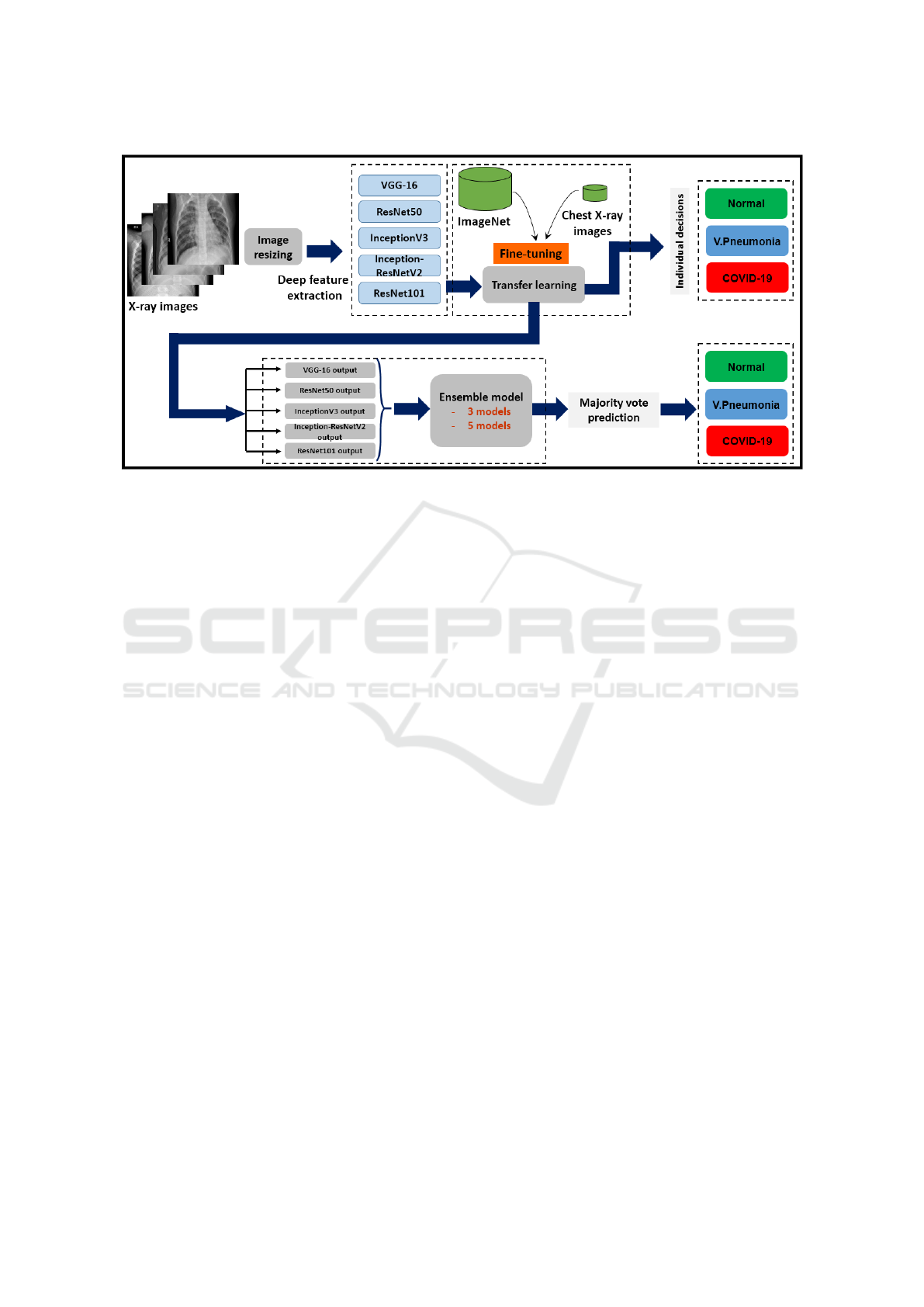
Figure 1: Overview of the different steps of our proposal.
ond step where their outputs will be combined using
an ensemble learning technique (also called ensemble
model) using the majority vote strategy. This combi-
nation enhances the classification performance of the
non-learned samples compared to the obtained rates
using each pre-trained model separately.
4 THE PROPOSED METHOD
4.1 Pre-Trained Models
In this Section, we present the architecture of the five
models that we used in our TL system. These mod-
els are pre-trained on ImageNet database (Krizhevsky
et al., 2012).
• VGG-16: (Simonyan and Zisserman, 2014) is
trained on the very large ImageNet dataset which
has over 14 million images and 1000 classes. It
contains 16 layers including 13 convolutional lay-
ers, 3 dense layers and 5 Max Pooling layers.
Each convolutional layer is 3×3 layer with a stride
size of 1 and the same padding. The pooling lay-
ers of VGG-16 are all 2×2 pooling layers with a
stride size of 2. Figure 2 presents modified VGG-
16 architecture as an example of TL.
• ResNet50: ResNet50 is a variant of ResNet pre-
trained model on ImageNet dataset which has 48
Convolution layers along with 1 MaxPool and 1
Average Pool layer. It has 3.8 × 10
9
Floating
points operations (He et al., 2016a).
• ResNet101: Anther variant of ResNet deep neu-
ral network series, trained on more than a million
images from the ImageNet database (Dai et al.,
2016). It consists of 101 deep layers with identity
connection between them.
• Inception-ResNetV2: A CNN that builds on the
Inception family of architectures (Szegedy et al.,
2017). Its architecture combines the Inception ar-
chitecture with residual connections. This CNN
contains 164 deep layers trained on ImageNet
dataset and is able to classify images into 1000
classes.
• InceptionV3: presented by google is the third
version of Inception DL convolution architec-
tures, with 42 deep layers contain Convolution
layer, AvgPool, MaxPool, Concat, Dropout, Fully
Connected layer and Softmax activation function
(Szegedy et al., 2016). The input layer size of
this model is different from the other models
(299 × 299 × 3 instead of 224 × 224 × 3).
4.2 Transfer Learning
We aim to transfer the acquired information from the
CNN models pre-trained on ImageNet dataset to our
specific task. The issue that needs to be addressed is
the highly dependance of these models on the initial
dataset, whereas our Chest X-ray images are differ-
ent. Consequently, the generalization of the network
will be poor since the extracted features from the large
amount of data are not inadequate to represent our
target images (to feed a classifier or softmax func-
tion). The solution consists of fine-tuning the pre-
trained models on our Chest X-ray images dataset that
is a very small dataset compared to the ImageNet. In
Advanced Deep Transfer Learning Using Ensemble Models for COVID-19 Detection from X-ray Images
357
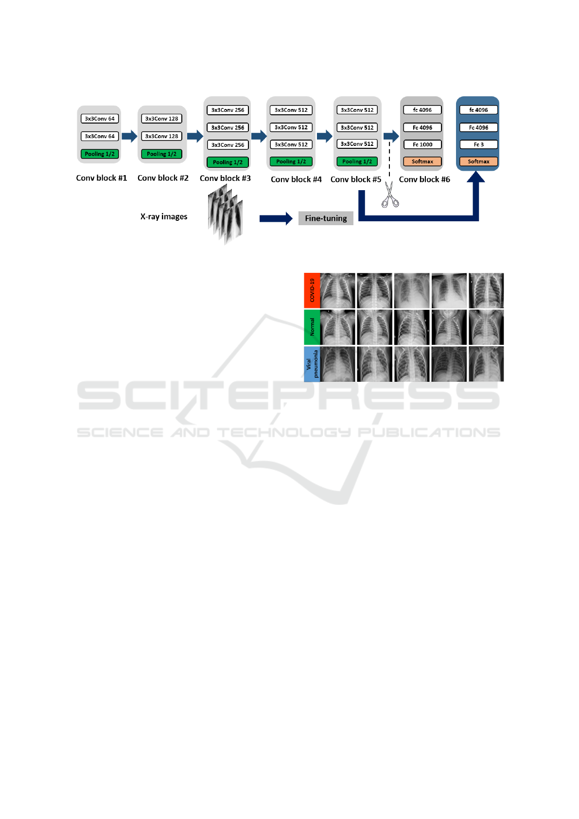
Figure 2: The modified VGG-16 network architecture.
other words, pre-trained CNN structures are updated
to suit our classification task. This strategy is gen-
erally much faster than the traditional training of the
CNN model from scratch with arbitrary weights. For
example, using VGG-16, the total number of parame-
ters after training is 14,789,955. The number of train-
able parameters is 75,267 which is very small com-
pared to 14,714,688 of non-trainable parameters.
4.3 Deep Ensemble Learning
To enhance the classification performance, we exploit
the different architectures of the five pre-trained mod-
els and we fuse their outputs to make a global deci-
sion. Thus, we propose to apply a majority voting
strategy as an ensemble learning stage. We then use
the combination of 5 and 3 models, respectively using
the output of the last epoch (30) of the 10
th
training
fold. The obtained results are presented and discussed
in detail in the following section.
5 EXPERIMENTAL RESULTS
All the experiments were performed on Windows
7 operating system 64 bits the TensorFlow/Keras
framework of python language. The implementation
of our proposal is provided by Google Colaboratory
Notebooks. The obtained results by each pre-trained
model, and using the deep ensemble model strategy
are presented in the following.
5.1 Database Description
COVID19 Radiography Database (Rahman, 2020):
contains 3616 COVID-19 positive cases along with
10,192 Healthy, 6012 Lung Opacity (Non-COVID
Figure 3: X-ray images from COVID19 Radiography
Database (Rahman, 2020).
lung infection), and 1345 Viral Pneumonia im-
ages. In this work, we carry out 3 class classification
(COVID-19, Healthy and Viral pneumonia). Some
scans from this database are shown in Figure 3.
5.2 Method Performance
The 10 fold-cross validation setting is applied using
the 4
th
version of COVID19 Radiography Database.
It is randomly split into training and test datasets. The
initial learning rate is of 0.0003 and cross entropy
loss. The models are trained for 30 epochs where the
batch size is 32.
In the following, we show the training perfor-
mances (accuracy and loss) as well as the validation
performances using the three best models including
Inception-ResNetV2, VGG-16 and InceptionV3, re-
spectively. The performance during the fold 1, 6 and
10 are displayed in detail in Figure 7. These curves
show that using VGG-16 architecture, the highest
training accuracy is observed 98.85% in epoch 7
where the highest validation accuracy is 99.65% in
epoch 24. The loss is 0.0324 and 0.024 at epoch 30 of
training and validation respectively. All the reported
results are put out from fold 10.
VISAPP 2023 - 18th International Conference on Computer Vision Theory and Applications
358
On the contrary, Inception-ResNetV2 achieved
higher training accuracy compared to VGG-16 by
99.46% in epoch 29. The highest validation accuracy
is 100%. Training and validation loss are respectively
0.026 and 0.005 (See Figure 8). Although the initial
loss value of Inception-ResNetV2 is very high com-
pared to that of VGG-16 (1.00 vs 0.2), the previous
values show that Inception-ResNetV2 is more effi-
cient during the training and validation stages com-
pared to VGG-16 over all the epochs of the 10
th
fold.
InceptionV3, however, is the third best model among
the five models (See Figure 9).
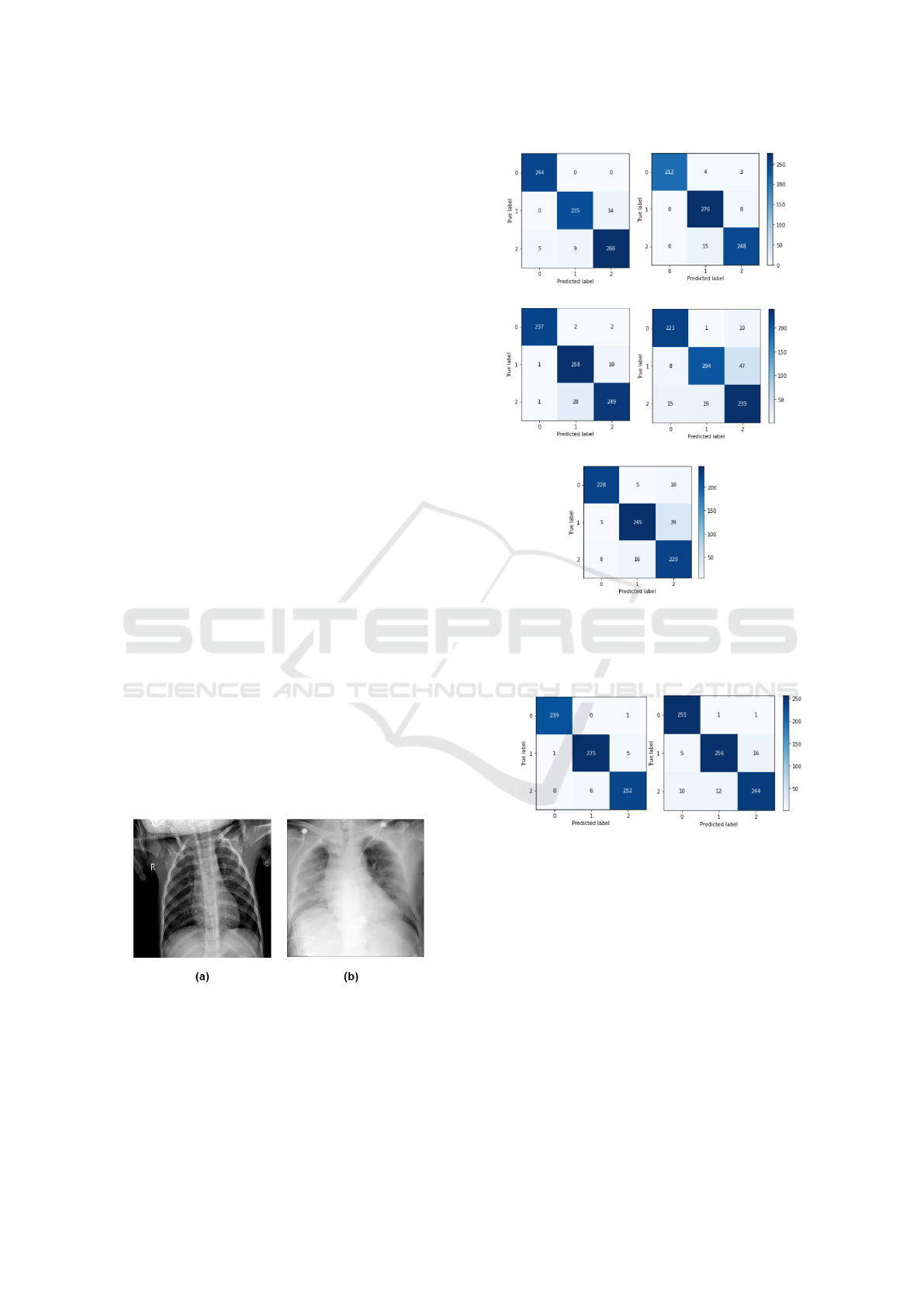
More details about the performance measures of
the best three models as well as the two remaining
ones (ResNet50 and ResNet101) are shown in Table
1. According to the displayed values of precision, re-
call, and F-score, ResNet101 is slightly better than
ResNet50. This performance can be clearly seen in
the confusion matrix of these models. Figure 5 shows
that ResNet101 has less false classifications. From
the same Figure, we can also notice that false classi-
fied samples of VGG-16 and Inception-ResNetV2 are
very limited compared to the two previous models.
This explains the very high rates registered as recall,
precision and F-score. Figure 4 presents two chal-
lenging samples of false positive and false negative
cases.
The use of ensemble learning, however, enhances
all the previous performances using the combination
of the three and five models’ outputs as shown in
the Table 1. Using this strategy, we achieve 1.00
of precision, recall and F-score when dealing with
the COVID-19 classes. Compared to recent state-
of-the-art methods, our proposed methods achieved
higher classification rate of the test set by 0.96 us-
ing the ensemble model of the combination (VGG-
16+ResNet50+ResNet101).
Figure 4: (a): False positive case, (b): false negative case.
5.3 Discussions
The experimental study proves that the TL is one of
the best deep learning techniques to efficiently detect
the COVID-19 using X-ray images.
(a) VGG-16 (b) IncResV2
(c) InceptionV3 (d) ResNet50
(e) ResNet101
Figure 5: Confusion matrix of the pre-trained models. La-
bel 0 refers to COVID-19 class, label 1 refers to Healthy
class, and label 2 refers to the VP.
(a) 5 models (b) 3 models
Figure 6: Confusion matrix of the majority vote prediction.
(a) refers to all the five models. (b) refers to the following
three models: VGG-16, ResNet50 and ResNet101.
The obtained results show that the ensemble mod-
els enhance the classification performance of the fine-
tuned CNNs. This paper proposes five pre-trained
models that give promising results which are still
competitive to those of the state-of-the-art method.
The TL strategy using the combination of the best
models outperforms the other methods and gives 0.98
accuracy of test images. The high performances
achieved are explained by the fact that TL is suitable
since the first CNN layers learn low-level features.
These features and mostly invariable from a classi-
Advanced Deep Transfer Learning Using Ensemble Models for COVID-19 Detection from X-ray Images
359
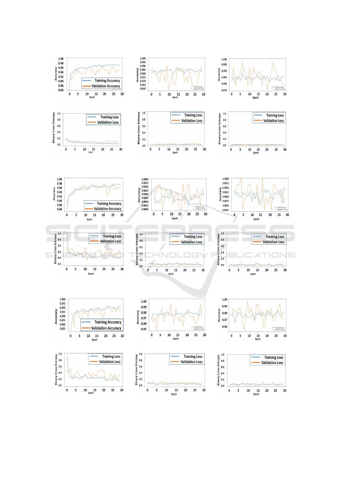
(a) Fold 1 acc (b) Fold 6 acc (c) Fold 10 acc
(d) Fold 1 loss (e) Fold 6 loss (f) Fold 10 loss
Figure 7: Training and validation accuracy / loss using VGG-16.
(a) Fold 1 acc (b) Fold 6 acc (c) Fold 10 acc
(d) Fold 1 loss (e) Fold 6 loss (f) Fold 10 loss
Figure 8: Training and validation accuracy / loss using Inception-ResNetV2.
(a) Fold 1 acc (b) Fold 6 acc (c) Fold 10 acc
(d) Fold 1 loss (e) Fold 6 loss (f) Fold 10 loss
Figure 9: Training and validation accuracy / loss using InceptionV3.
VISAPP 2023 - 18th International Conference on Computer Vision Theory and Applications
360
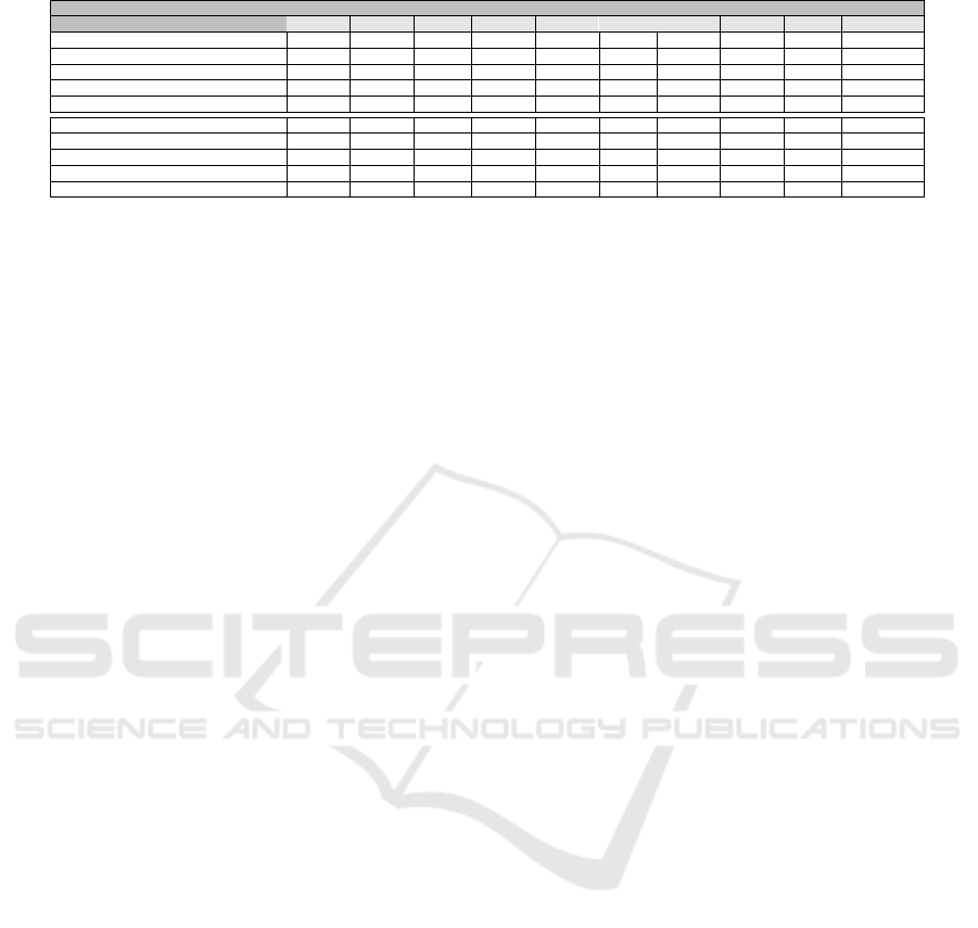
Table 1: Performance of 5 fine-tuned CNNs and ensemble learning with X-ray images of COVID-19, Healthy and VP cases.
Model / measure Precision Recall F-score Accuracy
Class COVID Healthy VP COVID Healthy VP COVID Healthy VP Test set
VGG-16 0.98 0.96 0.95 1.00 0.94 0.95 0.99 0.95 0.95 0.96
ResNet50 0.91 0.92 0.81 0.95 0.79 0.89 0.93 0.85 0.84 0.88
ResNet101 0.95 0.92 0.82 0.94 0.85 0.91 0.94 0.88 0.86 0.90
Inception-ResNetV2 1.00 0.94 0.96 0.97 0.97 0.94 0.98 0.95 0.95 0.96
InceptionV3 0.99 0.90 0.95 0.98 0.96 0.90 0.99 0.93 0.92 0.95
All five models 0.94 0.95 0.93 0.99 0.92 0.93 0.97 0.94 0.93 0.94
VGG-16+ResNet50+ResNet101 1.00 0.98 0.98 1.00 0.98 0.98 1.00 0.98 0.98 0.98
Pham et al. (Pham, 2021) / / / / / / / / / 0.96
Cavallo et al. (Cavallo et al., 2020) / / / / / / / / / 0.91
Rajpal et al. (Rajpal et al., 2020) 0.99 0.91 0.94 0.96 0.96 0.92 0.97 0.93 0.93 0.94
fication task to another (i.e. edges). The fine-tuning
provides specific features of the target domain such
as COVID-19 detection. It is worth noting that the
proposed method can be improved using class weight
algorithm to deal with the data imbalance issue which
is a common challenge in medical image diagnosis.
This is an important perspective since the majority
of medical datasets contains majority class of healthy
cases compared to positive cases.
6 CONCLUSION
Since the epidemic is still fast-spreading, the pro-
posed method seems to be a good solution to early di-
agnose the virus. We fine-tuned five pre-trained CNN
models to our COVID-19 dataset of X-ray images.
High classification performances have been achieved
especially with VGG-16 and Inception-ResNetV2.
Slightly lower performances have been achieved us-
ing the other models. This transfer learning technique
reduces considerably the training cost compared to
learning from scratch that becomes an amassed tech-
nique. To exploit the different features extracted by
each model, we proposed to combine their outputs to
find a global decision through a majority voting strat-
egy. This strategy further enhances the performance
of the proposed transfer learning-based method. In
future work, we look at the application of Graph neu-
ral network along with CNNs to improve the perfor-
mance and reduce the computation time. Also, the
visualisation using Grad-Cam technique can be made
to highlight the attention map of the classification.
ACKNOWLEDGEMENTS
The authors would like to thank the Agency for Re-
search Results Valuation and Technological Develop-
ment, Algeria (DGRSDT).
REFERENCES
Abbas, A., Abdelsamea, M. M., and Gaber, M. M. (2021).
Classification of covid-19 in chest x-ray images using
detrac deep convolutional neural network. Applied In-
telligence, 51(2):854–864.
Ahuja, S., Panigrahi, B. K., Dey, N., Rajinikanth, V., and
Gandhi, T. K. (2021). Deep transfer learning-based
automated detection of covid-19 from lung ct scan
slices. Applied Intelligence, 51(1):571–585.
Ali, H. A., Hariri, W., Zghal, N. S., and Aissa, D. B. (2022).
A comparison of machine learning methods for best
accuracy covid-19 diagnosis using chest x-ray images.
In 2022 IEEE 9th International Conference on Sci-
ences of Electronics, Technologies of Information and
Telecommunications (SETIT), pages 349–355. IEEE.
Altan, A. and Karasu, S. (2020). Recognition of covid-19
disease from x-ray images by hybrid model consist-
ing of 2d curvelet transform, chaotic salp swarm algo-
rithm and deep learning technique. Chaos, Solitons &
Fractals, 140:110071.
Bargshady, G., Zhou, X., Barua, P. D., Gururajan, R., Li, Y.,
and Acharya, U. R. (2022). Application of cyclegan
and transfer learning techniques for automated detec-
tion of covid-19 using x-ray images. Pattern Recogni-
tion Letters, 153:67–74.
Cavallo, A. U., Troisi, J., Forcina, M., Mari, P., Forte, V.,
Sperandio, M., Pagano, S., Cavallo, P., Floris, R., and
Geraci, F. (2020). Texture analysis in the evaluation
of covid-19 pneumonia in chest x-ray images: a proof
of concept study.
Chollet, F. (2017). Xception: Deep learning with depthwise
separable convolutions. In Proceedings of the IEEE
conference on computer vision and pattern recogni-
tion, pages 1251–1258.
Cohen, J. P., Morrison, P., Dao, L., Roth, K., Duong, T. Q.,
and Ghassemi, M. (2020). Covid-19 image data col-
lection: Prospective predictions are the future. arXiv
preprint arXiv:2006.11988.
Dai, J., Li, Y., He, K., and Sun, J. (2016). R-fcn: Object de-
tection via region-based fully convolutional networks.
arXiv preprint arXiv:1605.06409.
Das, N. N., Kumar, N., Kaur, M., Kumar, V., and Singh,
D. (2020). Automated deep transfer learning-based
approach for detection of covid-19 infection in chest
x-rays. Irbm.
Hariri, W. and Narin, A. (2021). Deep neural networks
for covid-19 detection and diagnosis using images and
Advanced Deep Transfer Learning Using Ensemble Models for COVID-19 Detection from X-ray Images
361

acoustic-based techniques: a recent review. Soft com-
puting, 25(24):15345–15362.
He, K., Zhang, X., Ren, S., and Sun, J. (2016a). Deep resid-
ual learning for image recognition. In Proceedings of
the IEEE conference on computer vision and pattern
recognition, pages 770–778.
He, K., Zhang, X., Ren, S., and Sun, J. (2016b). Iden-
tity mappings in deep residual networks. In Euro-
pean conference on computer vision, pages 630–645.
Springer.
Huang, M.-L. and Liao, Y.-C. (2022). A lightweight cnn-
based network on covid-19 detection using x-ray and
ct images. Computers in Biology and Medicine, page
105604.
Ismael, A. M. and S¸eng
¨
ur, A. (2021). Deep learning
approaches for covid-19 detection based on chest
x-ray images. Expert Systems with Applications,
164:114054.
Krizhevsky, A., Sutskever, I., and Hinton, G. E. (2012). Im-
agenet classification with deep convolutional neural
networks. Advances in neural information processing
systems, 25:1097–1105.
Kumar, S. and Mallik, A. (2022). Covid-19 detection from
chest x-rays using trained output based transfer learn-
ing approach. Neural Processing Letters, pages 1–24.
Loey, M., Manogaran, G., and Khalifa, N. E. M. (2020). A
deep transfer learning model with classical data aug-
mentation and cgan to detect covid-19 from chest ct
radiography digital images. Neural Computing and
Applications, pages 1–13.
Luz, E., Silva, P., Silva, R., Silva, L., Guimar
˜
aes, J.,
Miozzo, G., Moreira, G., and Menotti, D. (2022). To-
wards an effective and efficient deep learning model
for covid-19 patterns detection in x-ray images. Re-
search on Biomedical Engineering, 38(1):149–162.
Nayak, S. R., Nayak, D. R., Sinha, U., Arora, V., and Pa-
chori, R. B. (2020). Application of deep learning tech-
niques for detection of covid-19 cases using chest x-
ray images: A comprehensive study. Biomedical Sig-
nal Processing and Control, 64:102365.
Pham, T. D. (2021). Classification of covid-19 chest x-
rays with deep learning: new models or fine tuning?
Health Information Science and Systems, 9(1):1–11.
Rahimzadeh, M. and Attar, A. (2020). A new modified deep
convolutional neural network for detecting covid-19
from x-ray images. arXiv preprint arXiv:2004.08052.
Rahman, T. (2020). COVID-19 Radiography Database.
https://www.kaggle.com/tawsifurrahman/
covid19-radiography-database/. [Online;
accessed 05-April-2021].
Rajpal, S., Agarwal, M., Rajpal, A., Lakhyani, N., and Ku-
mar, N. (2020). Cov-elm classifier: an extreme learn-
ing machine based identification of covid-19 using
chest x-ray images. arXiv preprint arXiv:2007.08637.
Simonyan, K. and Zisserman, A. (2014). Very deep con-
volutional networks for large-scale image recognition.
arXiv preprint arXiv:1409.1556.
Szegedy, C., Ioffe, S., Vanhoucke, V., and Alemi, A.
(2017). Inception-v4, inception-resnet and the impact
of residual connections on learning. In Proceedings
of the AAAI Conference on Artificial Intelligence, vol-
ume 31.
Szegedy, C., Vanhoucke, V., Ioffe, S., Shlens, J., and Wo-
jna, Z. (2016). Rethinking the inception architecture
for computer vision. In Proceedings of the IEEE con-
ference on computer vision and pattern recognition,
pages 2818–2826.
Turkoglu, M. (2021). Covidetectionet: Covid-19 diagnosis
system based on x-ray images using features selected
from pre-learned deep features ensemble. Applied In-
telligence, 51(3):1213–1226.
Vaid, S., Kalantar, R., and Bhandari, M. (2020). Deep
learning covid-19 detection bias: accuracy through
artificial intelligence. International Orthopaedics,
44:1539–1542.
Zaabi, M., Smaoui, N., Derbel, H., and Hariri, W. (2020).
Alzheimer’s disease detection using convolutional
neural networks and transfer learning based methods.
In 2020 17th International Multi-Conference on Sys-
tems, Signals & Devices (SSD), pages 939–943. IEEE.
Zheng, C., Deng, X., Fu, Q., Zhou, Q., Feng, J., Ma, H.,
Liu, W., and Wang, X. (2020). Deep learning-based
detection for covid-19 from chest ct using weak label.
MedRxiv.
VISAPP 2023 - 18th International Conference on Computer Vision Theory and Applications
362
