
Arrythmia Classification Using MATLAB
®
Classification Learner App
Cesar N. Silva, Fernanda F. Lopes, Jefferson A. Matos
a
and Maria Claudia F. Castro
b
Electrical Engineering Department, Centro Universitario FEI, S
˜
ao Bernardo do Campo, Brazil
Keywords:
Electrocardiogram (ECG), Cardiac Arrhythmia, Classification, Machine-Learning.
Abstract:
Vital sign monitoring is becoming a part of our daily lives, emerging as a trend of smart wearable devices used
to manage health. Cardiac arrhythmia is any variation in the normal heartbeat rhythm, causing the heart to beat
improperly. This work presents a study on the classification of cardiac arrhythmias in 4 classes, Normal (N),
Supraventricular Ectopic (SVE), Ventricular Ectopic (LV), and Fusion of Normal and Ventricular (F). Using
the MIT-BIH Arrhythmia Database and the Classification Learner App from MATLAB
®
for training, it was
possible to investigate 24 models, where the Subspace KNN Ensemble obtained the best accuracy (74.4%)
and was later used for implementation in the suggested user interface application.
1 INTRODUCTION
According to the World Health Organization (WHO,
2021), cardiovascular diseases (CVDs) are the lead-
ing global cause of death, taking an estimated 17.9
million lives each year, with more than 75% occur-
ring in low- and middle-income countries (LMICs).
While intensive global efforts to prevent cardiovascu-
lar disease are underway, cardiac arrhythmias remain
neglected, especially in LMICs (Mkoko et al., 2020).
Cardiac arrhythmia refers to any variation in the
normal heartbeat rhythm, causing the heart to beat too
fast (Tachycardia), too slowly (Bradycardia), or errat-
ically. An arrhythmia occurs when the sinus node,
known as the natural pacemaker, develops an abnor-
mal rhythm, the normal conduction pathway is inter-
rupted, or when another part of the heart takes over
as the pacemaker (Humphreys et al., 2011; Ameri-
can Heart Association (AHA), 2016). When the heart
does not beat properly, it can not effectively pump
blood, and the organs, such as the brain, lungs, and
even the heart may be damaged or shut down. Thus,
arrhythmias should be diagnosed and treated as early
as possible to reduce the risk of sudden death.
Currently, vital sign monitoring is becoming a part
of our daily lives, emerging as a trend of smart wear-
able devices used to manage health. Their adoption
has further accelerated with the growth of telehealth
during the COVID-19 pandemic. The most widely
a
https://orcid.org/0000-0002-8800-713X
b
https://orcid.org/0000-0002-2751-0014
used tool for monitoring and diagnosing heart func-
tion, such as arrhythmia, is the Electrocardiogram
(ECG), a graphical representation of the heart’s elec-
trical activity. For an early diagnosis, an efficient, in-
telligent, and robust automated arrhythmia classifica-
tion system must be incorporated into smart wearable
devices (Bayoumy et al., 2021).
To cope with such challenges, several works
have been carried out on arrhythmia classification.
Machine-learning-oriented techniques are adopted,
requiring at least five steps: ECG signal condition-
ing such as amplification and denoising, feature ex-
traction, feature selection, classification, and perfor-
mance analysis (Mohebbanaaz et al., 2020).
ECG signal features mainly depend on time in-
terval, amplitude, and segment duration. The most
common are morphological information such as am-
plitudes and intervals identification of peaks P, R, T,
and QRS complex, as well as information about the
RR range/interspace, which is the distance between
peaks of two successive R waves in the ECG sig-
nal (de Albuquerque et al., 2018; Celin and Vasanth,
2018; Kuila et al., 2020; Mohebbanaaz et al., 2022).
Recently, a great interest has been in the applica-
tion of classification algorithms based on Deep Learn-
ing (Zhang et al., 2020; Hassan et al., 2022; Irfan
et al., 2022) with accuracies up to 99.35%. How-
ever, other techniques have also been used to clas-
sify arrhythmias, such as Decision Trees (Moheb-
banaaz et al., 2022), Random Forest (AbdElMoneem
et al., 2020), K-Nearest Neighbor (Mustaqeem et al.,
2018; AbdElMoneem et al., 2020), Ensemble Clas-
220
Silva, C., Lopes, F., Matos, J. and Castro, M.
Arrythmia Classification Using MATLAB
R
Classification Learner App.
DOI: 10.5220/0011666300003414
In Proceedings of the 16th International Joint Conference on Biomedical Engineering Systems and Technologies (BIOSTEC 2023) - Volume 4: BIOSIGNALS, pages 220-225
ISBN: 978-989-758-631-6; ISSN: 2184-4305
Copyright
c
2023 by SCITEPRESS – Science and Technology Publications, Lda. Under CC license (CC BY-NC-ND 4.0)
sifiers (Shalini et al., 2019; AbdElMoneem et al.,
2020), Support Vector Machines (de Albuquerque
et al., 2018; Mustaqeem et al., 2018; AbdElMoneem
et al., 2020), and others (de Albuquerque et al., 2018).
In these cases, accuracies range from 70 to 98%.
Most works comprise multiclass classification to
differentiate among up to 16 types of arrhythmias.
However, given that atrial fibrillation (AF) is the most
common heart arrhythmia, its detection has received
specific attention, either in a simple recognition sys-
tem or to classify it into subtypes (Celin and Vasanth,
2018; Horoba et al., 2019; Ganapathy et al., 2021;
Ramesh et al., 2021; Sager et al., 2021; da Silva et al.,
2021; Fuadah and Lim, 2022).
In this context, this work presents a study on the
detection and classification of cardiac arrhythmias us-
ing the MATLAB
®
Classification Learner applica-
tion. Four classes were defined according to the As-
sociation for the Advancement of Medical Instrumen-
tation (AAMI): Normal (N), Ectopic Supraventricular
(SVE), Ventricular Ectopic (VE), and Fusion of Nor-
mal and Ventricular (F). As a suggestion, a low-cost
device for ECG acquisition and a user interface for
communication with the health professional is also
presented.
2 MATERIALS AND METHODS
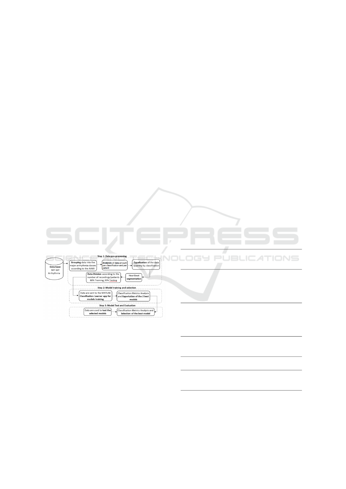
Figure 1: Data processing flowchart.
The flowchart for conducting the research is shown in
Figure 1. Firstly, the MIT-BIH dataset was prepared
for model training by performing signal extraction,
using the WFDB package, class preparation, data bal-
ance, and segmentation. Then ECG signals were par-
titioned into two sets of records in order to separate
the patients into training/validation (80%) and testing
(20%) groups. The structured data was exported to
the MATLAB
®
Classification Learner to the training
process and investigation of the best two models ex-
ported back to the MATLAB
®
algorithm to the test
phase and final metric analysis.
2.1 Dataset
The MIT-BIH Arrhythmia Database, which is pub-
licly available online at physionet.org (Goldberger
et al., 2000), is a well-known and worldwide used
standard dataset for arrhythmia detectors evaluation
(de Albuquerque et al., 2018; Celin and Vasanth,
2018; Kuila et al., 2020; Hassan et al., 2022; Ir-
fan et al., 2022; Mohebbanaaz et al., 2022). It was
collected by Boston’s Beth Israel Hospital (BIH) Ar-
rhythmia Laboratory between 1975 and 1979 (Moody
and Mark, 2001). The dataset contained 48 half-hour
excerpts of two-channel ambulatory ECG recordings,
obtained from 47 subjects, both male and female (25
and 22, respectively) of different age groups (between
23 and 89 years). The analog records were digi-
tized according to a sampling rate of 360 Hz, fil-
tered using a 0.1–100 Hz bandpass filter, the heart-
beats were marked and manually classified by experts
in 16 classes of arrhythmia.
Following the recommendation of ANSI/AAMI
standard EC57 (ANSI/AAMI, 2020), the 15 arrhyth-
mia classes reported in the database’s annotations
were grouped into 5 classes, as depicted in Table 1.
The 5th class (D), with an Unknown or with a pace-
maker, was discarded.
Table 1: MIT-BIH database classes grouped according to
AAMI Standard.
AAMI
Group
MIT-
BIH
Class
MIT-BIH Class (Description)
N
N Normal beat
L Left bundle branch block beat
R Right bundle branch block beat
e Atrial escape beat
j Nodal (junctional) escape beat
SVE
A Atrial premature beat
a Aberrated atrial premature beat
J Nodal (junctional) premature
beat
S Supraventricular premature beat
VE
V Premature ventricular contrac-
tion
E Ventricular escape beat
F F Fusion of ventricular and nor-
mal beat
D
f Fusion of paced and normal beat
/ Paced beat
Q Unclassifiable beat
However, as the database is unbalanced, with the
most typical classes having much more examples, the
results could be biased. Data balancing was per-
Arrythmia Classification Using MATLAB
R
Classification Learner App
221
formed through the resampling method with down-
sampling for the majority class. Each one of the con-
sidered classes had 2296 samples. Based on that, data
records were partitioned into two groups for training
and testing according to Table 2.
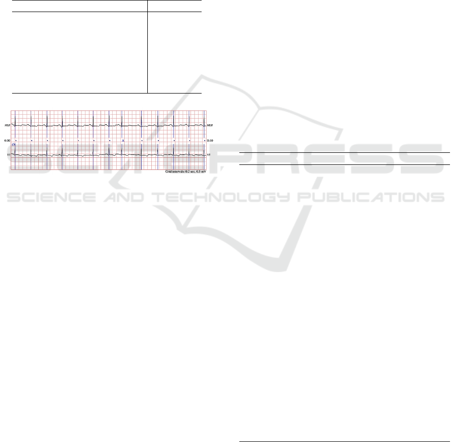
Before training, the annotated R wave peaks (Fig-
ure 2) were taken into consideration, a window of 300
samples around the peaks (P-149 to P+150 samples)
was segmented. No further pre-processing was done,
nor feature was extracted, other than the data window
(Singh et al., 2019).
Table 2: Data records partitionnig.
Training Testing
101 106 108 109 111 100 103
112 114 115 116 118 105 113
119 121 122 123 124 117 200
201 202 203 205 207 209 212
208 210 214 215 219 213
220 221 222 223 228
230 231 232 233 234
Figure 2: ECG example data.
2.2 Classification Learner App
The classification learner application provided by
MATLAB
®
is a toolbox that allows interactive data
analysis training classifiers with several machine-
learning models, such as Decision Trees, Discrim-
inant Analysis, Support Vector Machines (SVM),
Nearest Neighbor Classifiers (KNN), and Ensemble
Classifiers. The app provides classifier performance
metrics, such as validation accuracy, confusion ma-
trix, receiver operating characteristic curve (ROC),
and the area under the ROC curve (AUC), among
other resources.
In this research, a set of 24 classifiers were
adopted: Fine, Medium, and Coarse Decision Trees;
Linear and Quadratic Discriminant Analysis; Gaus-
sian and Kernel Naive Bayes; Linear, Quadratic, Cu-
bic, and Gaussian SVM; Fine Medium, Coarse, Co-
sine, Cubic, and Weighted KNN; Boosted Trees,
Bagged Trees, Subspace Discriminant, Subspace
KNN, and RUSBoost Trees Ensemble Classifiers.
Default classifier parameters, and the k-fold cross-
validation method, using k=10, were applied. Above
mentioned criteria were used to evaluate the perfor-
mances of different classifiers to assess the two classi-
fiers that had the highest validation accuracies. Those
models were exported for testing and final evaluation.
3 RESULTS
Table 3 shows validation accuracies for all classifiers.
Most SVM and KNN classifiers reached accuracies
above 90%. Cubic SVM and Subspace KNN Ensem-
ble Classifiers reached accuracies of 94%, being the
best, while Naive Bayes Classifiers presented had the
worst results. Figures 3 and 4 show the confusion ma-
trices of the best two classifiers, from which it can be
noted that the major misclassification occurred for the
F class.
These two trained models were exported back to
the MATLAB
®
algorithm for testing. Figures 5 and 6
show the resulting confusion matrices, with mean ac-
curacies of 67.1% for the Cubic SVM and 74.4% for
the Subspace KNN Ensemble. In both cases, ma-
jor misclassification occurred between the SVE e N
classes.
Table 3: Validation Accuracies.
Classifier Accuracy (%)
Cubic SVM 94.1
Subspace KNN Ensemble 94.0
Quadratic SVM 93.4
Fine KNN 93.2
Weighted KNN 92.2
Medium Gaussian SVM 91.8
Cosine KNN 91.7
Bagged Trees Ensemble 91.3
Fine Gaussian SVM 91.0
Medium KNN 90.8
Cubic KNN 90.8
Linear SVM 82.9
Boosted Trees Ensemble 81.6
Fine Tree 81.4
RUSBoost Trees Ensemble 81.0
Coarse KNN 78.5
Quadratic Discriminant 78.4
Coarse Gaussian SVM 78.3
Subspace Discriminant Ensemble 78.0
Linear Discriminant 77.6
Medium Tree 75.3
Coarse Tree 65.0
Kernel Naive Bayes 61.8
Gaussian Naive Bayes 55.9
BIOSIGNALS 2023 - 16th International Conference on Bio-inspired Systems and Signal Processing
222
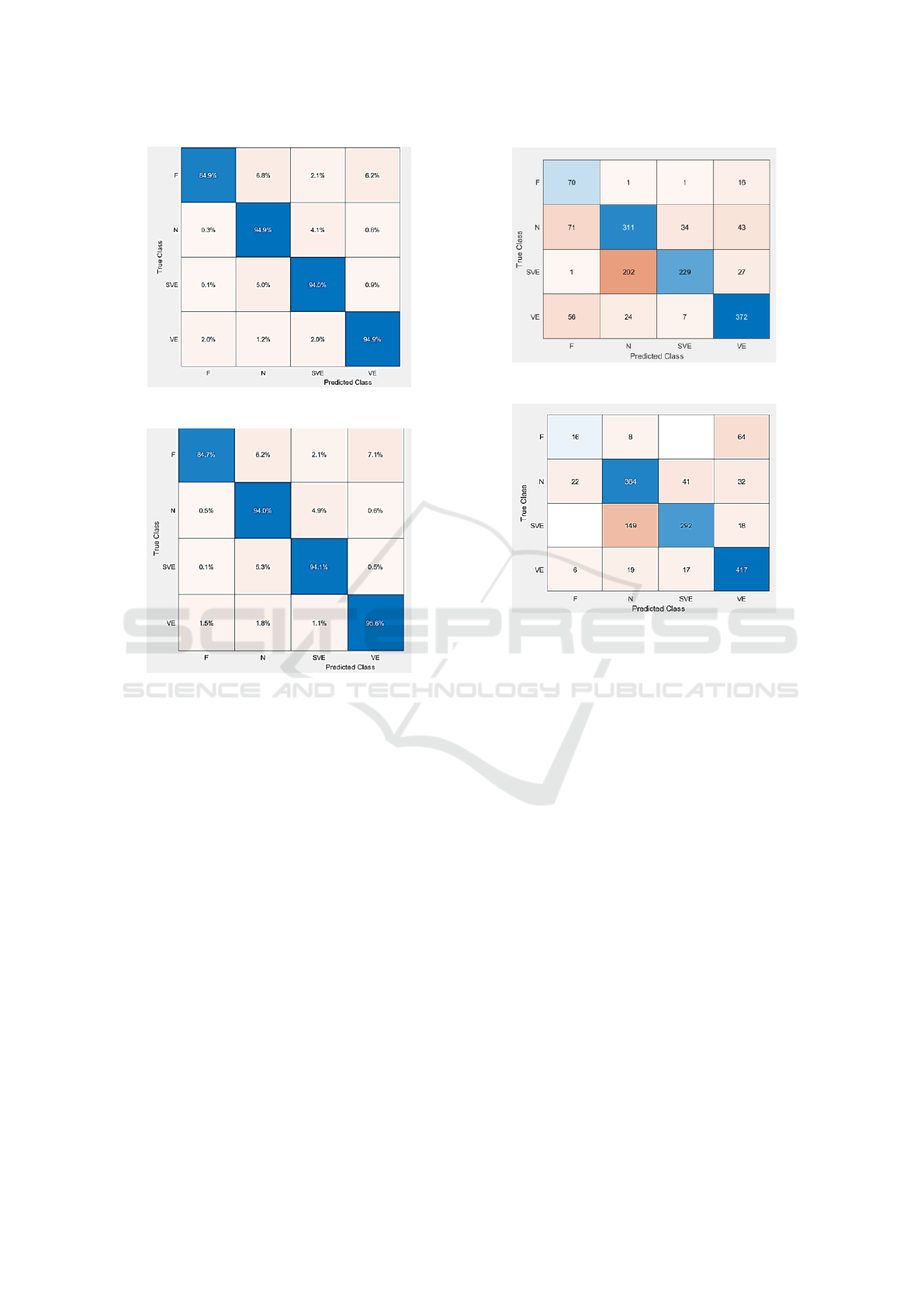
Figure 3: Validation Confusion Matrix for Cubic SVM.
Figure 4: Validation Confusion Matrix for Subspace KNN
Ensemble.
3.1 Hardware and User Interface
From a practical point of view, we propose the use of a
low-cost device for capturing the ECG signal, consist-
ing of an AD8233 module connected to an Arduino,
and a simple interface developed on the MATLAB
®
Appdesigner (Figure 7). After the acquisition, and
classification, the interface will inform, through an e-
mail, the detection of possible arrhythmia to a doctor
or accredited person.
4 DISCUSSION
The monitoring of vital signs by wearable devices
can contribute to the decentralization of health care,
allowing self-management and anticipation of emer-
gency care. Therefore, even if the final diagnosis is
the healthcare professional’s responsibility, machine-
learning techniques can automatically recognize and
classify specific patterns in these signals, indicating to
Figure 5: Test Confusion Matrix for Cubic SVM.
Figure 6: Test Confusion Matrix for Subspace KNN En-
semble.
the user. Several works in the literature are dedicated
to applying machine-learning techniques to recognize
cardiac arrhythmias, mostly with accuracies between
70% and 98%.
This work presented a study with 24 classifiers
using the Classification Learner application from
MATLAB
®
and the MIT-BIH Arrhythmia Database,
which is one of the most used databases. However,
although the validation results were promising, show-
ing accuracies of 94% for Cubic SVM and Subspace
KNN Ensemble, the test phase results showed lower
accuracies (74%), with most misclassifications be-
tween SVE e N classes.
The ECG data were digitized according to a sam-
pling rate of 360 Hz and filtered using a 0.1–100 Hz
bandpass filter. Hence, noises like power line inter-
ference, baseline drifts, motion artifacts, and elec-
tromyography noise can be added, and thus the lack
of pre-processing can be affected classification per-
formance. From a practical point of view, despite the
need for an acquisition system to implement some fil-
tering in the signal to eliminate noise, the literature
consulted did not clearly show this type of process-
ing.
Another fact was using the raw QRS complex in-
Arrythmia Classification Using MATLAB
R
Classification Learner App
223
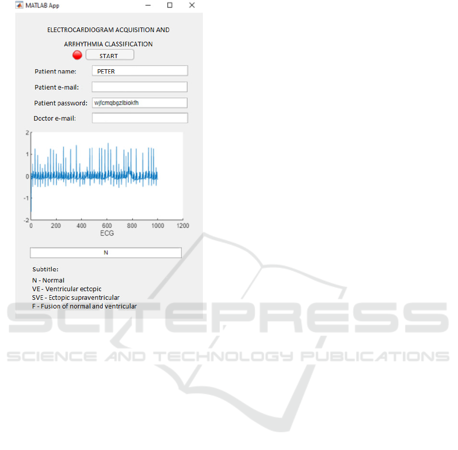
Figure 7: Hardware Design.
stead of a set of extracted features. Results showed
that the investigated models could not deal with it.
The literature showed that the only model able to deal
with a raw signal are those based on Deep Learning
due to a proper structure (Zhang et al., 2020; Has-
san et al., 2022; Irfan et al., 2022). Other models
need a set of features able to discriminate different
classes (de Albuquerque et al., 2018; Celin and Vas-
anth, 2018; Kuila et al., 2020; Mohebbanaaz et al.,
2022). Furthermore, despite reducing the number
of classes, arrhythmias groups may interfere in this
discrimination, especially without specific features as
applied in this study.
Moreover, datasets are usually unbalanced. The
results could be biased because the class with non-
ectopic data has much more samples than the others.
It is critical to balance the dataset or approximate it to
ensure that each class receives the same priority (Ab-
dElMoneem et al., 2020; Hassan et al., 2022). How-
ever, most of the literature work did not mention any
data balance. The use of a resampling approach can
accomplish balancing. Otherwise, as this work imple-
mented data balance only using the down-sampling
technique for the majority class, the number of re-
sulting samples could be not enough for the machine-
learning approach, being responsible for the reached
accuracies. The use of the up-sampling technique in
combination with down-sampling would increase the
number of available samples, improving results.
Despite that, for practical use, some improve-
ments are expected, such as the accuracy increase
for the arrhythmia classification, and for a smart de-
vice, the classifier must be embedded instead of being
through a PC interface.
5 CONCLUSION
This work showed a study of 24 models for cardiac
arrhythmia classification using the Classification App
from MATLAB
®
and suggested a low-cost device
for capturing the ECG signal with a simple interface
developed on the MATLAB
®
Appdesigner, allowing
rapid health professional communication for practical
use. Results were promising; however, more attention
should be given to the extraction of features in order
to increase classification accuracy and to the imple-
mentation of an embedded system.
ACKNOWLEDGEMENTS
The authors would like to thank Centro Universit
´
ario
FEI for project support.
REFERENCES
AbdElMoneem, S. S., Said, H. H., and Saad, A. A. (2020).
Arrhythmia disease classification and mobile based
system design. Journal of Physics: Conference Se-
ries, 1447(1):012014.
American Heart Association (AHA) (2016).
What is an arrhythmia? Available on-
line: https://www.heart.org/en/health-
topics/arrhythmia/about-arrhythmia.
ANSI/AAMI (2020). ANSI/AAMI EC57:2012/(R)2020 -
Testing and reporting performance results of cardiac
rhythm and ST segment measurement algorithms.
Bayoumy, K., Gaber, M., Elshafeey, A., Mhaimeed, O.,
Dineen, E. H., Marvel, F. A., Martin, S. S., Muse,
E. D., Turakhia, M. P., Tarakji, K. G., and Elshazly,
M. B. (2021). Smart wearable devices in cardiovascu-
lar care: where we are and how to move forward. Nat
Rev Cardiol., 18:581–599.
Celin, S. and Vasanth, K. (2018). Ecg signal classification
using various machine learning techniques. J Med
Syst, 42.
da Silva, L. F., Queiroz, J. A., Vanessa, C., Barros, A. K.,
Lopes, G. C., and Cabral, L. (2021). Separation
BIOSIGNALS 2023 - 16th International Conference on Bio-inspired Systems and Signal Processing
224

method of atrial fibrillation classes with high order
statistics and classification using machine learning. In
BIOSIGNALS.
de Albuquerque, V. H. C., Nunes, T. M., Pereira, D. R.,
da S. Luz, E. J., Menotti, D., Papa, J. P., and Tavares,
J. M. R. S. (2018). Robust automated cardiac arrhyth-
mia detection in ecg beat signals. Neural Comput &
Applic, 29:679–693.
Fuadah, Y. N. and Lim, K. M. (2022). Optimal classification
of atrial fibrillation and congestive heart failure using
machine learning. Frontiers in Physiology, 12.
Ganapathy, N., Baumg
¨
artel, D., and Deserno, T. M. (2021).
Automatic detection of atrial fibrillation in ecg us-
ing co-occurrence patterns of dynamic symbol assign-
ment and machine learning. Sensors, 21(10).
Goldberger, A., Amaral, L., Glass, L., JM, H., Ivanov,
P., Mark, R., Mietus, J., Moody, G., Peng, C.,
and Stanley, H. (2000). Physiobank, physiotoolkit,
and physionet: components of a new research re-
source for complex physiologic signals. Circulation.,
101(23):E215–20.
Hassan, S. U., Zahid, M. S. M., Abdullah, T. A., and
Husain, K. (2022). Classification of cardiac ar-
rhythmia using a convolutional neural network and
bi-directional long short-term memory. DIGITAL
HEALTH, 8:20552076221102766.
Horoba, K., Czabanski, R., Wrobel, J., Matonia, A., Mar-
tinek, R., Kupka, T., Kahankova, R., Leski, J. M., and
Graczyk, S. (2019). Recognition of atrial fibrilation
episodes in heart rate variability signals using a ma-
chine learning approach. In 2019 MIXDES - 26th In-
ternational Conference ”Mixed Design of Integrated
Circuits and Systems”, pages 419–424.
Humphreys, M., Warlow, C., and McGowan, J. (2011). Ar-
rhythmias and their Management, chapter 10, pages
132–155. John Wiley & Sons, Ltd.
Irfan, S., Anjum, N., Althobaiti, T., Alotaibi, A. A., Sid-
diqui, A. B., and Ramzan, N. (2022). Heartbeat classi-
fication and arrhythmia detection using a multi-model
deep-learning technique. Sensors, 22(15).
Kuila, S., Dhanda, N., and Joardar, S. (2020). Fea-
ture extraction of electrocardiogram signal using ma-
chine learning classification. International Journal
of Electrical and Computer Engineering (IJECE),
10(6):6598–6605.
Mkoko, P., Bahiru, E., Ajijola, O. A., Bonny, A., and
Chin, A. (2020). Cardiac arrhythmias in low- and
middle-income countries. Cardiovasc Diagn Ther.,
10(2):350–360.
Mohebbanaaz, Kumari, L. V. R., and Sai, Y. P. (2022). Clas-
sification of ecg beats using optimized decision tree
and adaptive boosted optimized decision tree. Signal,
Image and Video Processing, 16:695–703.
Mohebbanaaz, Sai, Y. P., and kumari, L. R. (2020). A
review on arrhythmia classification using ecg sig-
nals. In 2020 IEEE International Students’ Confer-
ence on Electrical,Electronics and Computer Science
(SCEECS), pages 1–6.
Moody, G. B. and Mark, R. G. (2001). The impact of the
mit-bih arrhythmia database. IEEE Engineering in
Medicine and Biology Magazine., 20(3):45–50.
Mustaqeem, A., Anwar, S. M., and Majid, M. (2018). Mul-
ticlass classification of cardiac arrhythmia using im-
proved feature selection and svm invariants. Com-
putational and Mathematical Methods in Medicine,
2018:7310496.
Ramesh, J., Solatidehkordi, Z., Aburukba, R., and Sagahy-
roon, A. (2021). Atrial fibrillation classification with
smart wearables using short-term heart rate variabil-
ity and deep convolutional neural networks. Sensors,
21(21).
Sager, S., Bernhardt, F., Kehrle, F., Merkert, M., Potschka,
A., Meder, B., Katus, H., and Scholz, E. (2021).
Expert-enhanced machine learning for cardiac ar-
rhythmia classification. PLoS One, 16(12):e0261571.
Shalini, B., Nandini, V., Sandhya, M., and R, B. (2019).
Prediction and classification of cardiac arrhythmia.
International Research Journal of Engineering and
Technology (IRJET), 6(6):572–576.
Singh, V., Tewary, S., Sardana, V., and Sardana, H. K.
(2019). Arrhythmia detection - a machine learning
based comparative analysis with mit-bih ecg data. In
2019 IEEE 5th International Conference for Conver-
gence in Technology (I2CT), pages 1–5.
WHO (2021). Cardiovascular diseases (cvds). fact sheets.
Available online. https://www.who.int/en/news-
room/fact-sheets/detail/cardiovascular-diseases-
(cvds). Accessed Oct/2022.
Zhang, J., Liu, A., Gao, M., Chen, X., Zhang, X., and
Chen, X. (2020). Ecg-based multi-class arrhythmia
detection using spatio-temporal attention-based con-
volutional recurrent neural network. Artificial Intelli-
gence in Medicine, 106:101856.
Arrythmia Classification Using MATLAB
R
Classification Learner App
225
