
Application of Magnetic Resonance Imaging Technology in the
Detection of Brain Diseases
Yuke Shen
School of Medicine, University of Leeds, Leeds, LS2 9JT, U.K.
Keywords:
Functional Magnetic Resonance Imaging (Fmri), Diffusion Tensor Imaging (DTI), Brain Tumor, Cerebral
Ischemia, Brain Injury.
Abstract: The brain is the nerve center of human beings which is one of the most important organs of the human body.
Once a brain disease occurs, it will bring a heavy blow to the patient, and even endanger the patient's life. At
present, brain diseases mainly include brain tumor, brain injury, cerebral ischemia, and so on. Computed
tomography (CT) and X-ray photography both have different levels of radiation. In contrast, magnetic
resonance imaging (MRI) technology is considered a safe detection method without radiation hazards, so it is
suitable for the detection of brain diseases. However, pure MRI has limitations and cannot detect some fine
structures or neurological states. Magnetic resonance imaging such as functional magnetic resonance imaging
(fMRI) and Diffusion tensor imaging (DTI) can segment brain imaging, detect microbleeds, and judge the
state of brain function, and are widely used in the detection of brain diseases. This paper mainly introduces
the application of fMRI and DTI in the detection of brain diseases such as brain tumor, brain injury and
cerebral ischemia, aiming to provide some ideas for the diagnosis of brain diseases.
1 INTRODUCTION
The brain is the nerve center of human beings which
is one of the most important organs of the body. Once
a brain disease strikes, it can be a heavy or even
devastating blow to the patient. (JIANG, 2011)
According to available data, there will be nearly 800
billion euros of brain patients by 2010. (JIANG,
2011) Encephalopathy affects 127 million Europeans,
and the annual cost of treatment is €385 billion.
(LAWRENCE, 2017) Mental illness accounts for
62% of the cost and neurological disorders, including
dementia, for 38%. According to research, brain
diseases are more costly than heart disease or cancer.
(LAWRENCE, 2017) Also, such a predicament is not
unique to Europe and people all over the world are at
risk of brain disease. (LIANG, 2020) The brain is the
nerve center of the human being, brain damage can
lead to a silent, rapidly developing disease that
ultimately leads to death. (LIU, 2015) Therefore,
diagonalize brain disease has become extremely
important.
Brain tumors, damage, ischemia, and other
illnesses are all prevalent. Modern diagnostic
treatments include Computed tomography (CT), x-
rays, and magnetic resonance imaging (MRI).
(MACDONALD, 2015) However, there are some
brain lesions that may easily be missed with CT
examinations. For example, because the lesions are
too small or close to the base of the skull, the skull is
dense and the resection is too thick, which makes it
difficult to diagnose due to "volume effects".
(MACDONALD, 2015) In other early-stage disease,
including cerebral infarction, the density difference
between the lesion and normal brain tissue is
significant, making CT difficult to diagnose subacute
and chronic intracranial hemorrhage (JIANG, 2011).
(PATEL, 2011) X-rays can have diagnostic
limitations. For starters, X-rays are 2D pictures, and
there is overlap in the diagnosis of brain tissue,
making illness localization difficult. (SOTAK, 2002)
Then, the brain is mostly soft tissue and fluid, which
is absorbed by X-rays, making it impossible to
distinguish disease from normal tissue,
(UNDERHILL, 2010) but X-rays are superior in
determining skull fractures. (VILELA, 2017)
Compared with X-ray, MRI is more effective at
detecting brain tumors. It is a cutting-edge, non-
invasive technique that generates cross-sectional
pictures of the patient's anatomy using magnetic and
radio waves. (YUAN, 2004) Therefore, MRI is
considered to be a safe detection method without
198
Shen, Y.
Application of Magnetic Resonance Imaging Technology in the Detection of Brain Diseases.
DOI: 10.5220/0012018300003633
In Proceedings of the 4th International Conference on Biotechnology and Biomedicine (ICBB 2022), pages 198-203
ISBN: 978-989-758-637-8
Copyright
c
2023 by SCITEPRESS – Science and Technology Publications, Lda. Under CC license (CC BY-NC-ND 4.0)

radiation hazards. This is due to the fact that MRI uses
radiofrequency pulses. This pulse is a long
wavelength, low intensity electromagnetic wave that
does not damage the body's hydrocarbon bonds.
(CAZALIS, 2006) In terms of soft tissue resolution,
MRI allows better visibility of the brain's grey matter,
nucleus accumbens, nephron cortex and medulla.
(CAZALIS, 2006) In addition, MRI can be used in a
variety of ways to reflect multi-parametric
information about the tissue, including T1 and T2
values, proton density, flux, water molecule diffusion
and other parameters. (MACDONALD, 2015) This
way of obtaining multiparametric information is not
only beneficial for the display of lesions, but also for
their qualitative diagnosis. (LAWRENCE, 2017)
However, MRI has several limitations. First and
foremost, it takes a significant amount of time to
picture. Typically, a cranial examination takes about
20 minutes, and a spinal examination takes about 15
minutes. (MCDONALD, 2012) If enhancement is
required, the waiting time will be more than 10
minutes. (NARAYANA, 2017) Then, due to the lack
of H protons, low-signal calcifications are difficult to
detect in MRI pictures, making them insensitive to
calcifications. (NARAYANA, 2015) For
intramedullary lesions such oedema and tumor
invasion, MRI displays high contrast soft tissues but
low concentrations of H protons in bone structures.
(ROSS, 2011) The most crucial item is that MRI
artefacts are more common, as there are numerous
contributing elements in MRI imaging. (SHENTON,
2012) Finally, owing to time limits, MRI is not suited
for patients in severe condition or with metal
implants. (LI, 2020)
This article mainly discusses the use of MRI in the
treatment of brain tumors, cerebral ischemia, and
brain damage. Then, it makes some reference ideas
for diagnosing brain disorders.
2 BRAIN SEGMENTATION
METHOD
The two most critical stages in the treatment of brain
tumours are segmentation of the brain and
identification of the tumour. MRI is a widely used
technique for the segmentation and detection of
tumours. (WANG, 2017) MRI, which mainly
includes fMRI and DTI, can identify the functionality
of the brain, especially with increased visualization of
the grey and white matter of the brain. (WU, 2020)
FMRI is a neuroimaging detection method. MRI
was used to quantify hemodynamic changes induced
by neural activity. (AMIN, 2020) Due to its non-
invasiveness and minimal radiation exposure, fMRI
establishes an important position in the field of
functional brain localization. (GUPTA, 2010) In the
1890s, researchers discovered that changes in blood
flow and oxygen saturation were intricately linked to
neuronal activity. The activation of nerve cells
requires oxygen. Oxygen is transported by
microvessels near nerve cells using hemoglobin in red
blood cells. (GUPTA, 2010) Thus, when a neuron
activates, blood flow increases to replenish lost
oxygen. There is typically a 1-5 second delay between
neural activation and hemodynamic changes,
followed by a 4-5 second peak before returning to
baseline. (MOHD, 2014) This results in changes in
cerebral blood flow not only in areas of neuronal
activity but also in local blood deoxyhemoglobin and
oxyhemoglobin concentrations and cerebral blood
volume. (MYRONENKO, 2019) When subjects are
given several types of radioactive chemicals during
positron emission tomography (PET) scans, the
radioactive chemicals are taken up by activated brain
cells. (LAWRENCE, 2017; TIWARI, 2020) MRI
uses magnetic fields and radiofrequency radiation to
generate pulses of energy in the brain. The pulses may
be tuned to certain frequency ranges, inducing atomic
couplin. (WADHWA, 2019) When the magnetic
pulse is removed, these atoms vibrate and return to
their original state. A specialized radio frequency
receiver detects these resonances and transmits the
information to a computer, which then generates a
picture of the location of individual atoms within the
brain region. (YANG, 2007) As a result, fMR is more
widely used to identify brain disorders.
Magnetic resonance diffusion functional imaging
was first introduced by Peter Basser in 1994. (AMIN,
2020) It is an improved version of traditional MRI.
DTI uses the diffusion of water as a probe to
determine the anatomy of brain networks, providing
static anatomical information that is not affected by
brain function. (GUPTA, 2010) Because the obstacles
along the fibers are relatively small and cannot
restrict the movement of water molecules, the water
molecules should move faster along the axonal fibers,
rather than moving upright toward the fibers.
(MOHD, 2014) In axon-based directions, anisotropic
diffusion can generate entirely new image contrasts,
and this anisotropy is used in DTI to determine the
organization of nerve cells in the brain. A 3D
diffusion model is estimated by repeating this process
in multiple directions. (MYRONENKO, 2019) This
approach may result in signal degradation due to
diffusing molecules, resulting in darker volumetric
pixels or voxels. For instance, white matter fibres
Application of Magnetic Resonance Imaging Technology in the Detection of Brain Diseases
199

running parallel to the magnetic field gradient
direction will generate a dark diffusion-weighted
picture of that direction. (WADHWA, 2019) The
diffusion tensor is then measured by comparing the
signal loss to the original signal. The two main
parameters to define the orientation of neurons from
tensor calculations are fractional anisotropy (FA) and
mean diffusivity (MD). (YANG, 2007) FA quantifies
the directionality of diffusion, whereas MD quantifies
the horizontal average diffusivity. (NARAYANA,
2015) In summary, the above analysis of water
diffusion is performed by applying a magnetic field
gradient to produce images sensitive to diffusion in a
specific direction. A DTI technique is then
performed. The DTI technique consists of delivering
external magnetic pulses that apply a random phase
shift to the diffusing water molecules. And this
technique allows for detection and diagnosis. (LI,
2020)
Image processing is another key step in applying
MRI to detect tumours. For brain image
segmentation, mixed population-learning vector
quantization (LVQ) is often employed to find tumour
regions in abnormal brain images. (TIWARI, 2020)
This approach utilizes Flair, T1C, and T2-weighted
imaging, providing an entropy-based strategy. This
novel technique employs the formation of
reconstruction filters to assist radiologists in rapidly
localizing image brightness fluctuations and poorly
defined tumour regions. (AMIN, 2020) In addition,
LVQ can correct tissue with non-uniform gain and the
difficulty of identifying tiny lesions in images.
Compared with traditional computer detection
systems, 3D magnetic resonance image (MBA) is a
semantic segmentation network based on the
encoder-decoder structure to segment tumour
subregions. (MYRONENKO, 2019) Because brain
tumours are relatively difficult to classify, different
tumours can exhibit similar appearances, which poses
a barrier to traditional computer terminology.
(MYRONENKO, 2019) Nonetheless, 3D MRI
pictures adhere to the encoder-decoder structure of
convolutional neural networks (CNNs) to extract
deep visual information through asymmetric large
encoders. (MYRONENKO, 2019) The decoder part
reconstructs the dense split encoding. At the
simultaneous time it adds the variable autoencoder
(VAE) branch to the network. In this way the input
image can be reconstructed together with the
segments, thus normalizing the shared encoder. The
method is able to improve the accuracy to about 97%.
(TIWARI, 2020)
3 APPLICATION OF MRI IN
BRAIN DISEASES
3.1 Application of MRI in the
Detection of Brain Tumor
Among the brain segmentation methods described
above, FMR and DTI are the most widely used.
FMR is often used for preoperative planning.
These results help guide whether to perform surgery,
assess risk and prognosis, plan the surgical route, and
maximize tumour resection. (GUPTA, 2010) Gupta et
al. have demonstrated that FMR imaging mapping of
the central sulcus is resistant to potential limiting
artefacts from head movement, patient anxiety, and
abnormal vasculature. (GUPTA, 2010) Furthermore,
since anatomical predictors alone cannot determine
whether a specific language region is affected by
tumour, FMR is also important in language function
and laterality localization. (AMIN, 2020) At the New
York Cancer Center, a large proportion of brain
tumour patients underwent FMR imaging, mostly
preoperative imaging. (MOHD, 2014) This means
that a large number of studies underscore the
importance of neurosurgeons in obtaining
preoperative FMR.
Diffusion-Weighted Imaging (DWI) or DW-MRI
is software that generates pictures using a certain
magnetic resonance imaging sequence. (WADHWA,
2019) This procedure produces contrast in magnetic
resonance pictures by exploiting the diffusion of
water molecules. (MOHD, 2014) DTI, a subtype of
DWI, has been widely used to map white matter tracts
in the brain. (GUPTA, 2010) Traditionally, applying
three gradients in one direction is sufficient to
determine the "average diffusivity" of the diffusion
tensor or trace, a measure of oedema in DWI.
(TIWARI, 2020) In clinical practice, trace-weighted
images have been shown to be effective in identifying
vascular strokes in the brain with early (within
minutes) detection of hypoxic oedema.
(MYRONENKO, 2019) Furthermore, the principal
directions of the diffusion tensor can be used to infer
white matter connectivity in the brain. TIWARI This
treatment approach is particularly critical in brain
tumour surgery, where inadvertent damage to
normally functioning white matter pathways can lead
to severe neurological damage. (MOHD, 2014).
ICBB 2022 - International Conference on Biotechnology and Biomedicine
200
3.2
MRI Brain Injury Detection
Applications
Over the past 30 years, fMRI has been increasingly
used to study various neuropsychiatric disorders. But
relatively few fMRI studies have examined the
cognitive and behavioral sequelae of Mild traumatic
brain injury (mTBI), its course over time, and its
utility as a biomarker for potential treatments. (WU,
2020) McDonald et al scanned 11 patients with mTBI
1 year after injury to assess changes in brain
activation patterns over time. (NARAYANA, 2017)
Although at one-year follow-up, the mTBI group no
longer reported significant post-concussion sequelae
(PCS), they continued to exhibit mild depression in
response speed compared to the control group. (LI,
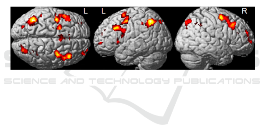
2020) In addition, mTBI patients exhibited task-
related increased activation of the right frontal lobe,
manifested by the highest white matter (WM) load
relative to controls 1 month to 1 year after injury see
Figure 1. fMRI in brain injury. (CAZALIS, 2006;
MCDONALD, 2012) In both groups, patients showed
activation of the left prefrontal cortex over time. This
finding suggests that despite the high resolution of
PCS, persistent brain dysfunction is still possible 1
year after mTBI. (SHENTON, 2012) McAllister et al.
then used fMRI to investigate whether there are
problems with episodic memory encoding and
retrieval after mTBI. Patients participated in the test,
listening to novel and familiar words separately.
Results showed increased activation in the right
dorsolateral prefrontal cortex (DLPFC) when hearing
familiar words. (ROSS, 2011) When new words were
heard, the activation of the middle temporal lobe
increased. Both the intensity and spatial extent of
activation were reduced in mTBI patients compared
to controls. (CAZALIS, 2006)
Figure 1. fMRI in brain injury.
DTI is a sensitive imaging tool for the detection
of diffuse axonal injury (DAI) and can be used to
detect mild traumatic brain injury (mTBI), also
known as concussion. (NARAYANA, 2017) As
previously described, DTI is sensitive to subtle
changes in white matter fiber tracts and can reveal
microstructural axonal damage. Arfanakis used DTI
for the first time to study diffuse axonal injury in
mTBI, evaluating the two main dependent measures
fractional anisotropy (FA) and mean diffusivity (MD)
in DTI. (LAWRENCE, 2017) The results showed that
there was no difference in MD between mTBI
patients and controls. However, researchers observed
group differences in the corpus callosum and internal
capsule, where FA was reduced in the mTBI group
compared to the control group. (CAZALIS, 2006)
Importantly, this result was consistent with
histopathology. This conclusion makes DTI an
important early indicator of brain injury in mTBI.
(MCDONALD, 2012) Dr. Alexander Lin et al. used
DTI to conclude that repetitive concussions and
concussion injuries occur in the etiology of chronic
traumatic encephalopathy in sports-related injuries
such as professional football. (MCDONALD, 2012)
In the report, FA and MD changes were the greatest
in one of the athletes. Moreover, the FA and MD of
all data changed in both directions of increase and
decrease. (MCDONALD, 2012) Johnston et al
showed that increased MD and decreased FA may
indicate vasogenic edema, which may resolve over
time. (NARAYANA, 2017) Whereas an increase in
FA and a reduction in MD may imply cytotoxic
edoema, which manifests as axonal swelling and
more constrained water transport. Thus, higher FA
and accompanying reduced MD may suggest a bad
prognosis in the early stages of brain damage.
(SHENTON, 2012)
3.3
Application of MRI in the Detection
of Cerebral Ischemia
Basic physiologic parameters for imaging in acute
ischemic stroke include assessment of
neuroparenchyma, vascular lumen patency, and
ischemic penumbra. (Hakimi, 2019)
Thromboembolism in vessels is markedly visualized
Application of Magnetic Resonance Imaging Technology in the Detection of Brain Diseases
201

on susceptibility-weighted imaging (SWI) due to high
levels of iron and increased deoxyhemoglobin
content in the thrombus. (Hui, 2021) According to
clinical experiments, SWI has higher sensitivity and
better contrast resolution in detecting
thromboembolism in the anterior and posterior
circulations. (Hui, 2021) Additionally, SWI is
sensitive in identifying the presence of fragmented
thrombi and their respective locations. (JIANG,
2011) This is because routine angiography requires
the detection of fragmented thrombi in the presence
of primary vessel occlusion or poor collateral
circulation. (JIANG, 2011) Because SWI is well
suited for assessing the intracranial vertebrobasilar
circulation, it is critical for assessing thrombus and
for neuron Intervention planning. (LAWRENCE,
2017)
DTI is critical in assessing ischemic brain
damage. Yang et al. published a preliminary DTI
study in experimental stroke and human stroke. DTI,
in comparison to other MR measures, gives
information on autopsy and geographic evolution of
illness. (LIANG, 2020) The unique ability of DTI to
differentiate between white and grey matter allows
quantitative assessment of ischemic injury in these
tissues. Andrew et al. have demonstrated that this
feature is useful in explaining spatially heterogeneous
changes in water diffusion during the temporal
evolution of clinical stroke. (LIU, 2015) In addition,
DTI can independently assess the therapeutic
response of white and grey matter to neuroprotective
therapy. (MACDONALD, 2015) Finally, diffusion
anisotropy measurements can be combined with other
MR parameters to provide a way to assess cerebral
ischemia in a time-independent manner. (PATEL,
2011) This feature is particularly important in a
clinical setting because autopsy of stroke onset is
often unknown.
4 CONCLUSION
MRI is a good non-invasive means of detection. In
addition to the absence of radiation, MRI provides
more detailed images than other diagnostic imaging
tests, and scans tend to be clearer. (SOTAK, 2002)
And allows medical professionals to quickly spot
structures or tumors that may be too small to show on
an X-ray or CT scan. In particular, it is more widely
used in the brain. (YANG, 2007) This article focuses
on describing segmentation methods and applications
of fMRI and DTI in brain injury, cerebral ischemia,
and brain tumors.
With the development of medical imaging
technology, magnetic resonance imaging technology
has become an important diagnostic tool in clinical
neuroradiology, neurology, and neurosurgery today.
(MOHD, 2014) Clinically, surgical teams have begun
to use fMRI and DTI imaging techniques to plan
surgical protocols to minimize the impact on the
function of important brain regions. (UNDERHILL,
2010) In addition, with the popularity of magnetic
resonance imaging equipment and the improvement
of data processing methods, fMRI and DTI have also
played a greater role in clinical decision-making.
(GUPTA, 2010) Using fMRI to study comatose
patients, decisions can be made about the level of
consciousness and the probability of recovery in
patients with persistent vegetative states. (AMIN,
2020) DTI also provides important value in analyzing
the effects of cerebral microbleeds on cognitive
impairment and functional impairment. (TIWARI,
2020)
REFERENCES
AMIN, J., SHARIF, M., YASMIN, M. & FERNANDES,
S. L. 2020. A distinctive approach in brain tumor
detection and classification using MRI. Pattern
Recognition Letters, 139, 118-127.
CAZALIS, F., FEYDY, A., VALABREGUE, R.,
PELEGRINI-ISSAC, M., PIEROT, L. & AZOUVI, P.
2006. fMRI study of problem-solving after severe
traumatic brain injury. Brain Inj, 20, 1019-28.
GUPTA, A., SHAH, A., YOUNG, R. J. & HOLODNY, A.
I. 2010. Imaging of brain tumors: functional magnetic
resonance imaging and diffusion tensor imaging.
Neuroimaging Clin N Am, 20, 379-400.
Hakimi, R. and S. Sivakumar, Imaging of Carotid
Dissection. Curr Pain Headache Rep, 2019. 23(1): p. 2.
Hui Jia, Z.L., Jun Yuan, Progress in application of high
resolution mri in middle cerebral atherosclerosis. 2021.
JIANG, Q., EWING, J. R. & CHOPP, M. 2011. MRI of
Blood–Brain Barrier Permeability in Cerebral
Ischemia. Translational Stroke Research, 3, 56-64.
LAWRENCE, T. P., PRETORIUS, P. M., EZRA, M.,
CADOUX-HUDSON, T. & VOETS, N. L. 2017. Early
detection of cerebral microbleeds following traumatic
brain injury using MRI in the hyper-acute phase.
Neurosci Lett, 655, 143-150.
Liang, S., Zhang, J., Zhang, Q., Li, L., Zhang, Y., Jin, T.,
ZHANG, B., HE, X., CHEN, L., TAO, J., LI, Z., LIU,
W. & CHEN, L. 2020. Longitudinal tracing of white
matter integrity on diffusion tensor imaging in the
chronic cerebral ischemia and acute cerebral ischemia.
Brain Res Bull, 154, 135-141.
Liu, C., Li, W., Tong, K. A., Yeom, K. W. & Kuzminski,
S. 2015. Susceptibility-weighted imaging and
ICBB 2022 - International Conference on Biotechnology and Biomedicine
202

quantitative susceptibility mapping in the brain. J Magn
Reson Imaging, 42, 23-41.
MacDonald, M. E. & Frayne, R. 2015. Cerebrovascular
MRI: a review of state-of-the-art approaches, methods
and techniques. NMR Biomed, 28, 767-91.
McDonald, B. C., Saykin, A. J. & McAllister, T. W. 2012.
Functional MRI of mild traumatic brain injury (mTBI):
progress and perspectives from the first decade of
studies. Brain Imaging Behav, 6, 193-207.
Mohd. Azhari, E.-E., Mohd. Hatta, M. M., Htike, Z. Z. &
WIN, S. L. 2014. Brain Tumor Detection And
Localization In Magnetic Resonance Imaging.
International Journal of Information Technology
Convergence and Services, 4, 1-15.
Myronenko, A. 2019. 3D MRI Brain Tumor Segmentation
Using Autoencoder Regularization. Brainlesion:
Glioma, Multiple Sclerosis, Stroke and Traumatic
Brain Injuries.
Narayana, P. A. 2017. White matter changes in patients
with mild traumatic brain injury- MRI perspective.
Narayana, P. A., Yu, X., Hasan, K. M., Wilde, E. A., Levin,
H. S., Hunter, J. V., Miller, E. R., Patel, V. K.,
Robertson, C. S. & McCarthy, J. J. 2015. Multi-modal
MRI of mild traumatic brain injury. Neuroimage Clin,
7, 87-97.
Patel, B. & Markus, H. S. 2011. Magnetic resonance
imaging in cerebral small vessel disease and its use as a
surrogate disease marker. Int J Stroke, 6, 47-59.
Ross, D. E. 2011. Review of longitudinal studies of MRI
brain volumetry in patients with traumatic brain injury.
Brain Inj, 25, 1271-8.
Shenton, M. E., Hamoda, H. M., Schneiderman, J. S.,
Bouix, S., Pasternak, O., Rathi, Y., Vu, M. A., Purohit,
M. P., Helmer, K., Koerte, I., Lin, A. P., Westin, C. F.,
Kikinis, R., Kubicki, M., Stern, R. A. & Zafonte, R.
2012. A review of magnetic resonance imaging and
diffusion tensor imaging findings in mild traumatic
brain injury. Brain Imaging Behav, 6, 137-92.
Shuangxin Li, H. B. 2020. Clinical application and research
progress of magnetic resonance diffusion peak imaging
(DKI) technique in central nervous system.
Sotak, C. H. 2002. The role of diffusion tensor imaging in
the evaluation of ischemic brain injury - a review. NMR
Biomed, 15, 561-9.
TIWARI, A., SRIVASTAVA, S. & PANT, M. 2020. Brain
tumor segmentation and classification from magnetic
resonance images: Review of selected methods from
2014 to 2019. Pattern Recognition Letters, 131, 244-
260.
Underhill, H. R., Hatsukami, T. S., Fayad, Z. A., Fuster, V.
& Yuan, C. 2010. MRI of carotid atherosclerosis:
clinical implications and future directions. Nat Rev
Cardiol, 7, 165-73.
Vilela, P. & Rowley, H. A. 2017. Brain ischemia: CT and
MRI techniques in acute ischemic stroke. Eur J Radiol,
96, 162-172.
Yuan, C. & Kerwin, W. S. 2004. MRI of atherosclerosis. J
Magn Reson Imaging, 19, 710-9.
Wanqian Wang, Q. Y., Kuncheng Li 2017. Clinical
application of high resolution magnetic resonance
imaging in cerebrovascular diseases.
Wadhwa, A., Bhardwaj, A. & Singh Verma, V. 2019. A
review on brain tumor segmentation of MRI images.
Magn Reson Imaging, 61, 247-259.
Yalin Wu, J. Y. 2020. Advances in magnetic resonance pH
imaging and its application in central nervous system.
Yang, M. S., Lin, K. C., Liu, H. C. & Lirng, J. F. 2007.
Magnetic resonance imaging segmentation techniques
using batch-type learning vector quantization
algorithms. Magn Reson Imaging, 25, 265-77.
Application of Magnetic Resonance Imaging Technology in the Detection of Brain Diseases
203
