
A Case of Congenital Giant Nevus with Generalized Small Congenital
Nevus Treated with Facial Laser
Aiai Xia, Chaohui Li
*
, Lan Ge and Liangjin Cheng
Department of Dermatology, First Affiliated Hospital of Army Military Medical University, Chongqing, 400038, China
Keywords:
Congenital Giant Nevus, Pancytopenia Congenita, Laser Treatment.
Abstract: The patient was a 16-year-old male with black spots on the trunk, extremities and face for 16 years.
Dermatologic examination: a large number of black macules ranging from corn to green bean in size,
scattered on the face and neck; a whole black macule on the trunk with an area of more than 1,000 cm2,
which is rare, with a markedly thickened surface, papillae and folds; a black macule of soybean to bean size
on the extremities. Diagnosis: congenital giant nevus with pancystic congenital nevus. Combined with the
combination of factors in this patient, laser treatment of her facial lesions was considered with good results.
1 INTRODUCTION
Congenital melanocytic nevi are defined as
congenital pigmented nevi >20 cm in diameter in
adulthood, and according to the Ruiz-Maldonao
(Ruiz-Maldonao, 2004) classification, congenital
melanocytic nevi (CMN) >20 cm in diameter are
referred to as GCMN, which are subdivided into
three categories, with G1 measuring 21-30 cm in
diameter, G2 measuring 31-40 cm in diameter and
G3 measuring >40 cm in diameter. If there are more
than 50 satellite foci, the classification is increased
by one level from the above. Congenital
macromegaly is a relatively rare skin tumour with
an incidence of approximately 1 in 20,000
(Vourc'h-Jourdain, 2013) but has a certain risk of
malignancy and significant cosmetic abnormalities.
The disease often affects the appearance of the skin
and places a huge psychological and psychiatric
burden on patients and their families. The disease is
currently treated surgically with various surgical
procedures including free skin grafts, fractionated
excision, direct excision + flap transfer repair, and
skin soft tissue expansion (Saida, 2006; Ibrahimi,
2012; Nacarelli, 2014; Zhan, 2016). Some studies
have also used ultra-pulsed CO2 fractional laser to
treat giant nevi (Fu, 2019), but some of them
showed scarring. Eggen et al. found that exfoliative
lasers were most commonly used in giant or large
nevi, but were prone to scarring. Q-switched lasers
combined with CO2 lasers showed low incidence of
hyperpigmentation and scarring and good cosmetic
improvement (Eggen, 2018). Funayama et al. treated
children with a combination of PDL fuel laser +
Q-switched laser, which showed a significant
reduction in the number of melanocytes and in the
incidence of scarring compared to post-excisional
skin implants (Funayama, 2012). In the present case,
the facial lesions were treated with a sequential
treatment of multiple lasers of CO2 laser, 585 dye
laser and CO2 fractional laser with remarkable
efficacy as reported below.
2 MEDICAL RECORD
INFORMATION
The patient is a 16-year-old male who presented to
our department with a 16-year history of black spots
on the trunk, extremities and face. The patient had no
apparent cause for the gradual appearance of black
spots on the face, trunk and extremities, which
increased over the years and have since spread all
over the body. On the face and neck, a large number
of black spots ranging in size from corn to green
beans were seen in a scattered pattern; on the trunk, a
whole black rash, >40 cm in diameter, with a
markedly thickened surface, papillae and folds; on
the extremities, a black rash the size of a soybean to
a bean was seen. The rash is not seasonal or sunlight
dependent, and there is no family history of similar
disease. Growth and mental development are normal.
Xia, A., Li, C., Ge, L. and Cheng, L.
A Case of Congenital Giant Nevus with Generalized Small Congenital Nevus Treated with Facial Laser.
DOI: 10.5220/0012015500003633
In Proceedings of the 4th International Conference on Biotechnology and Biomedicine (ICBB 2022), pages 167-170
ISBN: 978-989-758-637-8
Copyright
c
2023 by SCITEPRESS – Science and Technology Publications, Lda. Under CC license (CC BY-NC-ND 4.0)
167
Physical examination: good general condition,
normal development, responsive, clear, cooperative,
no trauma or deformity of the skull, normal eye
distance, normal eye shape, no yellowing or spotting
of the sclera bilaterally, normal hearing in both ears,
clear respiratory sounds, no murmur in the
auscultation area of the heart valves, no subcostal
palpation of the liver and spleen, no abnormality of
the external genitalia or anus, no deformity of the
spine of the limbs, no pathological reflexes elicited,
no abnormality of other systems. No other systemic
abnormalities were noted. The clinical diagnosis is:
congenital macro nevus with generalized congenital
micro nevus. The patient requested laser treatment of
the facial lesions and was advised of the possibility
of malignant transformation.
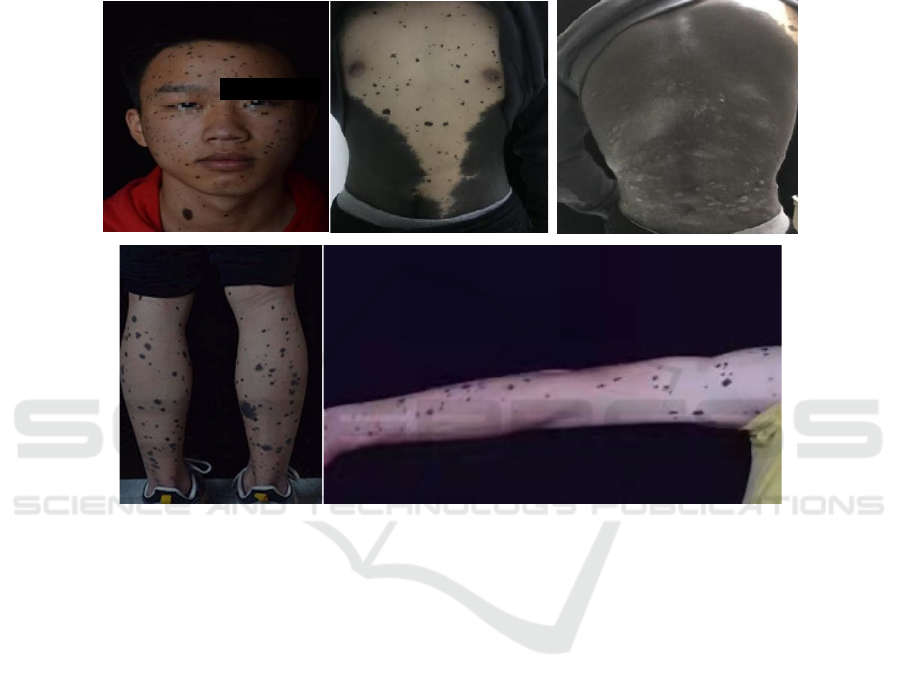
Figure 1: Clinical photographs 1A: preoperative face; 1B: abdomen; 1C: back; 1D: lower leg; 1E: arm.
3 LASER TREATMENT
The patient requested laser treatment for facial
lesions, and the first treatment was with a carbon
dioxide laser (Chongqing Jinyu), wavelength
10600nm, treatment parameters: energy 15mJ,
frequency 15HZ, pulse width 2ms. 2% lidocaine
local infiltration anesthesia for 1 hour before the
start of treatment, after cleaning, the lesions were
first disinfected with 75% alcohol, then 0.9% saline
wiped, the laser continuously and evenly swept the
lesions The lesion is then repeatedly scrubbed with a
0.9% saline swab to remove the carbonised tissue,
then continuous sweeping plus wiping, followed by
intermittent firing pattern plus repeated wiping
when there is less melanin, until the black tissue is
invisible to the naked eye. The patient is instructed
not to touch the lesions with water for 10-15 days
and not to scratch the lesions, the scabs are left to
peel off on their own and the combination of
recombinant bovine basic epidermal growth factor
gel is applied topically once daily after surgery. The
second treatment was performed with a 585 dye
laser (cynosure, USA) to treat scattered erythema
and scarring on the face with the following
parameters The second treatment was performed
with a 585 dye laser (cynosure, USA) with the
following parameters: PDL 7.5 J/cm
2
, pulse width
0.5 ms, spot 7 mm. 1 month after the second
treatment, the third treatment was performed. The
third treatment was performed with a CO
2
fractional
laser (Wuhan Hi-Tech Hengda) on the scar,
treatment parameters: scanning area 7mm×7mm,
fractional energy 39mJ. Postoperative combination
with recombinant bovine basic fibroblast growth
factor gel was applied topically once daily. After the
3rd treatment the facial lesions were significantly
improved and the patient was very satisfied with the
results, and the patient did not undergo further laser
treatment at a later stage. See Figure 2.
ICBB 2022 - International Conference on Biotechnology and Biomedicine
168
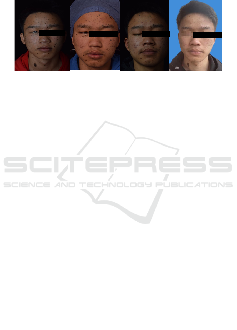
Figure 2: 2A: preoperative face; 2B: after 1st treatment with CO
2
laser; 2C: after 2nd treatment with 585 dye laser; 2D: after
3rd treatment with CO
2
fractional laser.
4 DISCUSSION
Surgery is currently the main treatment for
congenital nevus, including free skin grafting,
fractional excision, direct excision + flap transfer
repair, and soft tissue expansion (Saida, 2006;
Ibrahimi, 2012; Nacarelli, 2014; Zhan, 2016).
However, in this case, the lesion was particularly
large, with a diameter of 80 cm and an area of more
than 1,000 cm
2
, which was already in the G4 class,
so neither flap grafting nor fractionated excision was
appropriate, and the site of the giant nevus was
located at the trunk, so there was little therapeutic
significance and no treatment was done for the time
being. Although the treatment of congenital giant
nevus panchromatic congenital nevus is not
particularly satisfactory, it is often accompanied by
a heavy psychological burden and a severe lack of
self-confidence if the patient has severe skin lesions
in exposed areas, especially on the face (Wan,
2020). Therefore, the facial lesions of the disease
have some therapeutic implications. Treatment
options vary from patient to patient, taking into
account cosmetic, psychosocial and functional
factors (Li, 2020). In some cases, the facial lesions
are large flaps, and significant results have been
reported with flap plasty and skin expansion (Ye,
2014; Zhang, 2017). The treatment of congenital
nevi should be individualized and comprehensive,
taking into account the patient's age, the location
and size of the lesion, the presence of a large
number of small nevi around the nevus, the risk of
secondary melanoma, the expected outcome of the
surgery, the postoperative complications and
psychological guidance. Long-term follow-up is
also required to prevent recurrence and malignancy.
In this case, the facial lesions were heavily
granular, so laser treatment was considered, starting
with carbon dioxide laser treatment, but the melanin
was deep and it was difficult to avoid superficial
scar formation after carbon dioxide laser treatment
(Zhu, 2016), so sequential treatment of the later scar
was very important. There are various scar treatment
modalities, and dye laser and fractional laser have
some synergistic effects in the treatment of scarring,
especially in the inflammatory scarring phase where
PDL with dye laser is very effective (R. Rox
Anderson, 2014). 585 dye laser is used early after
the scab has fallen off to reduce the late scar shape,
and further CO2 fractional laser is used later to
improve the scar and promote healing (Tan, 2017).
Further repair after laser surgery combined with
recombinant bovine basic fibroblast growth factor
gel has been effective in preventing and improving
scarring (Zhang, 2021).
5 CONCLUSION
In summary, although there have been many reports
of congenital macromegaly, it is still rare to find a
macromegaly with severe lesions at the G4 level,
and even rarer to have a generalized nevus. Laser
treatment of the large number of scattered lesions on
the face has also been less frequently reported. In
order to increase the patient's self-confidence and
improve the quality of life, treatment of congenital
giant nevus with congenital small nevi is necessary.
REFERENCES
Eggen CAM, Lommerts JE, van Zuuren EJ, et al.Laser
treatment of congenital melanocytic naevi: a
systematic revew [J].Br J Dermatol,2018, 178(2):
369-383.
A Case of Congenital Giant Nevus with Generalized Small Congenital Nevus Treated with Facial Laser
169

Fu Qiutao, Meng Hui, Sheng Jufang et al. Efficacy of
ultra-pulsed CO 2 laser in the treatment of congenital
giant nevus in 40 cases [J]. Chinese Journal of Laser
Medicine,2019,28(3):175-176,180.
Funayama E, Sasaki S, Furukawa H, et al.Effectiveness of
combined pulsed dye and Q-switched ruby laser
treatment for large to giant congenital melanocytic
naevi [J].Br J Dermatol,2012,167(5):1085-1091.
Ibrahimi OA, Alikhan A, Eisen DB. Congenital
melanocytic nevi: Where are we now? Part II.
Treatment options and approach to treatment [J]. J Am
Acad Dermatol, 2012, 67(4):515. e1 - 13; quiz
528-530.
Li Yue. Advances in the treatment of congenital giant
nevus [J]. Chinese aesthetic medicine, 2020, 29(6):
181-185.
Nacarelli T, Azar A, Sell C. Inhibition of mTOR prevents
ROS production initiated by ethidium
bromide-induced mitochondrial DNA depletion [J].
Front Endocrinol (Lausanne), 2014, 5: 122.
Ruiz-Maldonao R. Measuring congenital melanocytic nevi
[J]. Pediatr Dermatol, 2004, 21(2): 178-179.
R. Rox Anderson, MD, Matthias B.Donelan, MD.Laser
Treatment of Traumatic Scars with an Emphasis on
Ablative Fractional Laser Resurfacing Consensus
Report [J]. JAMA Derm,2014,150(2): 187-193.
Saida T. Histogenesis of congenital and acquired
melanocytic nevi: A unifying concept [J]. Am J
Dermatopath, 2006, 28(4):377 - 379.
Tan Jun. Current status and outlook of laser treatment for
scarring [J]. Chinese aesthetic medicine, 2017,
26(2):1-4.
Vourc'h-Jourdain M, Martin L, Barbarot S, et al. Large
congenital melanocytic nevi: Therapeutic
management and melanoma risk: A systematic review
[J]. J Am Acad Dermatol, 2013, 68(3):493- 498
Wan Meihong, Ai Yong. Perioperative care of a pediatric
case of congenital giant nevus of the head [J].
Dermatology and Venereal Diseases, 2020,
42(6):896-897.
Zhan JY, Wang XF, Liu YH, et al. Andrographolide
sodium bisulfate prevents UV-induced skin
photoaging through inhibiting oxidative stress and
inflammation [J]. Mediators Inflamm,2016,
2016:3271451.
Ye Xiangbo, Chen Juan, Yan Xiaohui et al. Application of
large slice of skin graft in the treatment of pediatric
giant nevus [J]. Chinese Journal of Medical Aesthetics
and Beauty, 2014, 20(2):139-141.
Zhang Jiaqi, Zhang Jinming, Liang Weiqiang et al.
Application of skin expansion in the treatment of
congenital giant melanocytic nevus of the face[J].
Chinese Journal of Medical Aesthetics and
Beauty,2017,23(4):223-225.
Zhu Wei, Shi Yan, Tan Longfeng et al. Clinical efficacy
of different methods in the treatment of 738 cases of
pigmented nevi [J]. Dermatology and Venereal
Diseases, 2016, 38(4):288-290.
Zhang Surui, Liu Wei, Chen Feng et al. Effectiveness and
safety of ultra-pulsed CO2 fractional laser combined
with recombinant bovine basic epidermal growth
factor gel in the treatment of postoperative depressed
facial scarring [J]. Journal of Hebei Medical
University, 2021, 42(2):201-204.
ICBB 2022 - International Conference on Biotechnology and Biomedicine
170
