
Double Root and Root Canals in Right Mandibular Cusp Teeth and
Double Root Canals in Lateral Incisor Teeth: A Case Report
Jingli Zhu
1
, Tianzhu Song
2
, Lili Ding
1
and Miao Li
1,*
1
Hospital of Stomatology, Lanzhou University, Lanzhou, Gansu, 730000, China
2
Key Laboratory of Oral Diseases of Gansu Province, Northwest Minzu University, Lanzhou, Gansu, China
Keywords:
Mandibular Canines, Lateral Incisors, Double Root Canals, X-ray, CBCT, Microscope.
Abstract:
Root canal therapy is an important treatment for pulp disease and periapical disease. Root canal omission is
one of the main reasons for the failure of root canal therapy. Most of the mandibular apical and lateral incisors
are single root canals, and the number of double root canals is less. The case of double root canals of
mandibular canines is even less. This case will report the root canal treatment in which the right mandibular
canines and lateral incisors are double root canals at the same time.
1 INTRODUCTION
Root canal treatment is an important treatment for
endodontic and periapical diseases. Perfect root canal
preparation and root canal filling are the key factors
for successful root canal treatment. However, in the
clinic it is found that some patients still have
discomfort after root canal treatment, such as occlusal
pain and radicular pain, which are mostly caused by
root canal leakage due to root canal anatomical
variation (Siqueira, 2001). Routine imaging before
treatment is the primary factor for successful root
canal treatment, but the limited view of parallel
projection radiographs often makes it easy to miss
root canals routinely. Therefore, Therefore, CBCT
can be used in difficult cases or cases where root canal
omission is considered possible. The mandibular
cuspids have long roots in stomatognathic system and
have an occlusal guidance role (Magne, 2015). The
mandibular cuspids are mostly single root with single
root canal. The double root with double root canal
have been rare in recent year compared with single
root with single root canal. In contrast, more
mandibular lateral incisors with double root canals
have been reported. This is a case of double root with
double canals in right mandibular cusp teeth and
double root canals in lateral incisor teeth. Therefore,
it is important to discuss the root canals anatomical
morphology of the mandibular cuspids and lateral
*
Corresponding author
incisors and clinical considerations for the success of
root canal treatment and the preservation of the
treated tooth.
2 CASE INFORMATION
The patient, a 45-year-old female, was treated in an
outside hospital for her lower right tooth ten years
ago. The original restoration was fractured three days
ago. She was then treated in our prosthodontics
department, and is now referred to our department for
root canal treatment.. The examination found 42 was
residual roots, with the edge flush with the gingiva or
1-2mm above the gingiva. 43 was residual crowns
with distal residual tooth tissue and exposed root
canal orifice which have no probing pain;42 and 43
teeth without loosening and periodontal pocket were
not suitable for percussion and was slightly painful
for palpation, and no obvious abnormality was found
in buccal mucosa 42 and 43 cases were diagnosed as
chronic apicitis and tooth tissue defect. The treatment
plan was post core crown repair after 42 and 43 root
canal treatment. The treatment process was that 42
and 43 teeth cleared rotten matter under the
microscope with rubber barrier.42 probed two root
canals and 43 one root canal. After root canal length
measurement, M3 cleared root canal with ultrasonic
swing washing and 1% sodium hypochlorite. Before
138
Zhu, J., Song, T., Ding, L. and Li, M.
Double Root and Root Canals in Right Mandibular Cusp Teeth and Double Root Canals in Lateral Incisor Teeth: A Case Report.
DOI: 10.5220/0012014900003633
In Proceedings of the 4th International Conference on Biotechnology and Biomedicine (ICBB 2022), pages 138-141
ISBN: 978-989-758-637-8
Copyright
c
2023 by SCITEPRESS – Science and Technology Publications, Lda. Under CC license (CC BY-NC-ND 4.0)
glass ion temporary sealing ,42and 43 was dried with
paper tip suction and then used calcium hydroxide in
root canal. The root canal filling was prepared at the
second visit, but it was found that glue tips of 42 tooth
could reach the root canal length in the buccal root
canal, while the lingual could not reach the root canal
length completely. After X-ray, it was found that 42
tooth test tips were suitable, while 43 were double
roots and there were root canal omissions. Then 43
was rediscovered the lingual root canal under the
microscope and was cleaned .42 and 43 teeth were
filled with hot gum after ultrasonic swing washing.
X-ray showed that the root was filled in place and the
tooth was sealed with glass ion. It was suggested that
the prosthetic department should carry out further
prosthetic treatment after one week of observation.
(Fig.1-4).
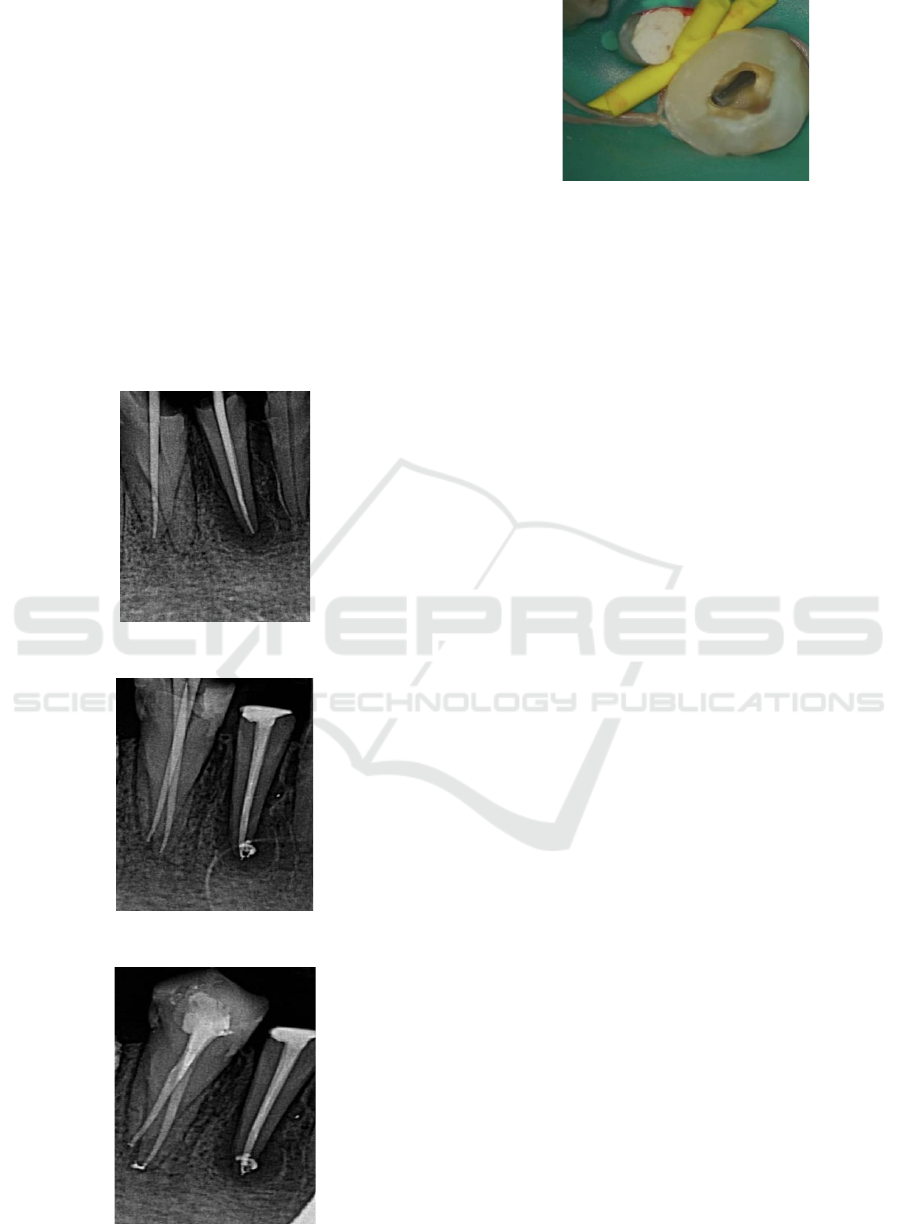
Figure 1: First root canal test tip.
Figure 2: Second root canal test tip.
Figure 3: Root filling.
Figure 4: omitted root canals Under microscope.
3 DISCUSSION
3.1 Root Canal Typing
Root canal therapy will greatly reduce the success
rate of root canal therapy if root canal omission
occurs. Mastering the anatomical morphology of root
canal will greatly reduce the occurrence of root canal
omission. Scholars have carried out a series of
research and experiments in recent years. Weine et al.
divided the whole path change from dental pulp
cavity to apical foramen into four types by truncation
and radiology methods. Type I: one root canal from
pulp cavity to apical foramen (type 1-1); Type II: two
canals left the medullary cavity and fused into one
canal before reaching the apical foramen (type 2-1);
Type III: two independent canals from medullary
cavity to apical foramen (type 2-2); Type IV: one
canal leaves the medullary cavity and is divided into
two canals with two apical foramen (type 1-2)
(Weine, 1969). Vertucci et al. studied the transparent
specimens of the second premolar by using the
staining method and found that the complexity of root
canal morphology is far more than that of
weineclassification. He proposed eight types: 1-1, 2-
1, 1-2-1, 2-2, 1-2-1-2, 3-3 (Vertucci, 1974). This
method includes most root canal morphology in
clinic. Some researchers have shown that the peak
incidence of multiple root canals of mandibular
canines is concentrated over the age of 30, and shows
a downward trend after the age of 60. This may be
related to different ethnic groups, regional
populations or sample sizes, as well as root canal
cross-sectional morphology and secondary dentin
deposition (Fig. 5). Therefore, for middle-aged and
elderly patients, in addition to mastering the number
of root canals per tooth, we should also consider the
impact of secondary dentin deposition on the
morphology and number of root canals with age.
Especially for single root canal flat root anterior teeth,
it is possible to form isthmus formed by secondary
dentin in the middle of root canal. The occurrence of
Double Root and Root Canals in Right Mandibular Cusp Teeth and Double Root Canals in Lateral Incisor Teeth: A Case Report
139
this situation may cause the clinician to lose the root
canal during the root canal preparation or fail to
complete the root canal cleaning due to the
appearance of steps in the root canal. The 43 teeth of
the cases were vertucciiv (2-2) root canals. The
mandibular lateral incisors root is flat with mostly
single root canal, but also double root canal mostly
distributed in the labial and lingual direction. At
present, there are many cases of mandibular lateral
incisors with double root canals. It is generally
believed that about 10% of mandibular lateral incisors
have double root canals. Laws et al. Respectively
used X-ray films to study the samples inside and
outside the mouth. It was found that the incidence of
double root canals of mandibular lateral incisors was
more than 40%. VertucciⅡ (2-1) root canal accounted
for 5.33%. The double root canals of the central
incisor in this case were vertucciⅡ (2-1).
3.2 Application of CBCT
In order to avoid root canal omission, it is necessary
to master the anatomical morphology of root canal. In
addition, the imaging examination before individual
tube treatment is also particularly important.
Conventional parallel projection X-ray film is one of
the conventional imaging examination methods
before the treatment of dental periapical disease. It
can show the shape and length of root canal, and
roughly evaluate whether there is root canal
calcification. However, the specific number and
shape of root canal and tooth root are often unclear
because the X-ray film is the two-dimensional image.
It will be inaccurate to judge whether there is root
canal omission completely based on X-ray
examination (Eisner, 1998). With the development of
science and technology, the emergence of CBCT
makes this problem simple (Kaasalainen, 2021).
Three-dimensional imaging of CBCT can accurately
judge the number, shape and variation of root canals.
Therefore, CBCT examination of difficult root canals
is very important. However, CBCT is not routinely
used in root canal therapy due to its high cost. In this
case, the patient did not take CBCT at the initial
diagnosis due to economic reasons. When CBCT
cannot be taken, we should also fully interpret the X-
ray information. If the root canal image suddenly
becomes thinner or disappears, there may be root
canal branches at this location, which needs to be paid
attention to in actual operation.
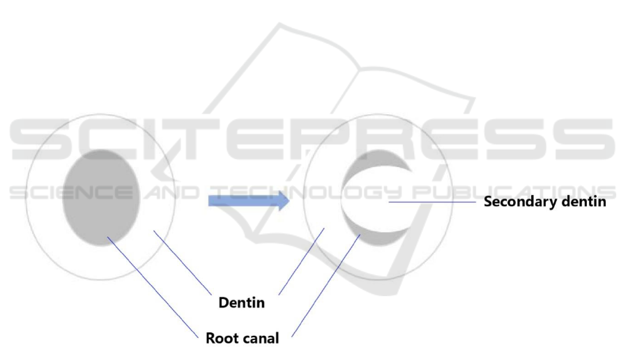
secondary dentin formation in root canal leads to the evolution of single root canal into multiple root canals with the increase
of age; The gray area is the root canal, and the white area protruding into the root canal is secondary dentin
Figure 5. Single root canal evolving into multiple root canals.
3.3 Intraoral Manipulation
3.3.1 Oral Operation
In clinical practice, because the root of the tongue is
more inclined to the lingual side, the root canal orifice
is often blocked by the top of the pulp chamber or the
dentin collar, which accounts for the main reason for
the root canal omission of the lower anterior teeth
(Sener, 2009). 43 was generally considered as a single
tube at the initial diagnosis, but after changing the X-
ray projection angle in the test tip film, it can be
clearly seen that 43 has two tubes and one tube is
missing. Later, when looking for the missing root
canal, it was found that 43 the missing root canal on
the lingual side was found after uncovering the
medullary chamber roof on the lingual side. In
practice, it should also be noted that when the root
canal deviates to one side, it should carefully look for
another root canal on the opposite side. If there is
ICBB 2022 - International Conference on Biotechnology and Biomedicine
140

calcification blocking the root canal orifice, it should
carefully remove the calcification with ultrasonic
instruments under the microscope to look for another
root canal.
3.4 Application of Microscope
The application of microscope is of great significance
for root canal therapy. The use of microscope in root
canal therapy can directly look into the medullary
cavity and avoid root canal omission to a greater
extent (Bonsor, 2015). For mandibular anterior teeth,
most of them are single root canals and it is difficult
to find multiple root canals. The 42 teethin this case
was single root and there was no obvious double root
image on the X-ray film.Butthen two root orifices
were obviously visible at the enamel cementum
boundary of the medullary cavity under the
microscope during the treatment. The42 teeth length
measurement and root canal preparation were carried
out, but the buccal gum tip could reach the root canal
length during the tip test while the lingual gum tip did
not fully reach the root canal length about 5mm away
from the root tip. Therefore, it is judged that 42 is the
2-1 type root canal. The results showed that the
detection rate of double root canals increased from
16.08% to 27.27% before and after microscope
. The
microscope can enlarge the operation area and
provide a clear field of vision for the operator. The
removal of calcified substances under the
microscope can effectively avoid the occurrence of
pulp chamber floor penetration and root canal
lateral penetration caused by unclear field of
vision, so as to improve the detection rate of root
canal and the success rate of root canal treatment.
4 CONCLUSION
This case suggests that we should be alert to the
existence of root canal variation and fully understand
the anatomical morphology of root canal of each
tooth before root canal treatment. In root canal
therapy, X-ray multi angle photography or CBCT
photography should be carried out to find the variant
root canal in time. Before root canal preparation, the
pulp chamber top should be uncovered and combined
with microscope to prevent root canal omission and
improve the success rate of root canal treatment.
ACKNOWLEDGEMENTS
This work was supported by Research Support Fund
of School of Stomatology Lanzhou University (No.
lzukqky-2020-t05).
REFERENCES
Bonsor SJ. The use of the operating microscope in general
dental practice. Part 2: If you can see it, you can treat it!
Dent Update. 2015 Jan-Feb;42(1):60-2, 65-6. doi:
10.12968/denu.2015.42.1.60. PMID: 26062280.
Eisner ER. Oral-dental radiographic examination
technique. Vet Clin North Am Small AnimPract. 1998
Sep;28(5):1063-87, v. doi: 10.1016/s0195-
5616(98)50103-9. PMID: 9779541.
Kaasalainen T, Ekholm M, Siiskonen T, Kortesniemi M.
Dental cone beam CT: An updated review. Phys Med.
2021 Aug; 88:193-217. doi:
10.1016/j.ejmp.2021.07.007. Epub 2021 Jul 17. PMID:
34284332.
Magne P. A new approach to the learning of dental
morphology, function, and esthetics: the "2D-3D-4D"
concept. Int J Esthet Dent. 2015 Spring;10(1):32-47.
PMID: 25625126.
Sener S, Cobankara FK, Akgünlü F. Calcifications of the
pulp chamber: prevalence and implicated factors. Clin
Oral Investig. 2009 Jun;13(2):209-15. doi:
10.1007/s00784-008-0212-x. Epub 2008 Jul 30. PMID:
18665398.
Siqueira JF Jr. Aetiology of root canal treatment failure:
why well-treated teeth can fail. Int Endod J. 2001
Jan;34(1):1-10. doi: 10.1046/j.1365-
2591.2001.00396.x. PMID: 11307374.
Vertucci, et al. Oral Surgery, Oral Medicine and Oral
Pathology, 1974, 58, 589-99.
Weine, et al. Oral Surgery, Oral Medicine and Oral
Pathology, 1969, 28, 419-25; Weine FS. Endodontic
therapy, 3rd edn (1982). St. Louis: Mosby.
Double Root and Root Canals in Right Mandibular Cusp Teeth and Double Root Canals in Lateral Incisor Teeth: A Case Report
141
