
Unsupervised Image-to-Image Translation from MRI-based
Simulated Images to Realistic Images Reflecting Specific Color
Characteristics
Naoya Wada
*
and Masaya Kobayashi
*
KYOCERA Corporation, 3-7-1 Minatomirai Nishi-ku, Yokohama, Japan
Keywords: Generative Adversarial Networks (Gans), Image-to-Image Translation, Domain Adaptation, Unsupervised
Learning, MRI.
Abstract: In this paper, a new domain adaptation technique is presented for image-to-image translation into the real-
world color domain. Although CycleGAN has become a standard technique for image translation without
pairing images to train the network, it is not able to adapt the domain of the generated image to small domains
such as color and illumination. Other techniques require large datasets for training. In our technique, two
source images are introduced: one for image translation and another for color adaptation. Color adaptation is
realized by introducing color histograms to the two generators in CycleGAN and estimating losses for color.
Experiments using simulated images based on the OsteoArthritis Initiative MRI dataset show promising
results in terms of color difference and image comparisons.
1 INTRODUCTION
Image synthesis can now achieve sufficient quality
for practical use thanks to the progress of generative
adversarial networks (GANs) (Goodfellow et al.,
2014). GANs have also been used in various tasks,
including image-to-image translation (Zhu et al.,
2017; Isola et al., 2017), image interpolation (Yu et
al., 2018), and data synthesis other than images (Yu
et al., 2017). While GANs now have a wide range of
applications, image synthesis to realize specific
characteristics is one of the main tasks for practical
use, and the demand for image synthesis has grown
as the quality of the generated images has increased.
One example is the task of generating images that
reflect the specific color or other characteristics of a
person.
This paper presents a new image-to-image
translation method that reflects specific color
characteristics, with the aim of generating a realistic
image reflecting a person’s characterics from
simulated images based on MRI data. In conventional
image translation methods, there are three main
considerations. The first is how to capture specific
characteristics of a domain (Karras et al., 2019). The
*
https://www.kyocera.co.jp
second is the necessary amount of training data (Liu
et al., 2019). The third is the labeling cost for
supervised learning, which is required for several
methods that involve pairing images across the
domains (Isola et al., 2017). To address these issues,
we propose a CycleGAN-based network model to
achieve unsupervised learning that requires a smaller
amount of data. However, the original CycleGAN
architecture cannot accept the characteristics of a
specific target as input and is unable to adapt the
generated image accordingly. Therefore, we import
the color characteristics as color histograms to the
middle layer between the encoder–decoder structures
of the two generators in the cyclic architecture of
CycleGAN. Then, color loss is independently
estimated and combined with other losses such as
cycle consistency loss and adversarial loss. We also
performed experiments to demonstrate image-to-
image translation across two domains. One domain
consisted of 3D simulated knee images obtained from
MRI data provided by the OsteoArthritis Initiative
(OAI). The other domain consisted of real-world knee
images. Image-to-image translation from MRI to
realistic knee images would make it easier for non-
healthcare professionals to understand MRI images.
184
Wada, N. and Kobayashi, M.
Unsupervised Image-to-Image Translation from MRI-based Simulated Images to Realistic Images Reflecting Specific Color Characteristics.
DOI: 10.5220/0010916300003123
In Proceedings of the 15th International Joint Conference on Biomedical Engineering Systems and Technologies (BIOSTEC 2022) - Volume 2: BIOIMAGING, pages 184-189
ISBN: 978-989-758-552-4; ISSN: 2184-4305
Copyright
c
2022 by SCITEPRESS – Science and Technology Publications, Lda. All rights reserved
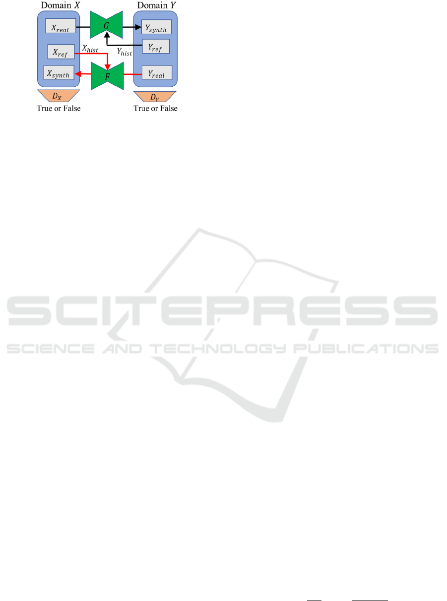
Figure 1: Schematic of the image-to-image translation
network.
It could also support communication between patients
and healthcare professionals by aiding in
explanations of MRI images from other patients.
In the remainder of this paper, we discuss related
work in Section 2, introduce our proposed method in
Section 3, present our experimental methods and
results in Section 4, and give our conclusions in
Section 5.
2 RELATED WORK
GANs have been used previously for image-to-image
domain adaptation. Pix2pix (Isola et al., 2017) is an
early, well-known method for image-to-image
translation that uses conditional GANs to learn paired
images. However, Pix2pix uses supervised learning
and a large number of paired images, so some
labeling cost is required. CycleGAN (Zhu et al.,
2017) is a method for image-to-image translation
without learning paired images. The model has a
cycle architecture that consists of two generators and
two discriminators and cycle consistency loss for
unsupervised learning. CycleGAN requires a
relatively small number of unpaired images for
training. However, it is difficult to adapt the
generated image to specific characteristics (color,
illumination, etc.) in a dataset. Recently, StarGAN-v2
(Choi et al., 2018; Choi et al., 2020) has been
proposed for image-to-image translation across
domains and can reflect styles by obtaining style
codes from datasets automatically. However, it
requires a large dataset for training, and it is difficult
to specify a specific style in a non-biased dataset
because style codes are automatically obtained by
capturing bias in a training dataset. DeepHist (Avi-
Aharon et al., 2020) can reflect specific color
characteristics in generated images by using a
differentiable network with kernel density estimation
of color histograms from the generated image.
However, paired images are required for training.
3 PROPOSED METHOD
Here we propose a network model for image-to-image
translation that is based on the cyclic architecture of
CycleGAN and is able to reflect target color
characteristics. For training, our method requires only
a relatively small amount of data and does not require
pairing images. Furthermore, we introduced an archite-
cture to accept color histograms for the target domain.
This section describes the details of this architecture
and the estimation of losses during training.
3.1 Overview of the Network
The network consists of two generators and two
discriminators as shown in Figure 1. 𝑋 and 𝑌 denote
the domains in image-to-image translation. 𝐺 and 𝐹
denote the generator from 𝑋 to 𝑌 and 𝑌 to 𝑋
respectively. Each of them generates a synthetic
image (𝑋
and 𝑌
) in the target domain from
a real image (𝑋
and 𝑌
) in the source domain.
𝐷
and 𝐷
denote the discriminators and 𝑋
and
𝑌
denote the color histograms for 𝑋 and 𝑌,
respectively. These histograms are input into 𝐹 and
𝐺, respectively, so that the generated synthetic images
reflect the color characteristics obtained from
reference images (𝑋
and 𝑌
) in the respective
target domains. Spectral normalization (Miyato et al.,
2018) is adopted for both generators and
discriminators to stabilize training of this network.
3.2 Importing Color Characteristics
The architecture adopted for 𝐺 and 𝐹 is shown in
Figure 2. The color distribution is imported with
reference to previous methods. First, an RGB
histogram is obtained from an image in the target
domain. Histograms for each color are concatenated
and imported to the middle layer between the encoder
and the decoder of the generator. The purpose of
importing the histograms is to import color
information after spatial features have been
convoluted. A translated image is output from the
decoder. To evaluate the color of the output image,
L2 loss between histograms of the source and output
images is obtained. The histograms of the output
image are obtained by kernel density estimation
because it enables backpropagation and updating of
the network. Kernel density estimation is done using
the following probability density function:
𝑓
𝑔
1
𝑁𝐵
Κ
𝐼
𝑥
𝑔
𝐵
∈
,
(1)
Unsupervised Image-to-Image Translation from MRI-based Simulated Images to Realistic Images Reflecting Specific Color Characteristics
185
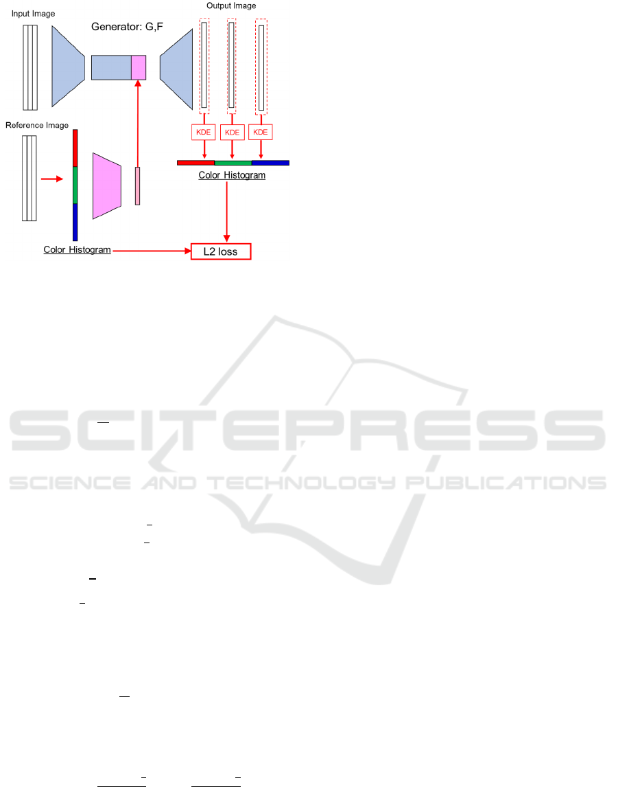
Figure 2: Architecture of the generators (KDE: kernel
density estimation).
Here, 𝑥 denotes the pixel position, 𝑔∈
1,1
denotes the pixel value, 𝐵 denotes the bandwidth,
𝑁
|
Ω
|
denotes the number of pixels, 𝐼
𝑥
∈
1,1 denotes the luminance, and Κ
∙
denotes the
kernel function, which is defined as
Κ
𝑧
𝜎
𝑧
𝜎
𝑧
𝜎
𝑧
.
(2)
Here, K(z) is the derivative of the sigmoid function
𝜎(z). Using 𝑓
𝑔
, each pixel in an image is assigned
to a histogram bin according to
𝑃
𝑘
𝑓
𝑔
𝑑𝑔
,
(3)
where 𝐿
denotes the bin width and 𝜇
1 𝐿
𝑘
denotes the center of the 𝑘-th bin
when normalized pixel values [−1, 1] are divided into
𝐾 bins 𝐵
. Equation (3) can be developed by
using (2) to give
𝑃
𝑘
1
𝑁
Π
𝐼
𝑥
∈
(4)
where Π
𝑧
is defined as
Π
𝑧
≜ 𝜎
𝜎
. (5)
Thus, 𝑃
𝑘
gives the value of the k-th bin of the color
histograms.
3.3 Loss Function
The loss function for the entire network is defined as
ℒ
𝜆
ℒ
𝜆
ℒ
𝜆
ℒ
𝜆
ℒ
,
(6)
where ℒ
denotes the adversarial loss, ℒ
denotes the cycle consistency loss, ℒ
denotes the
identity mapping loss, and ℒ
denotes the color
loss. ℒ
is defined by using L2 losses as
ℒ
𝒉
𝒉
𝒉
𝒉
𝒉
𝒉
,
(7)
where 𝒉
, 𝒉
, and 𝒉
denote the histograms
of each RBG color estimated from the generated
image and 𝒉
, 𝒉
and 𝒉
denote the
histograms of each color estimated from the imported
reference images.
4 EXPERIMENTS
Image-to-image translation experiments were
performed to evaluate the proposed method. The task
is to translate images across two domains.
4.1 Dataset
One domain consisted of 3D simulated knee images
generated from OAI MRI data. MRI data were
converted to 3D structure and a front-view image was
obtained as a 3D simulated knee image by using the
3D medical imaging software InVesalius 3.1. The
other domain consisted of real-world knee images
(the color source) obtained from several datasets
including the Fashion Product Images Dataset
provided by Kaggle. The resolution of both input and
output images was 256 × 256 pixels. The training
dataset consists of 477 3D simulated images and 438
real-world images because we sought to consider the
situation with a relatively small training dataset. For
learning, the network was trained for 400 epochs. The
test dataset consists of 28 images from each domain,
and image translation was performed from the MRI-
based 3D simulation domain to the real-world domain
to reflect the color characteristics of an input real-
world knee image. As a result, 784 images were
generated for each input combination comprising a
3D simulated image and a real-world image (28 × 28
images) in round-robin manner.
BIOIMAGING 2022 - 9th International Conference on Bioimaging
186
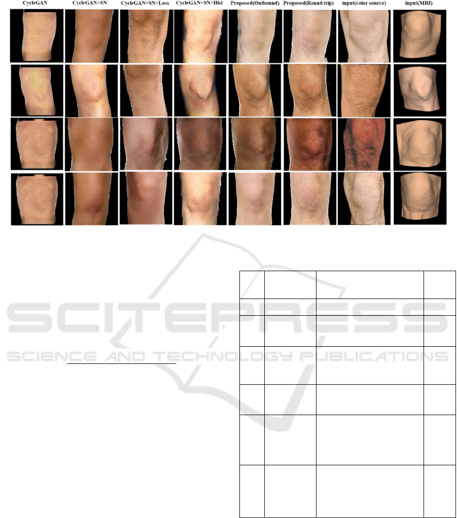
Figure 3: Comparison of images generated by the six methods shown in Table 1.
4.2 Evaluation
To evaluate how well the color characteristics in the
generated output images reflected those in the input
source images, their mean colors were compared.
The color difference ∆𝐸
∗
was calculated using
CIE76 to compare the mean colors in the L*a*b*
color space:
∆𝐸
∗
∆𝐿
∗
∆𝑎
∗
∆𝑏
∗
.
(8)
We compared the color difference among
conventional CycleGAN, the proposed method, and
some incomplete methods based on the proposed
method but with the omission of some of its
techniques. These incomplete methods are intended
to evaluate the effect of each technique. The
evaluated methods are summarized in Table 1.
4.3 Results
Figure 3 shows representative examples of images
generated by the evaluated methods for five
combinations of source MRI-simulation and real-
world images, and their color differences are
summarized in Table 1. Note that Methods 1-3 do not
have an architecture to accept input color histograms;
thus, unspecified color characteristics extracted from
whole dataset during training are reflected in the
generated images and cause the differences from the
color source images. The images generated by
methods 1 and 2 all have the same color
Table 1: Summary of methods and color differences.
No. Method Description Color
Diff.
1 CycleGAN Original CycleGAN 14.1
2 CycleGAN
+SN
Spectral normalization is
applied to method 1.
13.7
3 CycleGAN
+SN+Loss
Loss function considering
colors (6) is applied to
method 2.
13.0
4 CycleGAN
+SN+Hist
Color histograms are
applied to method 2.
8.9
5 Proposed
(outbound
only)
Loss function (6) and
color histograms are
applied to generator
𝐺
(MRI to real-world) only.
5.0
6 Proposed
(round trip)
Proposed method (the
method applied to
𝐺 in
method
5 is also applied
to
𝐹)
4.6
characteristics. The result for method 3 shows that it
is not very effective on its own. On the other hand,
methods 4-6 which accept input color histograms
improve the color differences. The effect of using
color histograms as input and evaluating losses for
color can be clearly seen. Especially, the result for
method 6 achieving ∆𝐸
∗
5.0 is promising because
this threshold is same as the color tolerance of printed
solids defined in ISO 12647-2. The improvement in
Unsupervised Image-to-Image Translation from MRI-based Simulated Images to Realistic Images Reflecting Specific Color Characteristics
187

color difference between methods 5 and 6 seems not
large. However, the images generated by method 6
show clear differences in brightness within each
image, whereas the images generated by method 5 are
slightly blurry. More examples of images generated
by method 6 are shown in Figure 4, which gives an
overview of the relationships between the source 3D
simulated images and the source colors. We can see
that the generated images change according to the
source model and color source.
4.4 Discussion
In this section, we discuss the importance on
incorporating color histograms and the loss function
considering color loss into CycleGAN. Method 5
incorporated loss function (6) and color histograms
into only generator 𝐺 (for translating MRI-simulation
images into realistic images), where the aim is to
reflect the color characteristics of an input image. On
the other hand, method 6 incorporated them into not
only 𝐺 but also 𝐹, where the color characteristics of
the MRI-simulation images are reflected. The
surfaces of MRI-simulation images are colorized by
the medical imaging software with a certain fixed
color and brightness as shown in Figure 3 because the
source MRI data does not include the surface colors
of the scanned individual. Therefore, color
histograms input to the generator 𝐹 mainly reflect the
contrast of brightness rather than the color of the
person’s skin. The detailed characteristics of the
Figure 4: Examples of generated images for 25
combinations of source 3D simulated images and source
real-world images. The first column and first row show the
respective source images.
images generated by method 6 are attributable to the
architecture of the proposed method. In other words,
incorporating color histograms into CycleGAN was
effective not only for reflecting color characteristics
of the source image, but also for better representing
contrasts, unevenness, and other characteristics.
5 SUMMARY
In this paper, a new technique for image-to-image
translation reflecting specific color characteristics
was presented. That technique was intended to
translate MRI-based 3D simulated images to realistic
images reflecting the characteristics of a specific
person’s appearance. In our image-to-image
translation network, which was based on CycleGAN,
color histograms of an input image were concatenated
to the input vector as reference color characteristics
and the generated image was evaluated with a loss
function considering color loss. The experimental
results showed that the presented technique was
effective not only for reflecting the color
characteristics of the input source image but also for
better representing contrasts, unevenness, and other
characteristics.
Topics for future work include realizing more
useful image-to-image translation with representation
of various lighting environments. In the 3D simulated
image domain, it is easy to obtain images under many
different lighting conditions. If this can be reflected
in the generated image along with color
characteristics, then image translation to the real-
world image domain would be more flexible and
useful.
ACKNOWLEDGEMENTS
Data and/or research tools used in the preparation of
this manuscript were obtained and analyzed from the
controlled access datasets distributed from the
Osteoarthritis Initiative (OAI), a data repository
housed within the NIMH Data Archive (NDA). OAI
is a collaborative informatics system created by the
National Institute of Mental Health and the National
Institute of Arthritis, Musculoskeletal and Skin
Diseases (NIAMS) to provide a worldwide resource
to quicken the pace of biomarker identification,
scientific investigation and OA drug development.
Dataset identifier: 2343.
BIOIMAGING 2022 - 9th International Conference on Bioimaging
188

REFERENCES
Goodfellow I., Pouget-Abadie J., Mirza M., Xu B., Warde-
Farley D., Ozair S., Courville A., Bengio Y. (2014).
Generative Adversarial Nets. In Neural Information
Processing Systems.
Zhu J., Park T., Isola P., Efros A. (2017). Unpaired image-
to-image translation using cycle-consistent adversarial
networks. In International Conference on Computer
Vision (ICCV).
Isola P., Zhu J., Zhou T., Efros A. (2017). Image-to-image
Translation with Conditional Adversarial Networks. In
International Conference on Computer Vision and
Pattern Recognition (CVPR).
Yu J., Lin Z., Yang J., Shen X., Lu X., Huang S. T. (2018).
Generative Image Inpainting with Contextual
Attention. In International Conference on Computer
Vision and Pattern Recognition (CVPR).
Yu, L.; Zhang, W.; Wang, J.; and Yu, Y. (2017). SeqGAN:
Sequence generative adversarial nets with policy
gradient. In Association for the Advancement of
Artificial Intelligence (AAAI) Conference on Artificial
Intelligence.
Karras T., Laine S., Aila T. (2019) A Style-based Generator
Architecture for Generative Adversarial Networks. In
International Conference on Computer Vision and
Pattern Recognition (CVPR).
Liu M., Huang X., Mallya A., Karras T., Aila T., Lehtinen
J., Kautz J. (2019) Few-Shot Unsupervised Image-to-
Image Translation. In International Conference on
Computer Vision (ICCV).
Choi Y., Choi M., Kim M., Ha J., Kim S., Choo J. (2018)
Stargan: Unified Generative Adversarial Networks for
Multi-domain Image-to-image Translation. In
International Conference on Computer Vision and
Pattern Recognition (CVPR).
Choi Y., Uh Y., Yoo J., Ha J. (2020) Stargan v2: Diverse
Image Synthesis for Multiple Domains. In International
Conference on Computer Vision and Pattern
Recognition (CVPR), pages 8188–8197.
Avi-Aharon M., Arbelle A., Raviv R. T. (2020). Deephist:
Differentiable Joint and Color Histogram Layers for
Image-to-image Translation. In arXiv preprint
arXiv:2005.03995.
Miyato T., Kataoka T., Koyama M., Yoshida Y. (2018)
Spectral Normalization for Generative Adversarial
Networks. In International Conference on Learning
Representations (ICLR).
Unsupervised Image-to-Image Translation from MRI-based Simulated Images to Realistic Images Reflecting Specific Color Characteristics
189
