
Research on Intelligent Analysis Model of Heart Sound based on
Deep Learning
Hui Yu
1,2,3 a
, Jing Zhao
1b
, Jinglai Sun
2,3 c
and Zhaoyu Qiu
2d
1
Academy of Medical Engineering and Translation Medicine, Tianjin University, Tianjin 300072, China
2
School of Precision Instrument and Opto-Electronics Engineering, Tianjin University, Tianjin 300072, China
3
Department of Biomedical Engineering, Tianjin Key Laboratory of Biomedical Detecting Techniques and Instruments,
Tianjin University, Tianjin 300072, China
Keywords: Heart Sound, Bispectrum Analysis, Convolutional Neural Networks.
Abstract: Heart sound auscultation is one of the most basic cardiac diagnosis techniques, but the traditional artificial
auscultation method requires experienced clinicians and is limited by environmental factors. In this study, an
intelligent analysis model of heart sound based on deep learning was designed to meet the daily public
screening. Firstly, two public data sets and clinical self-collected data sets were fused, and pretreatments were
carried out,such as normalization, denoising, overlapping cutting and subsampling. Then, the extraction and
quantitative analysis of heart sound features were completed using bispectrum analysis technology. Finally,
the features were input into the constructed improved convolutional neural network for classification. The
results show that the accuracy, sensitivity, specificity and F1 score of normal and abnormal heart sounds were
85.5%, 85.7%, 85.3% and 85.9%, respectively, and the performance of pathological heart sounds
classification was over 90%, reaching the highest level of this kind of research at present. This model provides
a standardized evaluation with high classification performance and can quickly complete the intelligent
analysis of heart sounds, which has important clinical significance.
1 INTRODUCTION
Heart sound is a mechanical wave phenomenon
caused by the movement of the heart. The digital
signals collected by sensors are called
phonocardiogram (PCG). Heart sounds are clinically
associated with many heart pathologies, common
among which are aortic stenosis (AS), mitral stenosis
(MS), mitral regurgitation (MR), and mitral valve
prolapse (MVP) (Reed 2004).
Heart sound auscultation is of great significance
in the diagnosis of cardiovascular diseases. It is one
of the most commonly used cardiac diagnostic
techniques because of its characteristics of non-
invasive, fast and low cost. However, the traditional
manual auscultation method requires experienced
clinicians and is limited by environmental factors,
making it highly subjective and easy to make
mistakes. According to statistics, cardiologists'
auscultation accuracy is about 80%, while that of
a
https://orcid.org/0000-0002-8511-7296
b
https://orcid.org/0000-0003-3231-5544
primary care physicians is only in the range of 20%
to 40%(Ma 2020). With the increasing demand for
heart sound auscultation, clinical patients are eager to
develop an accurate and rapid intelligent heart sound
detection algorithm suitable for public screening.
At present, the automatic diagnosis of heart sound
signal mainly has the following methods: Extracting
features efficiently and using traditional pattern
recognition methods such as support vector machine
(SVM), empirical parameters and k-nearest neighbor
(K-NN), the diagnosis is made, or the ability of the
neural network itself to extract features and classify,
such as convolutional neural network (CNN), deep
neural network (DNN), recurrent neural network
(RNN), deep confidence network (DBN)
(Dominguez-Morales 2018, Abduh 2019, Chen 2018,
Wu 2019).
Most of the current studies are based on foreign
open data sets, and it remains to be seen whether the
research results apply to Chinese clinical practice.
c
https://orcid.org/0000-0003-3683-1968
d
https://orcid.org/0000-0002-7728-7367
244
Yu, H., Zhao, J., Sun, J. and Qiu, Z.
Research on Intelligent Analysis Model of Heart Sound based on Deep Learning.
DOI: 10.5220/0011291400003444
In Proceedings of the 2nd Conference on Artificial Intelligence and Healthcare (CAIH 2021), pages 244-248
ISBN: 978-989-758-594-4
Copyright
c
2022 by SCITEPRESS – Science and Technology Publications, Lda. All rights reserved

Therefore, an intelligent heart sound analysis model
based on deep learning was proposed to be suitable
for clinical practice and higher classification
accuracy. Firstly, two available data sets and clinical
self-collected data sets were fused, and pretreatments
such as normalization, denoising, overlapping cutting
and sub-sampling were performed. Then, we used the
bispectrum analysis technology to complete the heart
sound feature extraction and quantitative analysis.
Finally, the features were input into the constructed
improved convolutional neural network for the
dichotomies of healthy and abnormal heart sounds
and four classifications of pathological heart sounds,
and the performance of the model was evaluated by
accuracy, sensitivity, specificity, accuracy rate and F1
score.
2 MATERIALS AND METHODS
2.1 Dataset
The first dataset used in this paper is the 2016
PhysioNet/CinC Challenge (Liu 2016), which
includes 3,126 heart sound in two classes of abnormal
and normal. The second used dataset is derived from
GitHub, which contains four pathological and normal
heart sound types uploaded by researcher Yaseen at
Sejong University (Yaseen 2018). The third available
dataset, including 29 kinds of the typical heart sound,
is collected by our laboratory and Tianjin Chest
Hospital of China.
2.2 Bispectrum Analysis
The feature engineering in this paper is based on the
bispectrum analysis. Bispectrum analysis is one of
the most commonly used higher order spectrum
analysis methods. It can sufficiently suppress the
signal's phase relationship and detect and quantify the
phase coupling of non-Gaussian signals (Alqudah
2020). Its formula is as follows:
𝑆
(
𝜔
,𝜔
)
=
∑∑
𝐶
(𝜏
,𝜏
)𝑒
((
))
(1)
2.3 Improved Convolutional Neural
Networks
In this study, a 19-layer improved convolutional
neural network was constructed. A submodule
contains a convolutional layer, BN layer, ReLU layer
and max pooling layer. After being processed by three
identical sub-modules, the pooling layer in the fourth
sub-module is changed into a full connection layer,
and then a layer of softmax layer is used to complete
logistic regression to get our classification results.
Figure 1 shows the overall network structure, and
specific parameters of the model are shown in Table
1.
Table 1: Model parameter selection.
# Layer Information #
1 Input layer Size 256*256
2 Conv_1
Number of filters 32
Kernel size 3*3
Activation ReLU
3 Batch_Norm_1 Number of channels 32
4 ReLU_1 Activation ReLU
5 Maxpol_1
Kernel size 2*2
Stride 2*2
6 Conv_2
Number of filters 16
Kernel size 3*3
Activation ReLU
7 Batch_Norm_2 Number of channels 16
8 ReLU_2 Activation ReLU
9 Maxpol_2
Kernel size 2*2
Stride 2*2
10 Conv_3
Number of
filters
8
Kernel size 3*3
Activation ReLU
11 Batch_Norm_3 Number of channels 8
12 ReLU_3 Activation ReLU
13 Maxpol_3
Kernel size 2*2
Stride 2*2
14 Conv_4
Number of filters 16
Kernel size 3*3
Activation ReLU
15 Batch_Norm_4 Number of channels 16
16 ReLU_4 Activation ReLU
17 Fully connected layer Number of channels 16
18 Softmax Activation Softmax
19 Output layer Size 2*1
3 EXPERIMENTAL RESULTS
AND DISCUSSIONS
3.1 Data Preprocessing
PhysioNet, Yaseen, and clinical self-collected data
sets were used in this study. First, the heart sound
signal was normalized to facilitate the subsequent
study of the heart sound signal, and then the soft
threshold denoising algorithm of the wavelet was
used to process the heart sound to eliminate the
influence
of environmental noise. The db6 wavelet
Research on Intelligent Analysis Model of Heart Sound based on Deep Learning
245
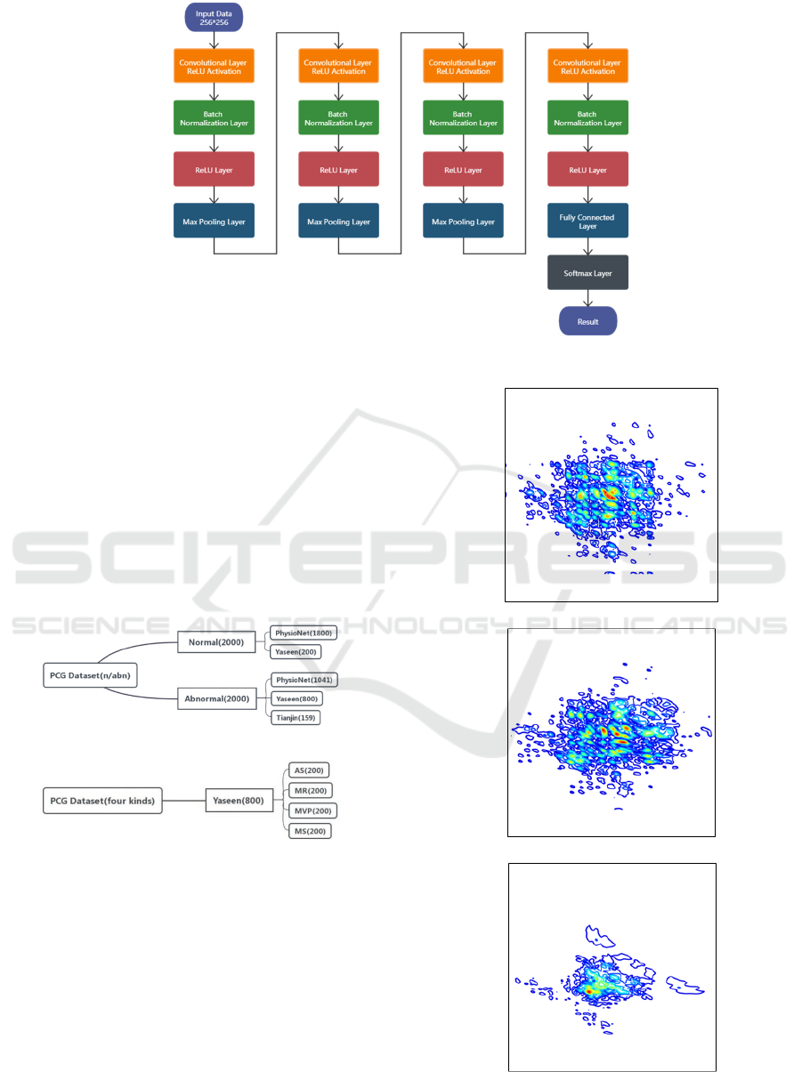
Figure 1: Diagram of the proposed network architecture.
was selected for four-layer wavelet decomposition,
and the threshold function was 'sqtwolog'. Finally, the
heart sound signal was sampled to 1000Hz, and the
heart sound sample was cut by 2.5s, with 50% overlap
each time, to realize samples were doubling while
reducing the calculation pressure. The preprocessed
heart sound samples were fused to build two fusion
data sets, as shown in Figure 2, a normal/abnormal
heart sound dataset was established, and an
AS/MR/MVP/MS heart sound dataset was also
established.
(a) Normal/Abnormal dataset
(b) AS/MR/MVP/MS dataset
Figure 2: Construction of dataset.
3.2 Experimental Results
According to the bispectrum analysis technology,
features were extracted from the heart sound signals
in the data set. Figure 3 is the contour map of
bispectrum plotted for two signals of aortic stenosis
and one signal of mitral stenosis, and each image is
the first quadrant image plotted. It can also be seen
that the images of the two cases of aortic stenosis are
similar but differ from the images of mitral stenosis.
(a) Aortic stenosis
(b) Aortic stenosis
(c) Mitral stenosis
Figure 3: Contour images of bispectrum.
CAIH 2021 - Conference on Artificial Intelligence and Healthcare
246
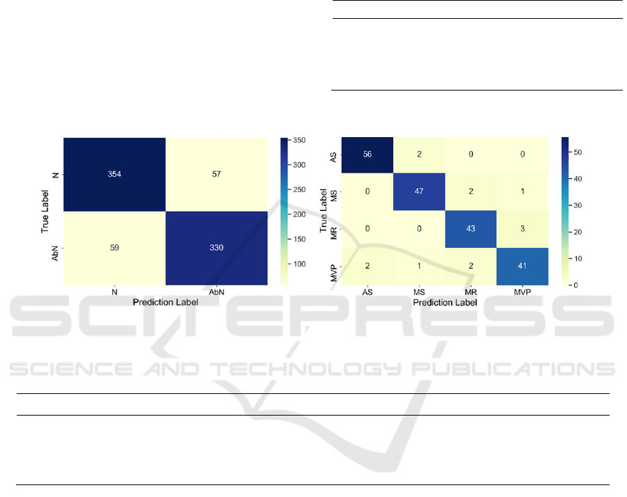
The extracted features were input into the 19-
layer convolutional neural network previously
established to distinguish between normal and
abnormal heart sounds. The learning rate was set to
0.001, the loss function was set to the cross-entropy
loss function, the optimizer was selected Adam, and
the batch size was set to 16. A total of 4000 samples
were divided into a training set and test set according
to 4:1. The classification performance is shown in
Table 2, and the confusion matrix is shown in Figure
4(a).
For the four categories of pathological heart
sounds, the setting was the same as that for the two
categories except batch-size 32. There were 800
samples for the four categories, and the number of the
four diseases was the same, but the training set and
test set were still allocated according to 4:1, and the
model training converged around 300 rounds. The
classification performance is shown in Table 3, and
the confusion matrix is shown in Figure 4(b).
Table 2: Performance of the binary model.
Indicators Score
Accuracy 0.980
Sensitivity 0.966
Specificity 0.986
F1 score 0.966
(a) Two categories (b) Four categories
Figure 4: Confusion Matrix
Table 3: Performance of the four classification models.
Category Accuracy Sensitivity Specificity F1 score
Aortic stenosis 0.980 0.966 0.986 0.966
Mitral stenosis 0.970 0.940 0.980 0.940
Mitral regurgitation 0.965 0.935 0.974 0.925
Mitral valve prolapse 0.955 0.891 0.974 0.901
3.3 Discussion
Although the model in this study has good
classification performance in the four categories,
there is still a particular gap compared with previous
studies in the two categories. We suspect this is
because the samples for the four categories come
from the same database, while the samples for the two
categories is a fusion of the three databases. Different
databases have different collection methods and noise
types, resulting in poor model performance in the
fusion data sets. We will also increase the research on
noise processing to narrow the differences between
different databases in future research. In addition,
sample collection will continue, especially the
collection of pathological samples of the four
categories, to expand the number of samples and
improve the robustness and generalization ability of
the model.
4 CONCLUSIONS
This article used three data sets: PhysioNet, Yaseen,
and the clinical self-collected data sets. Firstly,
normalization, wavelet denoising, subsampling and
overlapping cutting were used to complete the data
preprocessing, and then feature extraction was
completed based on bispectrum. Finally, the features
were put into the constructed neural network model
Research on Intelligent Analysis Model of Heart Sound based on Deep Learning
247

of dichotomies and quadrotors for the corresponding
classification, and the confusion matrix was
calculated, and several evaluation parameters were
obtained.
The results show that this model can provide a
standardized evaluation with high classification
performance and quickly complete the intelligent
analysis of heart sounds, which has important clinical
significance.
ACKNOWLEDGEMENTS
The authors express thanks to the doctors of Tianjin
Chest Hospital of China for providing full help in the
research process.
REFERENCES
Abduh, Z., Nehar, Y. E. A., Wahed, M. A., et al. (2019)
Classification of heart sounds using Fractional Fourier
Transform Based Mel-Frequency Spectral Coefficients
and Stacked Autoencoder Deep Neural Network. J.
Journal of Medical Imaging and Health Informatics,
9(1): 1-8.
Alqudah, A. M., Alquran, H. H., Abuqasmieh, I. (2020)
Classification of heart sound short records using
bispectrum analysis approach images and deep
learning. J. Network Modeling Analysis in Health
Informatics and Bioinformatics, 9(1):66.
Chen, L., Ren, J., Hao, Y., and Hu, X. (2018) The diagnosis
for the extrasystole heart sound signals based on the
deep learning. J. Journal of Medical Imaging and
Health Informatics, 8(5):959-986.
Dominguez-Morales Juan, P., Jimenez-Fernandez Angel,
F., Dominguez-Morales Manuel, J., Jimenez-Moreno
Gabriel. (2018) Deep neural networks for the
recognition and classification of heart murmurs using
neuromorphic auditory sensors. J. IEEE transactions on
biomedical circuits and systems, 12(1):1-11.
Liu, C., Springer, D., Li, Q., et al. (2016) An open access
database for the evaluation of heart sound algorithms.
J. Physiological Measurement, 37(12): 2181-2213.
Ma, L. Y., Chen, W. W., Gao, R. L., et al. (2020) China
cardiovascular diseases report 2018: an updated
summary. J. Journal of Geriatric Cardiology, 17(1): 1-
8.
Reed, T. R., Reed, N. E., Fritzson, P. (2004) Heart sound
analysis for symptom detection and computer-aided
diagnosis. J. Simulation Modelling Practice and
Theory, 12(2): 129-146.
Wu, M. T., Tsai, M. H., Huang, Y. Z., et al. (2019) Applying
an ensemble convolutional neural network with
Savitzky–Golay filter to construct a phonocardiogram
prediction model. J. Applied Soft Computing Journal,
78(1):29-40.
Yaseen, Son, G. Y., Kwon, S. (2018) Classification of heart
sound signal using multiple features. J. Applied
Sciences, 8(12):2344.
CAIH 2021 - Conference on Artificial Intelligence and Healthcare
248
