
Testing Gb3(CD77) Expression Level in Cancer Cells
Shuhao Chen
School of Biological Science, University of California Irvine, Irvine 92612, U.S.A.
Keywords: Shiga Toxins, CD77 Gb3, Cancer Targeted Therapy, Pancreatic Cancer, Colon Cancer, FACS, ATCC, DSMZ,
AnnexinV/PI.
Abstract: Purpose: Pancreatic cancer and colon cancer are common cancer types that are lethal. This study tries to find
the expression pattern of CD77 in cancer cells and investigate whether STxB-SN38 can be used for targeting
therapy in Pancreatic cancer and colon cancer. STxB was proved to have high specificity of binding to CD77,
and STxB is very efficient at cell killing. Methods: The experiments will use know human cell lines, ATCC
and DSMZ. Flow cytometry is used for monitoring cell proliferation and counting. AnnexinV/PI will be used
for killing measuring. Possible Results: There are 27 possible results Conclusion: The result of our study will
contribute to future clinical trials of Stxs-SN38 targeted therapy. Future studies should focus on eliminating
wrong pathways that Stxs-SN38 could kill normal cells. Detecting cancer at an early stage is still an important
study to research more.
1 INTRODUCTION
Colon and pancreatic cancer are common cancer
types in daily life. Pancreatic cancer is the third most
common in the United States. The five-year survival
rate for pancreatic cancer was 6% in 2003-2009 and
increased to 9% in 2009- 2015 (SEER 2019). Existing
methods for treating pancreatic include radiation
therapy, ablation or embolization treatments,
chemotherapy, targeted therapy, and immunotherapy
(SEER 2019). Appropriate therapies are chosen based
on the stage of cancer and other factors. Sometimes
these treatments are combined to obtain better effects.
Colon cancer is the second most common cause of
cancer in women and the third most common in men
(World Health Organization 2014). It also has the
third-highest cancer occurrence and death for people
in America (SEER 2019). Types of treatments for
colon cancer are the same, but different drugs may be
used. One famous drug used for the targeted
treatment of colon and pancreatic cancer is irinotecan
(IRT). Its analogs of the active metabolite, SN38 have
highly increased cytotoxicity than irinotecan (Geyer,
Maak, Nitsche, Perl, Novotny, Slotta-Huspenina,
Dransart, Holtorf, Johannes, Janssen 2016). Shiga
toxins produced by Enterohemorrhagic Escherichia
coli (EHEC), one food-borne pathogen, can cause
hemorrhagic colitis (HC) and hemolytic-uremic
syndrome (HUS) (Karmali 1989). Shiga toxins (Stxs)
were found to be the first ligands that proceed
endocytosis via clathrin-coated vesicles by using
glycolipid receptors (CD77) (Malyukova, Murray,
Zhu, Boedeker, Kane, Patterson, Peterson, Donowitz,
Kovbasnjuk 2009). To trigger the toxic effects of
Stxs, translocation of the A1(subunit) fragment into
the cytosol at ER is necessary (acewicz, Mobassaleh,
Gross, Balasubramanian, Daniel, Raghavan,
McCluer, Keusch 1994). However, the un-toxic B
subunit of Stxs specifically recognizes and binds its
cellular receptor Gb3(CD77) on the plasma
membrane, making it a potential tool for targeted
therapy. CD77 is one kind of glycolipid. It has been
proved that SHIGA toxin conjugate with SN38 can be
exploited for targeted therapy of cancer (Geyer,
Maak, Nitsche, Perl, Novotny, Slotta-Huspenina,
Dransart, Holtorf, Johannes, Janssen 2016). The
compound STxB-SN38 requires the receptor Gb3
(CD77) for intracellular uptake leading to a cytotoxic
effect (Geyer, Maak, Nitsche, Perl, Novotny, Slotta-
Huspenina, Dransart, Holtorf, Johannes, Janssen
2016). Thus, the expression of Gb3 (CD77) is very
important and should be investigated more. Until
now, Gb3 is reported to have an increased expression
on pancreatic and colon cancer cells in humans.
However, one paper that used carcinoma as a research
target found something interesting. They found that
the Gb3 expression falls down dramatically with the
increased tumor progression” (Maak, Nitsche, Keller,
Chen, S.
Testing Gb3(CD77) Expression Level in Cancer Cells.
DOI: 10.5220/0011242500003444
In Proceedings of the 2nd Conference on Artificial Intelligence and Healthcare (CAIH 2021), pages 203-208
ISBN: 978-989-758-594-4
Copyright
c
2022 by SCITEPRESS – Science and Technology Publications, Lda. All rights reserved
203

Wolf, Sarr, Thiebaud, Rosenberg, Langer, Kleeff,
Friess, et al. 2011). This finding contradicts previous
findings. Thus, it is necessary to examine the
expression pattern of Gb3 (CD77) of cancer cells.
Colon and pancreatic cancer will be tested in this
experiment. ATCC and DSMZ cell lines will be used.
Use STxB-Cy3 for staining and Shiga without
staining for staining control, mix them with cancer
cells and then check for fluorescent signal from
SHIGA on the cells by FACS. Killing measured by
MTT, AnnexinV/PI. The negative control is an
isotype-matched antibody for CD77, positive control
is a cell line that is already known to express high
levels of CD77. I predict the later passage pancreatic
cancer and colon cancer cells have increased CD77
and better binding to Shiga and increased killing with
Shiga-SN38 compared to earlier passage cells.
Measure CD77 by FACS as a function of passage
number (cell doublings).
2 METHODS AND MATERIALS
2.1 Cell Culture
This experiment will use two known cell lines (ATCC
and DSMZ),) Cultured cells in DMEM with FCS
(7%), 1% penicillin/streptomycin, and 1% glutamine
for 3 weeks. Take another set of cells with the same
passage number as the early passage. Pancreatic
(DanG and BxPC3) and colorectal (MKN-7,NCI-
N87 and HT29) cancer cells will be used. 2.5 μg/mL
of STxB-Cy3 will be added as the final concentration.
2.2 Reagents
STxB and STxB coupled with SN38
2.3 Staining
Making a covalent bonding between STxB and
fluorophore Cy3. Stain Gb3 on the 3%
paraformaldehyde fixed cryosections with STxB-Cy3
for 30 minutes at a final concentration of 10 μg/mL in
PBS containing 0.2% BSA (Geyer, Maak, Nitsche,
Perl, Novotny, Slotta-Huspenina, Dransart, Holtorf,
Johannes, Janssen 2016)
2.4 Flow Cytometry
Seed a total of 2000000 cells on one 10-cm cell
culture dish, then harvest and count the cells after 24
hours. Use 20 nmol/L STxB-Cy3 for staining for 15
minutes at 37°C. Use centrifugation for collecting
cells. Each experiment is repeated five times.
2.5 Cell Death Measured
AnnexinV/PI a: Add 5 μL Annexin V Alexa Fluor 488
to the target tubes, then incubate in the dark for 15
minutes at room temperature. Add 4 μL of PI that has
been diluted 1:10 in 1 x Annexin V binding buffer.
Incubate in the dark for 15 minutes at room
temperature. Centrifuge samples at 335 x g for 10
minutes. Resuspend cells in 500 μL 1 x Annexin V
binding buffer and 500 μL 2% formaldehyde. Add 1
mL 1 x PBS-/-. Centrifuge samples at 425 x g for 8
minutes. Add dd 16 μL of 1:100 diluted RNase A.
(Rieger, Aja M et al. 2011) Each experiment is
repeated five times.
2.6 Statistical Analysis
Use SPSS for analyzing data.
Table 1: Group A with passage number n (early passage).
Group CD77 staining STxB-Cy3 STx-B-SN38 STx-B-SN38 staining
1 yes no no no
2 yes yes no no
3 no no no no
4 yes no Yes no
5 yes no yes yes
CAIH 2021 - Conference on Artificial Intelligence and Healthcare
204
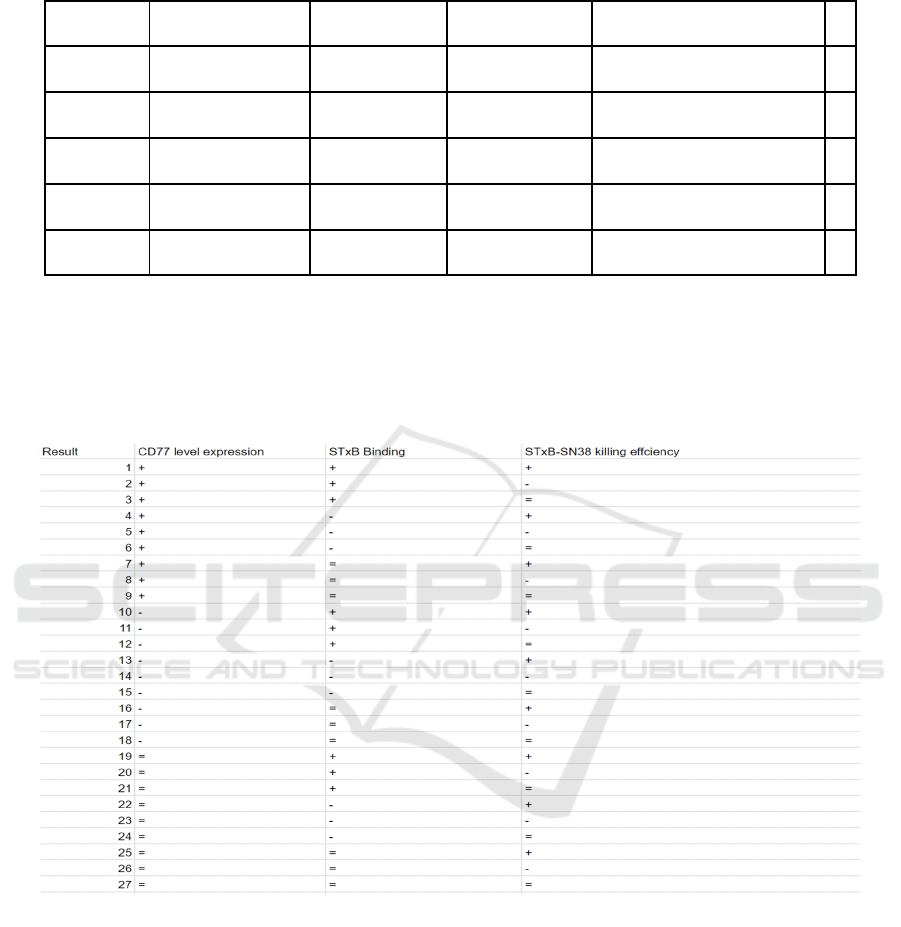
Table 2: Group B with passage number n+10 (late passage).
Group CD77 staining STxB-Cy3 STx-B-SN38 STx-B-SN38 staining
6 yes no no no
7 yes yes no no
8 no no no no
9 yes no Yes no
10 yes no yes yes
3 POSSIBLE RESULTS
The table is comparing groups A and B, “+” means
the value is higher in a later passage, “-” means the
value is lower in a later passage, “=” means the value
remains unchanged in a later passage.
Figure 1: A table represents all the possible results.
CD77 level expression is compared by group 1
and 6; STxB binding by group 2 and 7; STxB-SN38
killing efficiency by group 4 and 9. Group 3,5,8 and
10 are control groups.
Possible Result 1: The CD77 level expression is
increasing in later passage cancer cells (after one time
period of cell doublings), the percentage of cells
marked by STxB-Cy3 increased, more cell death
(percentage) is observed.
By comparing data from groups A and B, group B
has a higher percentage of CD77 and STxB present.
Possible Result 2: The CD77 level expression is
increasing in later passage cancer cells (after one time
period of cell doublings), the percentage of cells
marked by STxB-Cy3 increased, less cell death
(percentage) is observed. Possible Result 3: The
CD77 level expression is increasing in later passage
cancer cells (after one time period of cell doublings),
the percentage of cells marked by STxB-Cy3
increased, the number of cell death (percentage)
remains unchanged.
Testing Gb3(CD77) Expression Level in Cancer Cells
205

Possible Result 4: The CD77 level expression is
increasing in later passage cancer cells (after one time
period of cell doublings), the percentage of cells
marked by STxB-Cy3 decreased, more cell death
(percentage) is observed.
Possible Result 5: The CD77 level expression is
increasing in later passage cancer cells (after one time
period of cell doublings), the percentage of cells
marked by STxB-Cy3 decreased, less cell death
(percentage) is observed.
Possible Result 6: The CD77 level expression is
increasing in later passage cancer cells (after one time
period of cell doublings), the percentage of cells
marked by
STxB-Cy3 decreased, the number of cell death
(percentage) remains unchanged.
Possible Result 7: The CD77 level expression is
increasing in later passage cancer cells (after one time
period of cell doublings), the percentage of cells
marked by STxB-Cy3 remains unchanged, more cell
death (percentage) is observed.
Possible Result 8: The CD77 level expression is
increasing in later passage cancer cells (after one time
period of cell doublings), the percentage of cells
marked by STxB-Cy3 remains unchanged, less cell
death (percentage) is observed.
Possible Result 9: The CD77 level expression is
increasing in later passage cancer cells (after one time
period of cell doublings), the percentage of cells
marked by STxB-Cy3 remains unchanged, the
number of cell death (percentage) remains
unchanged.
Possible Result 10: The CD77 level expression is
decreasing in later passage cancer cells (after one
time period of cell doublings), the percentage of cells
marked by STxB-Cy3 increased, more cell death
(percentage) is observed.
Possible Result 11: The CD77 level expression is
decreasing in later passage cancer cells (after one
time period of cell doublings), the percentage of cells
marked by STxB-Cy3 increased, less cell death
(percentage) is observed.
Possible Result 12: The CD77 level expression is
decreasing in later passage cancer cells (after one
time period of cell doublings), the percentage of cells
marked by STxB-Cy3 increased, the number of cell
death (percentage) remains unchanged.
Possible Result 13: The CD77 level expression is
decreasing in later passage cancer cells (after one
time period of cell doublings), the percentage of cells
marked by STxB-Cy3 decreased, more cell death
(percentage) is observed.
Possible Result 14 The CD77 level expression is
decreasing in later passage cancer cells (after one
time period of cell doublings), the percentage of cells
marked by STxB-Cy3 decreased, less cell death
(percentage) is observed.
Possible Result 15: The CD77 level expression is
decreasing in later passage cancer cells (after one
time period of cell doublings), the percentage of cells
marked by STxB-Cy3 decreased, the number of cell
death (percentage) remains unchanged.
Possible Result 16: The CD77 level expression is
decreasing in later passage cancer cells (after one
time period of cell doublings), the percentage of cells
marked by STxB-Cy3 remains unchanged, more cell
death (percentage) is observed.
Possible Result 17: The CD77 level expression is
decreasing in later passage cancer cells (after one
time period of cell doublings), the percentage of cells
marked by STxB-Cy3 remains unchanged, less cell
death (percentage) is observed.
Possible Result 18: The CD77 level expression is
decreasing in later passage cancer cells (after one
time period of cell doublings), the percentage of cells
marked by STxB-Cy3 remains unchanged, the
number of cell death (percentage) remains
unchanged.
Possible Result 19: The CD77 level expression is
increasing in later passage cancer cells (after one time
period of cell doublings), the percentage of cells
marked by STxB-Cy3 increased, more cell death
(percentage) is observed.
Possible Result 20: The CD77 level expression
remains unchanged in later passage cancer cells (after
one time period of cell doublings), the percentage of
cells marked by STxB-Cy3 increased, less cell death
(percentage) is observed.
Possible Result 21: The CD77 level expression
remains unchanged in later passage cancer cells (after
one time period of cell doublings), the percentage of
cells marked by STxB-Cy3 increased, the number of
cell death (percentage) remains unchanged.
Possible Result 22: The CD77 level expression
remains unchanged in later passage cancer cells (after
one time period of cell doublings), the percentage of
cells marked by STxB-Cy3 decreased, more cell
death (percentage) is observed.
Possible Result 23: The CD77 level expression
remains unchanged in later passage cancer cells (after
one time period of cell doublings), the percentage of
cells marked by STxB-Cy3 decreased, less cell death
(percentage) is observed.
Possible Result 24: The CD77 level expression
remains unchanged in later passage cancer cells (after
one time period of cell doublings), the percentage of
cells marked by STxB-Cy3 decreased, the number of
cell death (percentage) remains unchanged.
CAIH 2021 - Conference on Artificial Intelligence and Healthcare
206

Possible Result 25 The CD77 level expression
remains unchanged in later passage cancer cells (after
one time period of cell doublings), the percentage of
cells marked by STxB-Cy3 remains unchanged, more
cell death (percentage) is observed.
Possible Result 26: The CD77 level expression
remains unchanged in later passage cancer cells (after
one time period of cell doublings), the percentage of
cells marked by STxB-Cy3 remains unchanged, less
cell death (percentage) is observed.
Possible Result 27: The CD77 level expression
remains unchanged in later passage cancer cells (after
one time period of cell doublings), the percentage of
cells marked by STxB-Cy3 remains unchanged, the
number of cell death (percentage) remains
unchanged.
4 DISCUSSION
Results 10-27 all overturn the hypothesis because the
CD77 expression level does not go up for later
passage cells. While results 1-9 only supports part of
the hypothesis. (The CD77 expression level increase
in later number) Result 1: It perfectly supports the
hypothesis, which means the later passage cells are
presenting more CD77 relative to earlier passage
cells, leading to the increased binding of STxB. Thus,
more STxB-SN38 kills more cells. This result
indicating STxB-SN38 has the potentials to limit
cancer grows up as targeted therapy. Moreover, due
to its high cytotoxicity, it may replace previous
medicine such as Irinotecan. Future experiments are
supposed to follow up. Animal research like mice
could be done using xenograft. Result 1,14 and 27: In
these three experiments, the STxB binding and
STxB-SN38 killing efficiency follows the CD77
level. They showed that STxB binding is positively
associated with CD77; the killing efficiency is
positively associated with STxB binding. For result
27, the CD77 expression level does not show any
relationship within later passage cells. It indicates
that the CD77 expression level seems not related to
the passage number of cells which does not support
the hypothesis. For result 14, the CD77 drops in later
passage cells. This result opposes that hypothesis.
Since much evidence point that colon and Pancreatic
cancer have some level of CD77 expression, a further
experiment should be followed up. The expression
pattern of CD77 could be complicated. The CD77
level possibly increases with the cells grow up and
drop while the passage number goes up. The reason
might associate with CD77’s function as a membrane
protein. It may also relate to the gene regulation
changes during mitosis.
For results that the STxB binding does not
correspond to STxB-SN38 killing efficiency, they can
be divided into two groups.
First group are result 2,3,8,11,12,17,20,26. In
these results, the killing efficiency of late passage is
reduced compared to early passage. In such case,
group 4 and 5 in group A and group 9 and 10 in group
B should be analyzed deeper. The difference between
group 4 and 5 or group 9 and 10 is the STxB-Sn38
staining. By comparing them, it can tell whether the
cell pathway of STxB-SN38 changes. If the pathway
changes, it is possible that the SN38 group of STxB-
SN38 are targeted by another molecule inside the
cells and eliminated. These results are unexpected
and won’t support or deny the hypothesis. The
unknown pathway is necessary to study more. If the
pathway does not change, then the STxB-SN38 may
have dose effects. A high concentration of STxB-
SN38 could downregulate the killing efficiency.
These results disprove the hypothesis that higher
binding of STxB with cells could increase cell killing
of STxB-SN38. A new experiment testing the
concentration of STxB-SN38 with its highest
efficiency could be processed.
Second group are results 4,6,7,13,15,16,22,24,25.
In these results, the killing efficiency of late passage
is higher compared to early passage. Test if the
pathway changes as talked about before in the paper.
If the pathway changes, the STxB-SN38 may bind to
something unexpected but still trigger the toxic
effects and thus kill the cells. Since STxB SN38 lost
its high specificity of binding to CD77, the
undifferentiated killing of all cells happened and
increase the kill numbers. These results are
unexpected and won’t support or deny the hypothesis.
Further experiments could be done by using cells that
have low or no expression of CD77 treated with
STxB-SN38 for control to see if STxB SN38 lost its
specificity. That may reveal another cell pathway of
how STxB-SN38 entering the cell. (As mentioned
before, STxB requires CD77 to get into cells) If the
pathway does not change, the STxB-SN38 may have
dose effects as mentioned before. It does not support
the hypothesis and more research should be done.
For result 10,11,12,16,17,18,19,20,21, STxB
binding is increasing while CD77 is not increasing or
remains the same while CD77 is dropping down. This
is strong evidence that STxB binds to another
receptor protein to get into targeted cells. That makes
it harder to let STxB mediate targeted therapy. These
results overturn the hypothesis. Later research and
study should focus on discovering and investigate the
Testing Gb3(CD77) Expression Level in Cancer Cells
207

new pathway. Then, the new pathways could be tested
(shut down it) to see if they can make the STxB CD77
pathway-specific again.
For results 4,5,6,7,8,9,22,23 and 24, STxB
binding is decreasing while CD77 remains the same
or increasing. If no huge artificial mistakes are made,
then these results
overturn the hypothesis. There might be a new
pathway presents as mentioned before. The STxB
could also be saturated if the STxB binding remains
the same. (result 7,8,9)
5 CONCLUSIONS
Known the expression pattern of CD77 will help a lot
in targeted cancer therapy. The result of the study will
give a detailed pattern of CD77 expression, which
will help further research.
REFERENCES
"Colorectal Cancer – Cancer Stat Facts". SEER. Retrieved
4 April 2019.
"Pancreatic Cancer – Cancer Stat Facts". SEER. Retrieved
4 April 2019.
10.1093/infdis/169.3.538
10.1152/ajpgi.90347.2008
acewicz M.S., Mobassaleh M., Gross S.K.,
Balasubramanian K.A., Daniel P.F., Raghavan S.,
McCluer R.H., Keusch G.T. Pathogenesis of Shigella
diarrhea: XVII. A mammalian cell membrane
glycolipid, Gb3, is required but not sufficient to confer
sensitivity to Shiga toxin. J. Infect. Dis. 1994; 169:538–
546. doi:
Geyer P.E., Maak M., Nitsche U., Perl M., Novotny A.,
Slotta-Huspenina J., Dransart E., Holtorf A., Johannes
L., Janssen K.P. Gastric Adenocarcinomas Express the
Glycosphingolipid Gb3/CD77: Targeting of Gastric
Cancer Cells with Shiga Toxin B-Subunit. Mol. Cancer
Ther. 2016; 15:1008–1017. doi: 10.1158/1535-
7163.MCT-15-0633
Karmali M.A. Infection by verocytotoxin producing
Escherichia coli. Clin. Microbiol.
Maak M., Nitsche U., Keller L., Wolf P., Sarr M., Thiebaud
M., Rosenberg R., Langer R., Kleeff J., Friess H., et al.
Tumor-specific targeting of pancreatic cancer with
Shiga toxin B-subunit. Mol. Cancer Ther. 2011; 10:
1918– 1928. doi: 10.1158/1535-7163.MCT-11-0006.
Malyukova I., Murray K.F., Zhu C., Boedeker E., Kane A.,
Patterson K., Peterson J.R., Donowitz M., Kovbasnjuk
O. Macropinocytosis in Shiga toxin 1 uptake by human
intestinal epithelial cells and transcellular transcytosis.
Am. J. Physiol. Gastrointest. Liver Physiol. 2009; 296:
G78–G92. doi:
Rev. 1989;2:15–38. doi: 10.1128/CMR.2.1.15.
Rieger, Aja M et al. “Modified annexin V/propidium iodide
apoptosis assay for accurate assessment of cell death.”
Journal of visualized experiments: JoVE ,50 2597. 24
Apr. 2011, doi:10.3791/2597
World Cancer Report. World Health Organization. 2014.
Chapter 5.7. ISBN 978-92-832-0429-9
CAIH 2021 - Conference on Artificial Intelligence and Healthcare
208
