
Influence of Endured Coronavirus Infection on Bioelectric Activity of
the Brain in Students
Elena M. Inyushkina
a
, Andrey A. Inyushkin
b
and Alexey N. Inyushkin
c
Samara National Research University, 443011 Samara, Russia
Keywords: Covid 19, Electroencephalogram, Spectral Power, Attention Tests, Students.
Abstract:
In the paper, differences in the spectral power (SP) of EEG rhythms of the main frequency ranges (alpha,
beta) in two groups of students, before and after the tests for attention are assessed. The first group consisted
of students, who did not have a coronavirus infection in their anamnesis (control), the second group consisted
of students recovered from a coronavirus infection no more than 3 months ago. EEG was recorded with NVX
36 digital DC EEG system according to the international scheme 10-20. The subjects were tested for
attentiveness. After passing the tests, the results were above the minimum indicating the strain of the attention
process. The registration time was 1 minute. It was found that students who underwent coronavirus infection,
compared with the control group, showed a statistically significant change in the SP of the alpha and beta
rhythms in several leads. In particular, there was an increase in the SP of the alpha rhythm in the frontal lead
Fz and the central lead C3. When comparing beta1 rhythm, an increase in SP in the frontal lead Fz was also
noted. Analysis of the beta 2 rhythm showed a decrease in the SP of the rhythm in students with coronavirus
infection in the occipital O2 lead. The results show the effects of coronavirus infection on the bioelectrical
activity of the brain in students. Despite the fact, that all the students at the time of the study were recovered,
there is still a difference between those who did not get sick and those who had recovered. This indicates the
effect of the coronavirus on the activity of the human brain.
1 INTRODUCTION
At present, it is generally accepted that sustainable or
harmonious development includes a whole range of
concepts, but most of it is aimed primarily at ensuring
the quality of life of people, which is impossible
without high-quality health care (Bobylev, Girusov,
Flight, 2004).
Recently, the health and well-being of people has
been declining due to the new coronavirus infection,
which, of course, leads to an imbalance in one of the
most important goals of sustainable development,
such as the health and well-being of the world's
population.
At the end of 2019, an outbreak of a new
coronavirus infection occurred in the People's
Republic of China, in the city of Wuhan. On February
11, 2020, the World Health Organization offered the
official name for the infection caused by the new
a
https://orcid.org/0000-0002-3390-962X
b
https://orcid.org/0000-0001-8564-4275
c
https://orcid.org/0000-0002-3678-2636
coronavirus – COVID-19 ("Coronavirus disease
2019"). In turn, the international committee on the
taxonomy of viruses on February 11, 2020 assigned
the official name to the causative agent of the
infection – SARS-CoV-2 (Conradi, Nedoshivin,
2020).
Emergence of COVID-19 has challenged
healthcare professionals to quickly diagnose it and
provide medical care to patients. Currently, an
intensive study of the clinical and epidemiological
characteristics of the disease continues, as well as the
development of new means of its prevention and
treatment. The most common clinical manifestation
of a new variant of coronavirus infection is bilateral
pneumonia. Some patients develop hypercoagulable
syndrome with thromboembolism and thrombosis,
and other organs and systems are also affected, and
large vessels may be damaged even in young people
(Klok, Kruip, VanderMeer, Arbous, Gommers, Kant,
Inyushkina, E., Inyushkin, A. and Inyushkin, A.
Influence of Endured Coronavirus Infection on Bioelectric Activity of the Brain in Students.
DOI: 10.5220/0011109700003439
In Proceedings of the 2nd International Scientific and Practical Conference "COVID-19: Implementation of the Sustainable Development Goals" (RTCOV 2021), pages 5-12
ISBN: 978-989-758-617-0
Copyright
c
2023 by SCITEPRESS – Science and Technology Publications, Lda. All rights reserved
5

Kaptein, vanPaassen, Stals, Huisman, 2020), (Oxley,
Mocco, Majidi, Kellner, Shoirah, Singh, De Leacy,
Shigematsu, Ladner, Yaeger, 2020). However,
damage to the lungs, kidneys, liver, or chronic heart
failure are not the only complications caused by the
coronavirus. Quite often, even several months after
recovery, patients experience cognitive impairments:
a decrease in mental performance, memory, and other
intellectual functions. It was found that neurological
consequences after COVID-19 can also be long-term
(Heneka, M.T., Golenbock, D., Latz, E., Morgan, D.,
Brown R.., 2020).
It was revealed that SARS-Cov-2 affects the brain
and nervous system in a completely different way
than other viruses. Researchers examined samples of
cerebrospinal fluid from patients with COVID-19
who had cognitive impairment and mental problems
and found a marked increase in inflammatory markers
that indicate activation of immune cells in the brain.
However, at the same time, scientists did not find
characteristic markers for viral damage to the central
nervous system (Egbert, A.R., Cankurtaran, S.,
Karpiak, S., 2020). Many researchers also note that
the mechanism of damage to the nervous system in
coronavirus differs from the effects of other
pathogens (Mao, L., Jin, H., Wang, M., Hu, Y., Chen,
S., He, Q., Chang, J., Hong, C., Zhou, Y., Wang, D.,
et al., 2020). Thus, the pathogenetic mechanisms of
the effect of SARS-CoV-2 on the central nervous
system require detailed study (Baig, A.M., 2020).
The aim of this paper was to study the effect of the
previous coronavirus infection on the bioelectrical
activity of the brain in students during tests for
attention.
2 METHODS
This paper was performed on 20 students aged 20 to
25 years. Ten students did not have a coronavirus
infection in their anamnesis (group 1), the other 10
had recovered from a coronavirus infection (group 2)
and were examined three months after recovery.
Group 1 included 4 males and 6 females, group 2
consisted of 5 males and 5 females. Students were
included in the study on the basis of informed
voluntary and confirmed written consent. All stages
of the experiment were carried out in accordance with
the rules of bioethics used in studies of physiological
functions in humans. The subjects were subjected to
electroencephalographic examination. The
electroencephalogram was recorded using NVX 36
digital DCEEG system. Lead electrodes were placed
on the subjects' heads in accordance with the
international scheme "10–20" in the following
standard leads: Fp1, Fpz, Fp2, F7, F3, Fz, F4, F8, T3,
C3, Cz, C4, T4, T5, P3, Pz, P4, T6, O1, Oz, O2. A
combined ear electrode (A1, A2) was used as a
reference. Each student's EEG was recorded twice: in
the initial state and after the tests for attention. In the
process of EEG registration, the subjects sat in a chair
in a dark room with their eyes closed. After a five-
minute period of adaptation to the experimental
setting, a background EEG was recorded from the
subjects for 1 minute. Re-registration of the EEG was
also carried out within 1 minute after performing the
tests for attention. Changes in the spectral power
(μV
2
) of the EEG rhythms within the main frequency
ranges were analyzed on the EEG: alpha (8-13 Hz),
beta (13-35 Hz), theta (4-8 Hz).
As the first test for attention, we used the
Bourdon-Anfimov proofreading test (crossing out the
specified letters in the form). This test took 5 minutes
to complete. As the next, the Landolt ring test was
used for 5 minutes. The final stage of testing was the
execution of the attention test from the KornFerry
company (Judge, T. A., Heller, D., Mount, M. K.,
2002). In accordance with the testing conditions, it
was required to check all the presented pairs of
numbers within 2 minutes and identify coinciding
ones. After passing the tests, all subjects had results
higher than the minimum level indicating the tension
of attention.
Since sample data distribution differed from
normal (P <0.05: Shapiro-Wilk test for normality),
the Mann-Whitney rank sum test was used to assess
statistically significant differences in the spectral
power of EEG rhythms between students who had
recovered from coronavirus infection and the control
group of students. The Wilcoxon signed rank test was
used to assess the differences before and after testing
for attention. Differences were considered
statistically significant at P < 0.05.
3 RESULTS
It was found that students who had recovered from
coronavirus infection showed changes in the EEG,
which were expressed in a change in the spectral
power of the alpha rhythm in some leads. In the initial
state (before the performance of attention tests), the
median power of the alpha rhythm in the median
frontal lead Fz in students, who had coronavirus
infection in their anamnesis, was significantly higher
than that in students of the control group by 7.30 μV
2
(P = 0.037: Mann-Whitney rank sum test). After
passing the tests for attention, an equally directed
RTCOV 2021 - II International Scientific and Practical Conference " COVID-19: Implementation of the Sustainable Development Goals
(RTCOV )
6
tendency to decrease in the spectral power of the
alpha rhythm in lead Fz was observed in students of
both groups, however, a statistically significant effect
of testing on the value of this parameter was found
only in students who had undergone coronavirus
infection (decrease in the median by 7.15 μV
2
; P =
0.039: Wilcoxon signed rank test), while in intact
students the change did not reach the level of
statistical significance (decrease in the median by
0.70 μV
2
; P = 0.203: Wilcoxon signed rank test).
After testing, no difference was found between the
power of alpha rhythm in the Fz lead in intact students
and students recovered from coronavirus infection (P
= 0.185: Mann-Whitney rank sum test), though the
median of this parameter was higher in those who had
recovered by 0.85 µV
2
. Statistical data on the values
of the spectral power of the alpha rhythm in the
median frontal lead Fz in the subjects of both groups
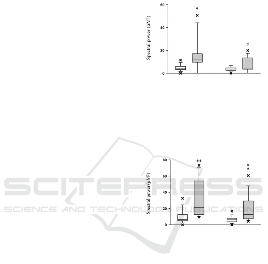
before and after testing are shown in Fig. 1.
Statistically significant differences in the spectral
power of the alpha rhythm in students who had
undergone coronavirus infection compared with
intact students were also detected in the central lead
C3. In this lead, the median of the power of the alpha
rhythm in students who had recovered from
coronavirus infection turned out to be significantly
higher than in intact students both before (by 15.05
μV
2
; P = 0.007: Mann-Whitney rank sum test) and
after testing for attention (by 6.60 μV
2
; P = 0.017:
Mann-Whitney rank sum test). Testing did not
significantly affect the spectral power of the alpha
rhythm in lead C3 of intact students (P = 0.129:
Wilcoxon signed rank test), in whom the median of
this parameter increased by 0.40 μV
2
after testing. In
students who had recovered from the coronavirus
infection, the spectral power of the alpha rhythm in
lead C3 after testing for attention decreased
comparing to the initial level by 8.85 μV
2
(P = 0.027:
Wilcoxon signed rank test). Statistical data on the
values of the spectral power of the alpha rhythm in
the central lead C3 in the subjects of both groups
before and after testing are demonstrated in Fig. 2.
We also carried out a comparative analysis of the
spectral power of EEG beta rhythms (β1 and β2
rhythms) in intact students and students who had
recovered from coronavirus infection before and after
performing attention tests.
Statistical analysis revealed differences in the
spectral power of beta rhythms in several leads.
Figure 1: The difference in the spectral power (μV
2
) of the
alpha rhythm in the median frontal lead Fz in intact subjects
(white boxes) and students who had endured a coronavirus
infection (gray boxes) before the test (left boxes) and after
the test (right boxes) for attention. An asterisk marks a
statistically significant difference between the two groups
of subjects before testing (* P <0.05: Mann-Whitney rank
sum test). The # symbol indicates a statistically significant
difference between the state before and after testing for
attention in students with endured coronavirus infection (#
P <0.05: Wilcoxon signed rank test).
Figure 2: The difference in the spectral power (μV2) of the
alpha rhythm in the central lead C3 in intact subjects (white
boxes) and students who had endured a coronavirus
infection (gray boxes) before the test (left boxes) and after
the test (right boxes) for attention. Asterisks denote
statistically significant differences between the two groups
of subjects (* P <0.05; ** P <0.01: Mann-Whitney rank sum
test). The # symbol denotes a statistically significant
difference between the state before and after testing for
attention in students with endured coronavirus infection (#
P <0.05: Wilcoxon rank test).
Before testing for attention, the median power of the
β1 rhythm in the median frontal lead Fz was higher in
the group of students with coronavirus infection by
6.05 μV
2
than in intact students (P = 0.011: Mann-
Whitney rank sum test). However, after testing, the
spectral power of the β1 rhythm in this lead in
students who had undergone coronavirus infection no
longer had a statistically significant difference from
that in intact students (P = 0.103: Mann-Whitney rank
Influence of Endured Coronavirus Infection on Bioelectric Activity of the Brain in Students
7

sum test), although the median of this parameter in
intact students at that moment was lower than that of
those who had recovered by 2.30 µV
2
. As a result of
testing, a weak tendency towards a decrease in the
spectral power of the β1 rhythm in the Fz lead in
intact students (by 0.50 μV
2
; P = 0.383: Wilcoxon
signed rank test) was found, while in the group of
subjects who had recovered from coronavirus
infection, the tendency of this parameter to the
reduction was more pronounced (by 4.25 μV
2
),
although the change also did not reach the level of
statistical significance (P = 0.055: Wilcoxon signed
rank test). Statistical data on the values of the β1
rhythm spectral power in the median frontal lead Fz
in the subjects of both groups before and after testing
for attention are shown in Fig. 3. Statistically
significant differences in the spectral power of the β1
rhythm in intact students and those had endured a
coronavirus infection were also found in the central
lead C3. The power of the β1 rhythm in persons who
had undergone coronavirus infection turned out to be
higher than in intact subjects both before and after
testing.
Figure 3: The difference in the spectral power (μV
2
) of the
β1 rhythm in the median frontal lead Fz in intact students
(white boxes) and students who had endured a coronavirus
infection (gray boxes) before the test (left boxes) and after
the test (right boxes) for attention. An asterisk marks a
statistically significant difference between the two groups
of subjects before testing (* P <0.05: Mann-Whitney rank
sum test).
Before testing, the spectral power of the β1
rhythm in this lead in students with endured
coronavirus infection, exceeded that in intact subjects
by 10.05 μV
2
(P = 0.013: Mann-Whitney rank sum
test); after testing, the value of this parameter in the
group of recovered patients also turned out to be
higher than in intact students by 6.00 μV
2
(P = 0.005:
Mann-Whitney rank sum test). Testing for attention
did not have a statistically significant effect on the
spectral power of the β1 rhythm neither in intact
students, nor in students who had undergone
coronavirus infection. Wherein, both those and others
showed a tendency towards a decrease in the spectral
power of the β1 rhythm in lead C3, respectively, by
1.45 μV
2
(P = 0.055: Wilcoxon signed rank test) and
7.40 μV
2
(P = 0.164: Wilcoxon signed rank test).
Statistical data on the values of the spectral power of
the β1 rhythm in the central lead C3 in the subjects of
both groups before and after testing for attention are
shown in Fig. 4.
Under these experimental conditions, no
statistically significant differences were found in the
spectral power of the β2 rhythm of students who were
intact and who had endured a coronavirus infection in
the median frontal lead Fz before and after testing for
attention. In the initial state (before testing), the
median power of this rhythm in students who had
endured a coronavirus infection exceeded that in
intact students (by 0.9 μV
2
), however, the difference
was not statistically significant (P = 0.344: Mann-
Whitney rank sum test). Similarly, there was no
difference in the spectral power of the β2 rhythm
between the two groups of subjects after testing (P =
0.265: Mann-Whitney rank sum test), when the value
of the median power of the β2 rhythm in students who
had undergone coronavirus infection turned out to be
by 0.65 μV
2
higher than that of intact students. As a
result of testing, the median β2-rhythm power tended
to decrease both in intact students (by 0.25 μV
2
) and
in students who had undergone coronavirus infection
(by 0.50 μV
2
), however, these changes were not
statistically significant (P = 0.250 and P = 0.156,
respectively: Wilcoxon rank sum test).
Figure 4: The difference in the spectral power (μV
2
) of the
β1 rhythm in the central lead C3 in intact students (white
boxes) and students who had endured a coronavirus
infection (gray boxes) before the test (left boxes) and after
the test (right boxes) for attention. Asterisks mark
statistically significant differences between the two groups
of subjects before testing (*P <0.05: Mann-Whitney rank
test) and after testing (** P <0.01: Mann-Whitney rank sum
test).
RTCOV 2021 - II International Scientific and Practical Conference " COVID-19: Implementation of the Sustainable Development Goals
(RTCOV )
8
In the central lead C3, there was also not
statistically significant difference in the spectral
power of the β2 rhythm in intact students and students
who had endured a coronavirus before and after
testing for attention. In students recovered from
coronavirus, the median β2 rhythm power before
testing was higher by 6.00 μV
2
than in intact students,
but this difference was not statistically significant (P
= 0.081: Mann-Whitney rank sum test). After testing,
there was also no statistically significant difference
between the spectral power of the β2 rhythm in
subjects who were intact and who had endured a
coronavirus (P = 0.054: Mann-Whitney rank sum
test), although in the latter group, the median of this
parameter was higher by 2.95 μV
2
. Testing had no
effect on the power of the β2 rhythm in lead C3
neither in intact students (P = 0.275: Wilcoxon signed
rank test), nor in those who had endured a coronavirus
infection (P = 0.203: Wilcoxon signed rank test),
despite the trend towards a reduction in the value of
this parameter in subjects of both groups (by 0.80 µV
2
and 3.85 µV
2
, respectively).
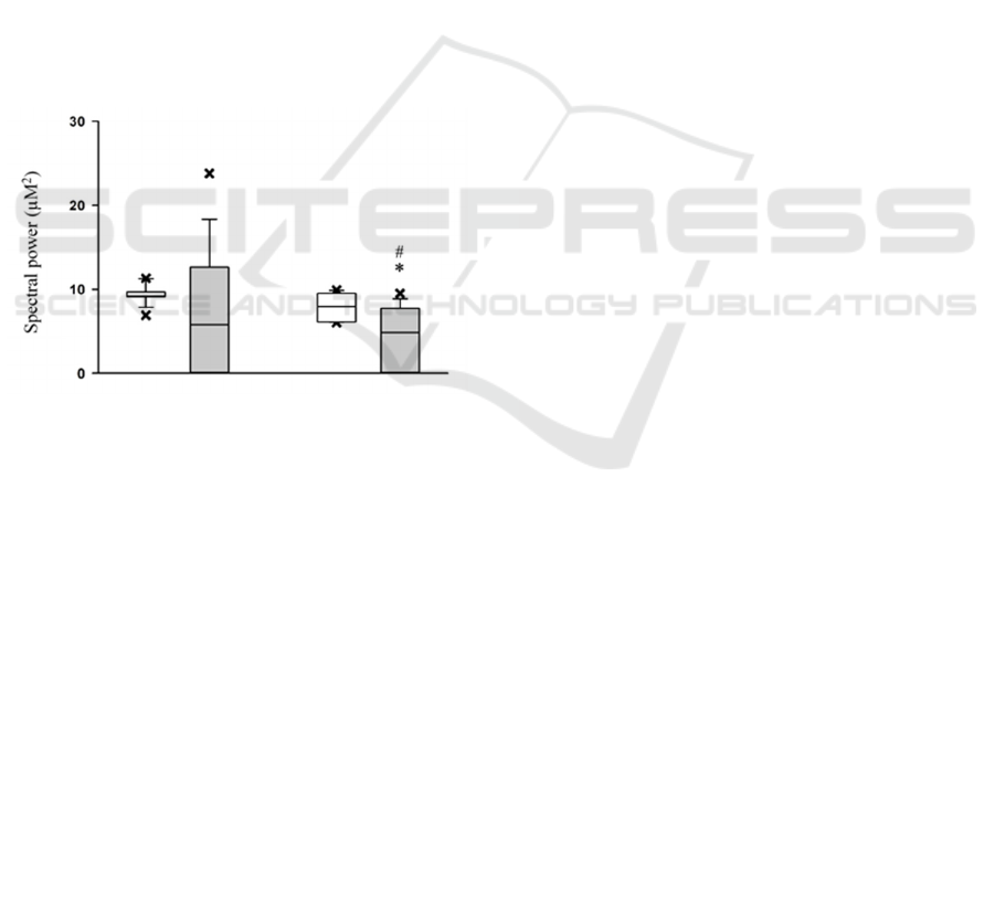
Figure 5: The difference in the spectral power (μV
2
) of the
β2 rhythm in the occipital O2 lead in intact
students (white
boxes) and students who had endured a coronavirus
infection (gray boxes) before the test (left boxes) and after
the test (right boxes) for attention. An asterisk denotes a
statistically significant difference between the two groups
of subjects after testing for attention (* P <0.05: Mann-
Whitney rank sum test). The # symbol denotes a statistically
significant difference between the state before and after
testing for attention in students who had endured a
coronavirus infection (# P <0.05: Wilcoxon signed rank
test).
The only EEG lead, in which statistically
significant differences between the groups of subjects
in the spectral power of the β2 rhythm were found
was the occipital O2 lead. In this lead, the median
power of the β2 rhythm in persons who underwent
coronavirus infection was lower by 3.10 μV
2
than that
of intact subjects after testing (P = 0.014: Mann-
Whitney rank sum test). The value of this parameter
before testing was also lower in students who had
recovered from coronavirus infection by 3.40 μV
2
,
however, at that moment, the differences between the
groups of subjects were not statistically significant (P
= 0.161: Mann-Whitney rank sum test). Testing for
attention had no effect on the spectral power of the β2
rhythm in the O2 lead in intact subjects (P = 0.131:
Wilcoxon signed rank test), despite the trend towards
a decrease in the median of this parameter (by 1.2
μV
2
). Wherein, as a result of testing, the median
spectral power of the β2 rhythm decreased by 0.90
μV
2
in subjects who had undergone coronavirus
infection (P = 0.031: Wilcoxon signed rank test).
Statistical data on the values of the spectral power of
the β2 rhythm in the occipital O2 lead in the subjects
of both groups before and after testing for attention
are presented in Fig. 5.
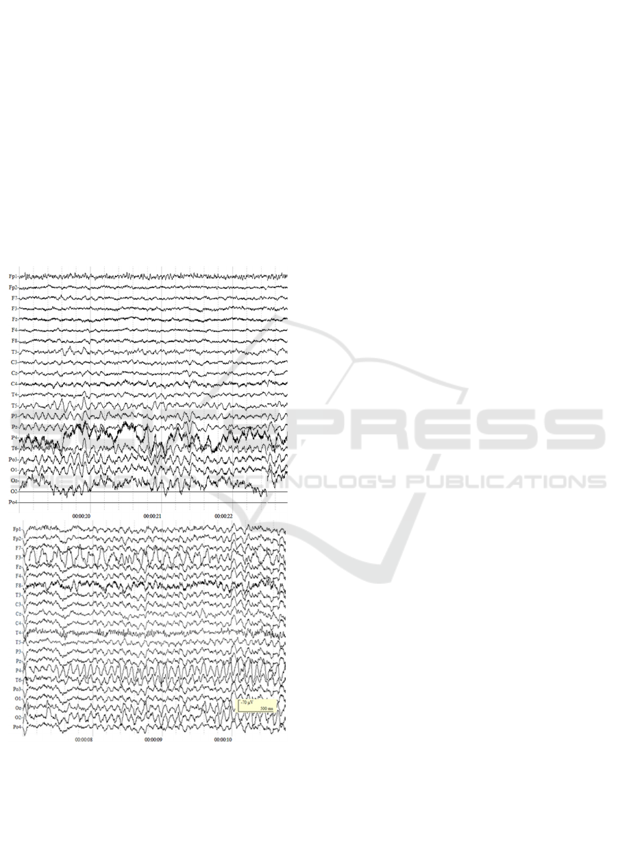
Typical examples of the EEG of a student who
had endured a coronavirus infection before and after
testing for attention are shown in Fig. 6.
4 DISCUSSION
During SARS-CoV-2 infection, mental disorders and
symptoms of stress disorders are recorded in almost
70 % of people, which leads to a decrease in the
quality of life and disrupts work productivity
(Shepeleva, I. I., Chernysheva, A. A., Kiryanova,
E.M., Salnikova, L.I., Gurin O. I. , 2020).
In this paper, we investigated the spectral power
of EEG rhythms before and after performing tests for
attention in intact students and students who had
suffered from coronavirus infection COVID-19 three
months ago. Three months after recovery, at least a
half of the patients retain neurological symptoms such
as dizziness, headache, and impaired consciousness
(Li, X., Geng, M., Peng, Y. et al., 2020; 16.
Niazkar, H.R., Zibaee, B., Nasim,i Q., Bahri, N.,
2020). In our opinion, it is necessary to investigate the
long-term effect of SARS-CoV-2 infection on the
central nervous system, especially on structures that
are easily attacked by the virus.
Use of the EEG technique is a traditional method
for studying the biopotentials of the brain, while it is
well known that for various levels of wakefulness, the
predominance of EEG signals of a certain frequency
with pronounced activity in the alpha range is
characteristic (Corsi-Cabrera, M., Guevara, M.A.,
Del Rio-Portilla, Y., Arce, C., Villanueva-Hernandez,
Y., 2000). Analysis of the main EEG rhythm, alpha
activity is one of the approaches to an objective
assessment of the disorganization of the functional
state of the cortical mosaic in the central nervous
Influence of Endured Coronavirus Infection on Bioelectric Activity of the Brain in Students
9
system (Ivanov, L. B., 2005), (Livanov, M.N., 1984).
The alpha rhythm is recorded mainly in the occipital
regions with closed eyes in a state of calm
wakefulness and is blocked by light stimulation,
concentration, and mental stress (Kostandov, Э. А.,
Cheremushkin, E. F., 2012). According to modern
concepts, the generation of the alpha rhythm is
associated with the reverberation of impulse activity
along the intercortical and thalamocortical neural
networks, and its severity reflects the synchronization
of the activity of various brain systems, namely, the
connection of information received from the afferent
system of the body with the mechanisms of working
memory. The EEG alpha range traditionally attracts
increased attention of researchers due to its high
sensitivity to various external influences and subtle
Figure 6: EEG of a student who had endured a coronavirus
infection before (above) and after (below) passing tests for
attention.
changes in the functional state of the cerebral cortex
accompanying sensory, motor, cognitive and mental
processes (Basar, E., Schurmann, M., Karakas, S.,
1997), (Itil, T.M., Le Bars, P., Eralp, E., 1994).
In the present study, we found that students who
had recovered from COVID-19 had an increase in the
spectral power of the alpha rhythm both before and
after passing the tests, compared to a group of intact
students. As for the influence of tests on attention on
the spectral power of the alpha rhythm, we can note
the similar tendency in the two groups. After passing
the tests, the spectral power of the alpha rhythm
decreased. This effect is quite natural, since the EEG
alpha rhythm is characteristic of the state of calm
wakefulness and disappears with increased attention
or mental activity (Ivanov, L. B., 2005), (Livanov,
M.N., 1984).
According to the results of the study, beta rhythms
(β1; β2) varied ambiguously in the subjects. In those
who had endured a coronavirus infection, the spectral
power of the β1 rhythm was higher than in those who
did not have it, in the central (C) and frontal (F) leads.
This fact may indicate that the previous coronavirus
infection can desynchronize the activity of cortical
neurons, in turn, increasing the level of cortical
excitation. In the dynamics of the β1 rhythm in two
groups, a tendency towards a decrease in the spectral
power of waves after the solution of the tests
prevailed, which may indicate an increased level of
brain activation (Livanov, M.N., 1984). These data
are confirmed by a decrease in the spectral power of
the β2 rhythm in occipital leads in subjects who had
endured the infection comparing to the control group,
as well as a decrease in this parameter as a result of
testing.
5 CONCLUSION
1. In the present study we found that the endured
coronavirus infection has a direct effect on the alpha
and beta rhythms of the EEG of students.
2. It was found that the spectral power of the alpha
rhythm in students who have recovered from
coronavirus infection is higher than in intact students.
A statistically significant increase in the spectral
power of the alpha rhythm was found in the frontal
lead Fz and central lead C3.
3. When studying the spectral power of the beta 1
rhythm in students who had endured a coronavirus
infection, there was a statistically significant increase
in the frontal lead Fz compared to the group of intact
students. Analysis of the beta 2 rhythm showed a
decrease in the spectral power of the rhythm in
students recovered from coronavirus infection in the
O2 lead.
RTCOV 2021 - II International Scientific and Practical Conference " COVID-19: Implementation of the Sustainable Development Goals
(RTCOV )
10

4. The results of the study indicate the influence
of previous coronavirus infection on the bioelectrical
activity of the brain in students passing tests for
attention. Despite the fact that all the students were
healthy at the time of the study, the difference in EEG
rhythms between intact and recovered subjects still
exists. This indicates the impact of COVID-19 on the
human brain, even in a delayed period after the
infection.
The study was carried out with the financial
support of the Russian Foundation for Basic Research
(scientific project No. 18-29-14073).
REFERENCES
Bobylev, S.N. et al., 2004. Economy of sustainable
development. Tutorial. Stupeni Publishing House,
Moscow , 303.
Conradi, A.O., Nedoshivin, A. O., 2020. Angiotensin II and
COVID-19. Secrets of interactions, Russian Journal of
Cardiology , 4 , pages 72–74.
Klok, F.A. et al., 2020. Incidence of thrombotic
complications in critically ill ICU patients with
COVID-19. Thromb Res. 191, 145.
Oxley, T.J. et al., 2020. Large-Vessel Stroke as a Presenting
Feature of Covid-19 in the Young. N. Engl. J. Med. 382.
Kostinov, M.P., 2020. Immunopathogenic properties of
SARS-COV-2 as a basis for the choice of pathogenetic
therapy. Immunology. 1 , pages 83–91.
Lvov, D.K., Alkhovsky, S. V., 2020. The origins of the
COVID-19 pandemic: the ecology and genetics of
Coronaviruses (Betacoronavirus: Coronaviridae)
SARS-COV, SARS-COV-2 (Subspect Sarbecovirus),
MERS-COV (SUBSPECT MERBECOVIRUS).
Virology issues. 2, pages 62–70.
Zhou, H. et al., 2020. The landscape of cognitive function
in recovered COVID-19 patients. J. Psychiatr. Res.129,
pages 98–102.
Solomon, I.H. et al., 2020. Neuropathological Features of
Covid. N. Engl. J. Med., 383, pages 989–992.
Heneka, M.T. et al., 2020. Immediate and long-term
consequences of COVID-19 infections for the
development of neurological disease. Alzheimer’s Res.
Ther., 12, 69.
Egbert, A.R. et al., 2020. Brain abnormalities in COVID-19
acute/subacute phase: A rapid systematic review. Brain
Behav. Immun., 89, pages 543–554.
Mao, L. et al., 2020. Neurologic Manifestations of
Hospitalized Patients with Coronavirus Disease 2019 in
Wuhan, China. JAMA Neurol, 77, pages 683–690.
Baig, A.M., 2020. Neurological manifestations in COVID-
19 caused by SARS-CoV-2. CNS Neurosci. Ther., 26,
pages 499–501.
Judge, T. A. et al., 2002. Fivefactor model of personality
and job satisfaction: A meta-analysis. J. App. Psychol.,
87, pages 530-541.
Shepeleva, I. I. et al., 2020. COVID-19: damage to the
nervous system and psychological and psychiatric
complications. Social and Clinical Psychiatry, 4 , pages
76-82.
Li, X. et al., 2020. Molecular immu ne pathogenesis and
diagnosis of COVID-19. J. Pharmaceut. Analysis. 10,
pages 102-108.
Niazkar, H.R. et al., 2020. The neurological manifestations
of COVID-19: a review article. Neurol. Sci. 41(7),
pages 1667-1671.
Corsi-Cabrera et al., 2000. EEG Bands During Wakefulness,
Slow-Wave and Paradoxical Sleep as a Result of
Principal Component Analysis in Man. Sleep. 23,
pages738–744.
Ivanov, L.B., 2005. Applied computer electroencephalogra
phy. 256.
Livanov, M.N., 1984. Electroencephalogram rhythms and
functional significance. Journal of Higher Nervous
Activity. AND. P. Pavlova. 4 , pages 611-621.
Kostandov, E. А., Cheremushkin, E. F., 2012. Changes in
the spatial synchronization of the electrical potentials of
the neocortex at different intervals between stimuli
during the development of a set for an emotionally
negative facial expression. Journal. higher. nerve.
active them. AND. P. Pavlova. 6 , pages 703-713.
Basar, E. et al., 1997. Alpha oscillations in brain
functioning: an integrative theory. Int. J.
Psychophysiol. 1, pages 5–15.
Itil, T.M. et al., 1994. Quantitative EEG as biological mar
ker. Neuropsychopharmacol.10, 310.
Barai, M. K. et al. Vietnam: achievements and challenges
for emerging as a fta hub. Transnational corporations
review, 9(2), pages 51–65.
Rowley, C. Rama, M., 2003 The changing face of
corruption in the asia pacific: current perspectives and
future challenges - tìm trên google. Elsevier.
Ding, C., 2003. Land policy reform in china: assessment
and prospects. Land use policy, 20(2), pages 109–120.
Li, H., Tang, M., Huhe, N., 2016. How does democracy
influence citizens’ perceptions of government
corruption? A cross-national study. Democratization,
23(5), pages 892–918.
López Jerez, M., 2020. The rural transformation of the two
rice bowls of vietnam: the making of a new asian
miracle economy? Innovation and development, 10(2),
pages 169–186.
Luan, N. T., 2020. Current law on general rights of
agricultural land users in vietnam: reality and issues
that need modification - tìm trên google. Juridical
tribune, 10(special issue), pages 122–141.
Nguyen, H. H., 2019. The persistence of a nonresponsive
political regime in vietnam. Asian politics and policy,
11(4), pages 527–543.
Pilarczyk, K. W., & Nuoi, N. S., 2005. Experience and
practices on flood control in vietnam. Water
international, 30(1), pages 114–122.
Powell, M., Wafa, D., & Mau, T. A., 2019. Corruption in a
global context: restoring public trust, integrity, and
accountability. Taylor and francis.
Smith, W., Williamson, I. P., Burns, A., Chung, T. K., Ha,
N. T. V., & Quyen, H. X., 2007. The impact of land
Influence of Endured Coronavirus Infection on Bioelectric Activity of the Brain in Students
11

market processes on the poor in rural Vietnam. Survey
review, 39(303), pages 3–20.
Thanh, L. N., 2019. Law on enterprises at using agricultural
land in vietnam nowadays: reality and petition for some
scopes to be changed. European researcher. Series a,
10, pages 66–74.
Van Der Burg, M., 2015. Cultural and legal transfer in
napoleonic europe: codification of dutch civil law as a
cross-national process. Comparative legal history, 3(1),
pages 85–109.
RTCOV 2021 - II International Scientific and Practical Conference " COVID-19: Implementation of the Sustainable Development Goals
(RTCOV )
12
