
Eye Abnormality Automatic Detection using Deep Learning based
Model
Audyati Gany
1,* a
, Meilan Jimmy Hasugian
1,2 b
, Erwani Merry Sartika
1c
,
Novie Theresia Br. Pasaribu
1d
and Hannah Georgina
1e
1
Department of Electrical Engineering, Maranatha Christian University, Surya Sumantri 65, Bandung, Indonesia
2
Department of Electrical Engineering, Chung Yuan Christian University, Zhongli, Taiwan
novie.theresia@eng.maranatha.edu, 1822007@eng.maranatha.edu
*
Corresponding author
Keywords: Ocular Disease, Fundus Retina, Deep Learning, Convolutional Neural Network.
Abstract: Early detection and diagnosis of ocular pathologies would enable to forestall of visual impairment. One
challenge that limits the adoption of a computer-aided diagnosis tool by the ophthalmologist is, the sight-
threatening rare pathologies such as central retinal artery occlusion or anterior ischemic optic neuropathy and
others are usually ignored. The aim of this research is to develop methods for automatic detection of eye
abnormality caused by the most common ocular disease along with the rare pathologies. For this purpose, we
developed the deep learning-based model trained with Retinal Fundus Multi-disease Image Dataset (RFMiD).
This dataset consists of a 1920 fundus retina images captured using three different fundus cameras with 46
conditions annotated through adjudicated consensus of two senior retinal experts. The model is built on the
top of some prominent pretrained convolutional neural network (CNN) models. From the experiment, the
model could achieve the accuracy level and recall 0.87, whereas precision and F1 score are 0.86, and area
under receiver operating characteristic (AUROC) is 0.90. The proposed model built in deep learning structure
could be a promising model in automatic classification of ocular disease based on fundus retina images.
1 INTRODUCTION
In the World Report on Vision 2019, (WHO, 2019)
stated that approximately 2.2 billion people
worldwide have visually impaired, of whom at least 1
billion have a vision impairment that could have been
prevented. The world faces considerable challenges
in terms of eye care, including inequalities in the
coverage and quality of prevention, treatment, and
rehabilitation services. Early detection and diagnosis
of ocular pathologies would enable to forestall of
visual impairment. One challenge that limits the
adoption of a computer-aided diagnosis tool by the
ophthalmologist is, the sight-threatening rare
pathologies such as central retinal artery occlusion or
a
https://orcid.org/0000-0002-7389-6667
b
https://orcid.org/0000-0003-0759-6663
c
https://orcid.org/0000-0003-3720-3584
d
https://orcid.org/0000-0001-7774-9675
e
https://orcid.org/0000-0002-5885-3503
anterior ischemic optic neuropathy and others are
usually ignored.
In the past two decades, many publicly available
datasets of color fundus images have been collected
with a primary focus on diabetic retinopathy,
glaucoma, and age-related macular degeneration, and
few other frequent pathologies. There are several
researches that use the fundus retina images as the
intake in their system (Qummar, et al., 2019)
(Soomro, et al., 2019) (Sarki, Ahmed, Wang, &
Zhang, 2020). However, most of them applied for
diabetic retinopathy disease.
This study is a preliminary research that aims to
develop methods in automatic detection of eyes
abnormality caused by not only a frequent ocular
disease but also along with the rare pathologies. For
370
Gany, A., Hasugian, M., Sartika, E., Pasaribu, N. and Georgina, H.
Eye Abnormality Automatic Detection using Deep Learning based Model.
DOI: 10.5220/0010754400003113
In Proceedings of the 1st International Conference on Emerging Issues in Technology, Engineering and Science (ICE-TES 2021), pages 370-375
ISBN: 978-989-758-601-9
Copyright
c
2022 by SCITEPRESS – Science and Technology Publications, Lda. All rights reserved
this purpose, we developed the deep learning-based
model trained with Retinal Fundus Multi-disease
Image Dataset (RFMiD).
Therefore, the contribution of this research is its
capability in auto detection of eye abnormality for not
only most common ocular disease such as diabetic
retinopathy, but also for other rare ocular diseases.
2 METHODS AND MATERIALS
2.1 Fundus Retina Datasets
In this research we deployed RFMiD dataset as
provided in Retina Image Analysis Challenge
(Pachade, et al., 2021). These datasets just published
in April 2021. Total images provided during our
experiment are 1920 fundus images. These images
were captured using three different fundus cameras
with 46 conditions annotated through adjudicated
consensus of two senior retinal experts. The
advantage of this dataset is its wide variety of diseases
that appear in clinical settings.
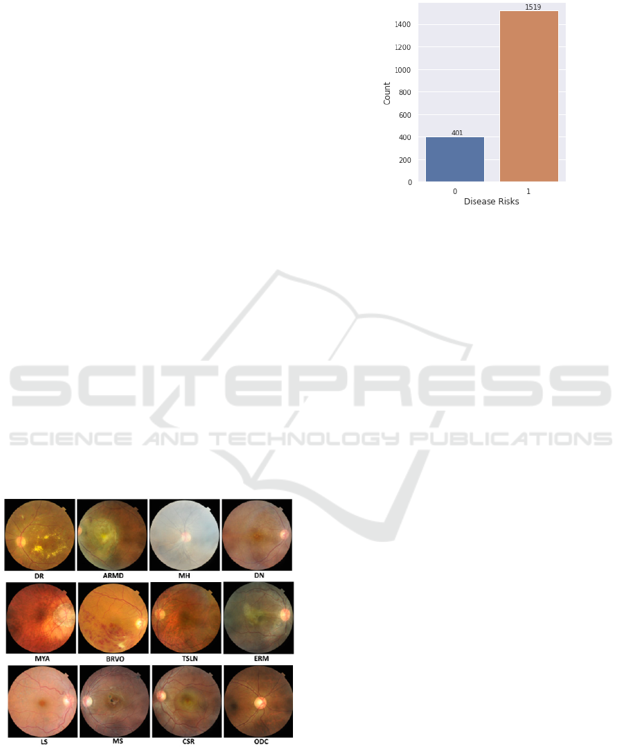
Several diseases/abnormalities that are included
in the dataset: diabetic retinopathy (DR), age-related
macular degeneration (ARMD), media haze (MH),
drusen (DN), myopia (MYA), branch retinal vein
occlusion (BRVO), tessellation (TSLN), epiretinal
membrane (ERM), laser scar (LS), macular scar
(MS), central serous retinopathy (CSR), optic disc
cupping (ODC), and many more total 42 types of eye
abnormalities. Visualization of these disease is shown
Figure 1.
Figure 1: Fundus retina images for several eye diseases
The data distribution between the normal eyes and
eyes with abnormalities is presented in the diagram
below.
Figure 2: Distribution between normal and abnormal eyes.
Around 20.9% (401 images) are normal eyes and
the rest of it are from eyes with several diseases.
2.2 Method
The usage of deep learning in computer vision
problem especially in biomedical imaging analysis
become more compelling (Liu, et al., 2018),
(Tajbakhsh, Shin, Gurudu, & Hurst, 2016) (Roy, et
al., 2020). In this paper, we report a deep-network
based model to develop a system for detecting eye
abnormality automatically. The model was built on
the top of some prominent pretrained convolutional
neural network (CNN) e.g., VGG-16 and Inception-
v3. These networks were trained by using ImageNet
dataset of over 14 million images belonging to 1000
classes. Therefore, it’s very suitable for common
computer vision problem. However, for fundus retina
images, we deployed the network in a transfer
learning fashion and without or with image-
augmentation to improve the generalization of the
model in detecting eyes abnormality.
The basic structure of VGG16 model (Simonyan
& Zisserman, 2015) is depicted in Figure 3. This
model consists of 16 layers, with 13 of convolutional
layers and 3 fully connected (FC) layers.
Eye Abnormality Automatic Detection using Deep Learning based Model
371
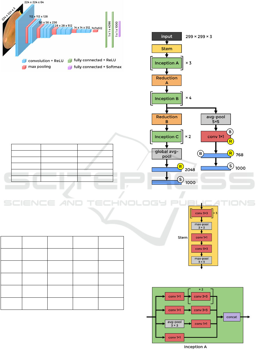
Figure 3: The basic structure of VGG16 model.
This fully connected layers parts of the model was
adjusted for the purpose of this research. The
complete configuration of the model, without and
with image-augmentation process is reported in Table
1 and Table 2 respectively.
Table 1: The configuration of the fully-connected layers in
VGG16, without image-augmentation.
Layers Model A1 Model A2
FC 1 1024, ReLU 1024, ReLU
Dro
p
out n.a. 0.5
FC 2 512, ReLU 512, ReLU
Drop out 0.5 0.5
Output 1, sigmoi
d
1, sigmoi
d
We deployed Rectifier Linear Unit (ReLU) as the
activation function in FC1 and FC2, and sigmoid
activation function at the output, for binary
classification system.
Table 2: The configuration of the fully-connected layers in
VGG16, with image-augmentation.
Layers Model
B1
Model
B2
Model
B3
Model
B4
FC 1 512,
ReLU
512,
ReLU
512,
ReLU
512,
ReLU
Drop
out
0.3 n.a. 0.3 n.a.
FC 2 512,
ReLU
512,
ReLU
512,
ReLU
512,
ReLU
Drop
out
n.a. 0.3 0.3 n.a.
Output 1,
sigmoi
d
1,
sigmoi
d
1,
sigmoi
d
1,
sigmoi
d
To avoid overfitting during the training, drop-out
technique was used as the regularization for the deep
learning model.
The basic structure of Inception-v3 network
(Szegedy, Vanhoucke, Ioffe, Shlens, & Wojna, 2016)
is illustrated in Figure 4. This model contains 42
layers. The detail structure in Stem, Inception A,
Reduction A, Inception B, Reduction B, and
Inception C blocks are depicted in Figure 5-10. This
model employs the Inception network (Szegedy, et
al., 2015).
Figure 4: The basic structure of Inception-v3 model.
Figure 5: The Stem block in Inception-v3 model.
Figure 6: The Inception A block in Inception-v3 model.
ICE-TES 2021 - International Conference on Emerging Issues in Technology, Engineering, and Science
372
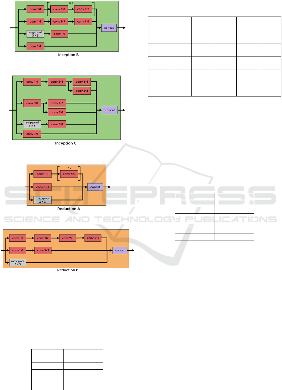
Figure 7: The Inception B block in Inception-v3 model.
Figure 8: The Inception C block in Inception-v3 model.
Figure 9: The Reduction A block in Inception-v3 model.
Figure 10: The Reduction B block in Inception-v3 model.
The fully connected layers parts of this model was
modified in order to be applicable in our purpose. The
complete configuration of the model, without and
with image-augmentation process is described in
Table 3 and Table 4, respectively.
Table 3: The configuration of the fully-connected layers in
Inception-v3, without image-augmentation.
La
y
ers Model C1
FC 1 4096, ReLU
Drop out 0.5
FC 2 512, ReLU
Drop out 0.3
Out
p
ut 1, si
g
moi
d
Table 4: The configuration of the fully-connected layers in
Inception-v3, with image-augmentation.
Layers Model
D1
Model
D2
Model
D3
Model
D4
Model
D5
FC 1 512,
ReLU
512,
ReLU
512,
ReLU
512,
ReLU
1024,
ReLU
Drop
out
n.a. n.a. 0.3 0.3. n.a.
FC 2 512,
ReLU
512,
ReLU
512,
ReLU
512,
ReLU
512,
ReLU
Drop
out
n.a. 0.3 n.a. 0.3 0.3
Output 1,
sigmoi
d
1,
sigmoi
d
1,
sigmoi
d
1,
sigmoi
d
1,
sigmoi
d
From the total 1920 fundus images, is split into
1305 for training images, 231 images for validation
data, and 384 for testing data. Stratified sampling
method is used for splitting the images, on order to
maintain the similar distribution for each class
(normal and abnormal eyes).
In order to improve the ability of the model to
preserve the generalization principles, we applied
data augmentation process. The parameter for this
augmentation is described in Table 5.
Table 5: Parameters for image-augmentation process.
Paramete
r
Value
rotation angle 30
width shift 0.2
height shift
range
0.2
shear range 0.1
horizontal fli
p
True
2.3 Evaluation Metrics
The performance of the model is measured in terms
of the ability of the model to correctly detect the eyes
with abnormality and the normal eyes as true positive
(TP) and true negative (TN) respectively. And
misjudge the normal eye to be abnormal as well as
abnormal eye classified as normal as false positive
(FP) and false negative (FN) respectively.
The metrics for evaluation are precision (Pre),
recall (Re), accuracy (Acc), and F1 score.
Pre = TP / (TP + FP) (1
)
Re = TP / (TP + FN) (2
)
Acc = (TP + TN) / (TP + TN + FP + FN) (3
)
F1-score = 2 * Pre * Re / (Pre + Re) (4
)
And another metrics that are prevalent in binary
classification is area under receiver operating curve
(AUROC) or sometimes called as AUC for
Eye Abnormality Automatic Detection using Deep Learning based Model
373
simplicity. That is the area under the curve between
false positive rate and true positive rate.
3 RESULTS AND DISCUSSION
The result for each model based on VGG16 structure
is reported in Table 6. It can be seen in the table that
model A1 outperformed all other models based on
VGG16 network in every evaluation metric.
Table 6: Model performance based on VGG16 network.
Model Acc Pre Re F1 AUC
A1 0.87 0.86 0.87 0.86 0.87
A2 0.85 0.84 0.85 0.84 0.84
B1 0.86 0.85 0.86 0.85 0.84
B2 0.84 0.83 0.84 0.82 0.83
B3 0.83 0.81 0.83 0.81 0.83
B4 0.84 0.83 0.84 0.83 0.84
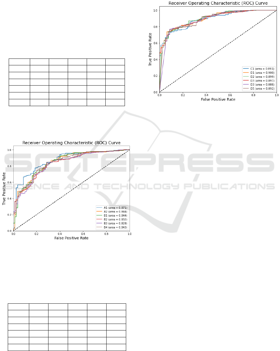
The comparation of ROC curve for each model is
presented in Figure 11. Figure 11 shows that the AUC
of model A1 is more superior than the other models.
Figure 11: The comparation of ROC curve result for each
model based on VGG16 structure.
Table 7 presents all metrics for each model based
on Inception-v3 structure.
Table 7: Model performance based on Inception-v3
network.
Model Acc Pre Re F1 AUC
C1 0.85 0.84 0.85 0.84 0.89
D1 0.86 0.85 0.86 0.85 0.90
D2 0.85 0.85 0.85 0.85 0.90
D3 0.86 0.85 0.86 0.85 0.90
D4 0.86 0.85 0.86 0.85 0.89
D5 0.83 0.84 0.83 0.84 0.89
In Table 7, model D1 and D3 shows a same result.
Both surpass the other models in every metric.
The ROC curve comparation between all models
based on Inception-v3 is shown in Figure 12.
Figure 12: The Reduction B block in Inception-v3 model.
Figure 12 shows that the AUC of each model is
different not too significant. Therefore, it can be said,
the variation of fully connected layers do not affect
the results essentially.
VGG16 structure is very simple compared to
Inception-v3. The number of neurons in FC layers
layer should be able to accommodate the features as
the output of the convolutional layer. Therefore, the
sudden change in numbers will affect the
performance. This causes the model A1 outperforms
more than the other model. The image augmentation
process seems not to help very much to improve the
performance.
On the other hand, Inception-v3 structure is
already complex. The variation of neuron numbers in
FC layers would not affect significantly. As it is seen
in Figure 12, all models have quite similar ROC.
However, the image augmentation gives more impact
to the performance.
To the best of our knowledge, since these datasets
were launched in April 2021, we haven’t found yet
any reported publication that used the same dataset in
their model. Therefore, at this current moment we
couldn’t provide any comparison from others’ model
that are trained with the same dataset.
4 CONCLUSIONS
From the experiment it can be concluded that VGG16
based model is not sensitive in image augmentation
but to the fully connected layers. On the other hand,
Inception-v3 based model is more impacted by image
ICE-TES 2021 - International Conference on Emerging Issues in Technology, Engineering, and Science
374

augmentation. However, by analysing the ROC
curve, both structures are still promising in
development of eye abnormality detection in further
research.
REFERENCES
Liu, J., Pan, Y., Li, M., Chen, Z., Tang, L., Lu, C., & Wang,
J. (2018, Mar). Applications of deep learning to MRI
images: a survey. Big Data Mining and Analytics, 1(1).
Pachade, S., Porwal, P., Thulkar, D., Kokare, M.,
Deshmukh, G., Sahasrabuddhe, V., . . . Mériaudeau, F.
(2021). Retinal fundus multi-disease image dataset
(RFMiD): a dataset for multi-disease detection
research. Data, 6(2).
doi:https://doi.org/10.3390/data6020014
Qummar, S., Khan, F. G., Shah, S., Khan, A.,
Shamshirband, S., Rehman, Z. U., . . . Jadoon, W.
(2019). A deep learning ensemble approach for diabetic
retinopathy detection. IEEE Access, 7, 150530-150539.
Roy, S., Menapace, W., Oei, S., Luijten, B., Fini, E., Saltori,
C., . . . Demi, L. (2020, Agt). Deep learning for
classification and localization of COVID-19 markers in
point-of-care lung ultrasound. IEEE Trans. on Medical
Imaging, 39(8).
Sarki, R., Ahmed, K., Wang, H., & Zhang, Y. (2020).
Automatic detection of diabetic eye disease through
deep learning using fundus images: a survey. IEEE
Access, 151133 - 151149.
Simonyan, K., & Zisserman, A. (2015). Very deep
convolutional networks for large-scale image
recognition. International Conference on Learning
Representations (ICLR). San Diego.
Soomro, T. A., Afifi, A. J., Zheng, L., Soomro, S., Gao, J.,
Hellwich, O., & Paul, M. (2019). Deep learning models
for retinal blood vessels segmentation: a review. IEEE
Access, 71696 - 71717.
Szegedy, C., Liu, W., Jia, Y., Sermanet, P., Reed, S.,
Anguelov, D., . . . Rabinovich, A. (2015). Going deeper
with convolutions. IEEE Conference on Computer
Vision and Pattern Recognition (CVPR).
Szegedy, C., Vanhoucke, V., Ioffe, S., Shlens, J., & Wojna,
Z. (2016). Rethinking the inception architecture for
computer vision. IEEE Conference on Computer Vision
and Pattern Recognition (CVPR), (pp. 2818-2826).
Tajbakhsh, N., Shin, J. Y., Gurudu, S. R., & Hurst, R. T.
(2016, May). Convolutional neural networks for
medical image analysis: full training or fine tuning.
IEEE Trans. on Medical Imaging, 35(5).
WHO. (2019). World report on vision. World Health
Organization.
Eye Abnormality Automatic Detection using Deep Learning based Model
375
