
Comparison of Two Dental Age Estimation Methods:
The London Atlas and the Schour & Massler Atlas
in 3-23 Years Old Indonesian
Aprianisa Obsidiany Daisy Tarigan
1
, Hendra Polii
2
and Rosalina Intan Saputri
2a
1
Undergraduate Program, Faculty of Dentistry, Maranatha Christian University, Jalan Surya Sumantri 65 Sukajadi,
Kota Bandung, Indonesia 40164
2
Faculty of Dentistry, Maranatha Christian University, Jalan Surya Sumantri 65 Sukajadi, Kota Bandung, 40164, Indonesia
Keywords: Dental, Age Estimation, Schour and Massler, London Atlas, Forensic Odontology.
Abstract: Age estimation is an important process in forensic identification, especially when there is insufficient
antemortem information. Tooth is one of the strong variables which could be used in estimating the age of
living or deceases. Non-invasive age estimation methods, including dental radiographs, have the advantage
of uncomplicated application without damaging the oral and surrounding tissues. The aim of this research
was to compare two radiographic dental age estimation methods, the London Atlas by Al Qahtani and the
Schour & Massler Atlas in 3-23 years old Indonesian population. Two hundred and fifty-three panoramic
radiographs from 156 females and 97 males with age ranged between 3-23 years old were retrospectively
collected from a Dental Hospital. Age estimation was performed on the radiographs using the London Atlas
and the Schour & Massler Atlas. Mann-Whitney U test was used to compare the chronological age and
estimated age from two methods. There was an insignificant difference between estimated age by both Atlases
(p> 0.05). Furthermore, there was also insignificant differences between estimated age of both Atlases and
the chronological age (p> 0.05). The performance of London Atlas and Schour & Massler Atlas were
equivalent in estimating 3-23 years old Indonesian in present study population.
1 INTRODUCTION
Age is an important identity. It is a basic human right
for an individual to know their dates of birth, hence
their ages (Cameriere et al., 2007). Age is required for
civil administration, such as school, registration,
work application, and retirement. Individual age also
plays a significant role in jurisdiction, such as cases
of employment, age falsification, marriage, athletes,
child custody, and immigration (Panchbhai, 2011).
However, there are possibility that the chronological
age of an individual is unknown because their
documented identities is not available or there is an
indication of falsification which required
examination for age estimation (Putri et al., 2015).
The chronological age can be estimated by
determining the physiological development of certain
organs (Adams et al., 2014). Teeth were being used
as a choice of age indicator because they are the
a
https://orcid.org/0000-0003-0811-6270
strongest parts of the human body, and can withstand
external influences such as high temperatures,
explosions, and other extreme conditions. Human
teeth also tend to be stable and barely affected by
other environmental factors such as socioeconomic
status, nutrition, diet, and even endocrine factors.
Therefore, teeth can be useful in the post-mortem
examination (Kaur et al., 2013; Adams et al., 2014).
Dental age can be determined by the development
of human teeth which occurs nearly one third of the
human life period. Radiograph was often used for
dental age estimation because of its non-invasive
method and does not involve tooth extraction (Putri et
al., 2015). One of the radiography techniques is atlas
method which consist of diagrammatic pictures of
developing teeth’s structure with related eruption
patterns (Senn, 2013). London Atlas by Al Qahtani
and Schour & Massler Atlas were the most popular
Atlas for dental age estimation which had not
122
Tarigan, A., Polii, H. and Saputri, R.
Comparison of Two Dental Age Estimation Methods: The London Atlas and the Schour Massler Atlas in 3-23 Years Old Indonesian.
DOI: 10.5220/0010745600003113
In Proceedings of the 1st International Conference on Emerging Issues in Technology, Engineering and Science (ICE-TES 2021), pages 122-125
ISBN: 978-989-758-601-9
Copyright
c
2022 by SCITEPRESS – Science and Technology Publications, Lda. All rights reserved

compared in Indonesia population. London Atlas by
Al Qahtani has 31 diagrams that describe the
development of teeth from 30 weeks of pregnancy to
the age of 23.5 years. Eight of these diagrams only
describe the development of third molars starting at
the age of 16.5 years. Whereas in Schour & Massler
Atlas, it describes 21 chronological steps of tooth
development from 5 months of pregnancy to 21 years.
Besides that, individual studies from these atlases in
Indonesian population only had small number of
samples, which was around 20 to 100 subjects. The
estimated age scope from both atlases is relatively
similar, which is around the prenatal age to 21 years,
which would supplement the comparison results of
both methods in different population (Senn, 2013;
Ciapparelli, 1992; Schour et al., 1941). Therefore,
this research aimed to compare the London Atlas by
Al Qahtani and the Schour & Massler Atlas in
Indonesian population.
2 METHODS (AND MATERIALS)
The panoramic radiographs of 253 individuals (males
= 97, females = 156) aged 3-22.99 years old were
retrospectively and anonymously collected from
Maranatha Dental Hospital. The selected radiographs
should meet the following inclusion criteria: data for
male or female patients aged 3-22.9 years and right
upper and lower jaw teeth were clearly visible on
panoramic radiographs. While the exclusion criteria
were missing tooth / no tooth seed and pathological
structure of tooth and surrounding tissue in the region
studied in the panoramic view, currently undergoing
orthodontic treatment, having systemic complication,
such as poor nutrition or congenital diseases.
Age estimation was performed on the radiographs
using the London Atlas by Al Qahtani and the Schour
& Massler Atlas (Al Qahtani et al., 2014; Schour et
al., 1941). The inter-rather reliability between two
examiners was 76%. Mann-Whitney U test was used
to compare the chronological age and estimated age
from two methods. Ethical clearance was granted by
Faculty of Medicine, Maranatha Christian University
Research Ethic Committee (006/KEP/II/2021).
3 RESULTS AND DISCUSSION
3.1 Study Population
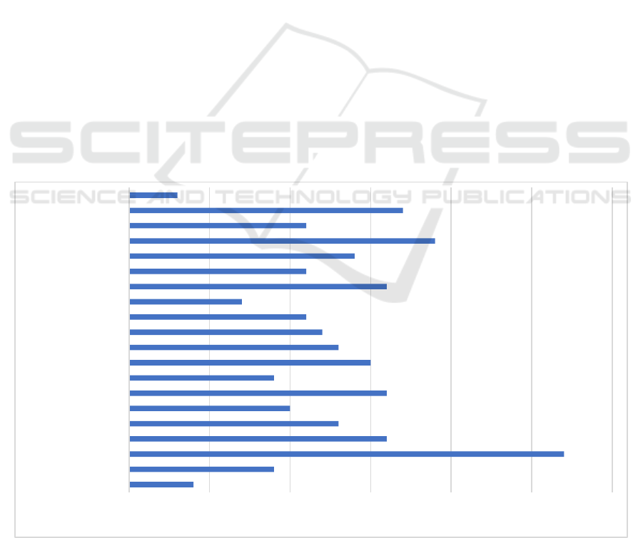
The percentage of sex in this study displayed in Table
I. From 253 respondents the highest chronological
age (11%) was between 5.0 and 5.99 years, then 8%
was between 19.0 to 19.99 years, and at least 1% of
respondents have the chronological age between 22.0
and 22.9 years (Figure 1).
Table 1: Number and Percentage of Study Population by
Sex.
Sex Amount (n) Percentage
Male 97 38%
Female 156 62%
Total 253 100%
3.2 Comparison between Chronological
Age and Two Dental Estimation
Methods
Analysis by Kolmogorov-Smirnov test showed that
chronological age, age estimation of London Atlas by
Al Qahtani method, and age estimation of the Schour
& Massler method were not normally distributed
(p<0.01). Therefore, Mann-Whitney U test was
performed. There was an insignificant difference
between the age estimation results of London Atlas
by Al Qahtani method and the age of the Schour &
Massler method. Moreover, there was also an
insignificant different between two methods with
chronological age (Table 3).
Table 2: Statistical p-value between Chronological Age, Al-
Qahtani Atlas Method, and Schour & Massler Method.
London Atlas by
Al Qahtani
Method
Schour and
Massler Method
Chronological
Age
0.982 0.575
London Atlas
by Al Qahtani
Method
0.574
3.3 Discussion
There are different methods in estimating the age in
forensic dentistry, including the London Atlas by Al
Qahtani method and the Atlas Schour & Massler
method. London Atlas by Al Qahtani method and the
Schour & Massler method are often used because
they are uncomplicated, costs effective, and mostly
not invasive, which do not damage dental tissue
(Alshihri et al., 2015). However, the Atlas methods
also have limitations, such as the inability to represent
all cases and the variability of development in tooth
Comparison of Two Dental Age Estimation Methods: The London Atlas and the Schour Massler Atlas in 3-23 Years Old Indonesian
123
formation time and tooth eruption stages and the
inability to differentiate between sexes (Alshihri et
al., 2015). In estimating dental age, crown and root
development, and eruption status in the specimens,
the diagram of London Atlas by Al Qahtani method
and the Schour & Massler Atlas method was adjusted.
This study was conducted to compare the accuracy in
estimating dental age with the London Atlas by Al
Qahtani method and Schour & Massler Atlas method
on panoramic radiographs (Alshihri et al., 2015).
A study by Al Qahtani et al. (2014) has compared
three methods in estimating age between the Schour
& Massler, Ubelaker and London Atlas (Al Qahtani)
methods in Portugal, Netherlands, United States,
Canada and France, which resulted that the Al
Qahtani Atlas method is the most accurate method
compared to other methods. The differences between
previous and this study was Al Qahtani et al. study
did not use the third molars for age estimation (Al
Qahtani et al., 2014), which included in this study. A
study conducted by Pavlovic et al (2017) who
examined the Al Qahtani Atlas method in 498 women
and 238 men in Portugal and found no significant
difference between chronological age and age
estimation using the London Atlas (Al Qahtani) in the
female sample only (Pavlović et al.,2017). The
drawback of this study is that it does not differentiate
between sex and the results only showed that there is
an insignificant difference in mixed sexes.
The differences in the results of previous studies
were also possible because of different study
population of each country. Racial differences could
lead to differences in the timing and sequence of
eruption of permanent teeth (Indriati E, 2001). Study
in Indonesia population had conducted by Fitri et al
(2016), which studied the age estimation using the Al
Qahtani method and identified 94 samples, and there
were 66 samples (70.21%) which showed similar
result between Al Qahtani age estimation method and
the chronological age (Rusydiana et al.,2016). It was
in agreement with present study result which
demonstrated that the Al Qahtani method has
insignificant differences with chronological age.
Another study which in the agreement with current
study was study by Nurfitria et al (2018) which used
the Atlas Al-Qahtani method, as a method of
estimating age and found that the difference in dental
age and chronological age was very small and could
be used in the population in Indonesia (Nurfitria et
al.,2018).
The exclusion criteria of this study were patients
should not have poor nutrition and a history of
systemic and congenital diseases, which in-line with
the study of Eshitha et al (2014) who estimated the
age of 25 children aged 5-16 years in good health in
a population in India using the Schour & Massler
method. From the study was found that the intraclass
correlation coefficient was 0.938 which indicates a
Figure 1: Number of study population per age category.
0 5 10 15 20 25 30
3.0 - 3.99 years
5.0 - 5.99 years
7.0 - 7.99 years
9.0 - 9.99 years
11.0 - 11.99 years
13.0 - 13.99 years
15.0 - 15.99 years
17.0 - 17.99 years
19.0 - 19.99 years
21.0 - 21.99 years
ICE-TES 2021 - International Conference on Emerging Issues in Technology, Engineering, and Science
124

high correspondence between chronological age and
dental age according to Schour & Massler, so it could
be concluded that the Atlas Schour and Massler
method can be applied to mentioned population
(Eshitha E et al., 2014). In addition, study by Trelia et
al. (2019) estimated age using the Atlas Schour &
Massler and Demirjian method with sample of 46
patients with the age range of 10-16 years at the
RSGM University of North Sumatra and showed that
age estimation results were similar to the actual age,
so there was no significant differences between two
methods (Trelia et al., 2019). This study is in
agreement with Trelia et al. research that the Schour
& Massler method is proven to be able to estimate
dental age in Indonesia population.
Besides there is no sex distinction, the
disadvantages of current study were uneven
distribution of the age range and there was no specific
analysis for each gender. Therefore, future research
should aim for enlarging the research population with
even sample per age range, and perform sex-specific
analysis.
4 CONCLUSIONS
It could be concluded that the performance of London
Atlas and Schour & Massler Atlas were equivalent in
estimating present study population.
ACKNOWLEDGEMENTS
We would like to thank drg.Citra Esperanza for the
time and effort as the second observer, as well as
Danny Prasetyo Hartanto, S.Si who helped in data
processing and analyzation of this study.
REFERENCES
Adams, C., Carabott, R., Evans, S. (2014). Forensic
odontology: an essential guide. Oxford: Wiley
Blackwell.
Alshihri, A.M., Kruger, E., Tennant, M. (2015). Dental Age
Assessment of Western Saudi Children and Adolescent.
Saudi Dent J. 27(3), 131-136.
Al Qahtani, S.J., Hector, M.P., Liversidge, H.M. (2014).
Accuracy of dental age estimation charts: Schour and
Massler, Ubelaker and the London Atlas. Am. J. Phys.
Anthropol. 154(1),70–78.
Cameriere, R., Ferrante, L., Belcastro, M.G., Bonfiglioli,
B., Rastelli, E., Cingolani, M. (2007). Age Estimation
by Pulp/Tooth Ratio in Canines by Mesial and
Vestibular Peri-Apical X-Rays. J. Forensic. Sci. 52(5),
1151–1155.
Ciapparelli, L. (1992). The chronology of dental
development and age assessment. In: Clark DH, editor.
Practical Forensic Odontology. Oxford: Wright
Butterworth‑Heinemann Ltd.
Eshitha, E., Prasanna, K.R., Laxmikanth C., Prashanth S.,
Veena, K.M., Rachana V.P., Shahin K.A., Tashika K.,
Prathima S., Shaul Hameed. (2014). Dental Age
Estimation Using Schour and Massler Method in South
Indian Children. Scholars. SJAMS. 2(5c), 1669–1674.
Indriati, E. (2013). Permanent Tooth Eruption in Javanese
Children. B. I. Ked. 33(4), 237-248.
Kaur, J., Rai, B. (2013). Evidence-Based Forensic Dentisty.
Berlin: Springer.
Lemeshow. S., David, W.H.Jr. (1997) Besar Sampel dalam
Penelitian Kesehatan (Terjemahan). Yogyakarta:
Gadjahmada University Press.
Nurfitria, D.T., Soedarsono, N., Yuniastuti, M., Nehemia,
B. (2018) Comparison of TCI–Benindra formula, Al-
Qahtani, and Blenkin-Taylor methods for age
estimation in 16–21 year olds. IOP Science.
1073:022012.
Panchbhai, A. (2011). Dental radiographic indicators, a key
to age estimation. Dentomaxillofac Radiol. 40(4),199–
212.
Pavlović, S., Pereira, C.P., Santos, R.F. (2017). Age
estimation in Portuguese population: the application of
the London atlas of tooth development and eruption. J.
Forensic. Sci. 272, 97-103.
Peretz, B., Gotler, M., Kaffe, I. (2012). Common errors in
digital panoramic radiographs of patients with mixed
dentition and patients with permanent dentition. Int. J.
Dent. 584138.
Putri, A.S., Nehemia, B., Soedarsono, N. (2015). Prakiraan
usia individu melalui pemeriksaan gigi untuk
kepentingan forensik kedokteran gigi. Jurnal PDGI.
62(3),55-63.
Rusydiana, F., Oscandar, F., Sam, B. (2016). Identifikasi
usia berdasarkan metode Al Qahtani melalui radiograf
panoramik di RSGM FKG UNPAD. J. Ked. Gi. Unpad.
28(3),1-6.
Senn, D.R., Weems, R.A. (2013). Manual of forensic
odontology, 5th edition. Boca Raton: CRC Press Taylor
& Francis Group.
Smrithi, D. (2014). Coronal Pulp biomarker: A lesser
known age estimation modality. JIAOMR
.26(4),398-
404.
Schour, I., Massler, M. (1941). Development of human
dentition. J. Am. Dent. Assoc.20, 379‑427.
Trelia, B., Tiara, A. (2019). Estimasi Usia Menggunakan
Metode Schour-Massler Dibandingkan dengan Metode
Demirjian. DENTIKA. 22(1), 15-19.
Willems, G.A. (2001). Review of the most commonly used
dental age estimation techniques. J. Forensic
Odontostomatol. 19(1),9-17.
Comparison of Two Dental Age Estimation Methods: The London Atlas and the Schour Massler Atlas in 3-23 Years Old Indonesian
125
