
Retinal Blood Vessel Segmentation using
Convolutional Neural Networks
Arun Kumar Yadav
1a
, Arti Jain
2b
, Jorge Luis Morato Lara
3c
and Divakar Yadav
1d
1
Department of Computer Science & Engineering, NIT Hamirpur, Himachal Pradesh, India
2
Department of Computer Science & Engineering, Jaypee Institute of Information Technology, Noida, Uttar Pradesh, India
3
Department of Computer Science, Universidad Carlos III de Madrid, Leganes, Madrid, Spain
Keywords: Blood Vessel Segmentation, Convolutional Neural Networks, CLAHE, Diabetic Patients, Retinal Images.
Abstract: Human beings often become victims to numerous diseases. Among these, diabetes stands out for its
impairment of quality of life and even potential mortality. The diabetes needs to be properly taken care of,
otherwise failure to detect its presence within proper time duration leads to a loss of life. According to the
World Health Organization, the worldwide number of diabetic patients were 463 million during 2019 and is
expected to cross 700 million by the 2045
i
. In the past, a lot of research has been carried out for retinal blood
vessel segmentation for identification of Diabetic Retinopathy using various machine learning and deep
learning models. In this research work, Convolutional Neural Network (CNN) and CLAHE are applied to
tackle the problem of retinal blood vessel segmentation. Experimental evaluation shows that the proposed
method outperforms with 0.9806 accuracy, quite competitive with respect to the state-of-art.
1 INTRODUCTION
Over the time, as age passes by, the human beings are
entangled in the clutches of some familiar or
unknown diseases. A major part of treatment for any
disease involves an identification and detection of
that disease. Although there are many techniques that
are responsible for the disease detection, however, in
most of the cases by the time the disease is detected,
it is already at an acute stage, and ultimately it
becomes un-curable, leading to the adverse
circumstances. On the other hand, there are diseases
like Diabetes where the mentioned tests are not so
effective to diagnose its presence at an earlier stage.
As is stated by (Fong et al., 2004) the Diabetes is
one of the prime reasons for blindness among the age
group of 20-74 years. This means that the human
retinas become blotted to such an extent, so that the
person’s vision goes down. During diabetes, the
blood sugar level increases in the body to a very high
level. Over a period of time can damage the retinal
blood vessels and make them swell up. In some cases,
a
https://orcid.org/0000-0001-9774-7917
b
https://orcid.org/0000-0002-3764-8834
c
https://orcid.org/0000-0002-7530-9753
d
https://orcid.org/0000-0001-6051-479X
the change is even visible to the naked eye. Also,
possibility arises that it can block the blood from
passing through the retinas, thus making the person
blind. There are Type-1 and Type-2 kinds of diabetic
patients. In the Type-1, initially destroys beta cells
from the human body which causes in the stoppage of
the insulin production. In the Type-2, the human body
still produces insulin but it is unable to use it
effectively. There are Type-1 (3.6%) patients, and
Type-2 (1.6%) patients that are even blind (Dabelea
et al., 2014) by blotting the retinal blood vessels. The
retinal layer contains many nerves or photo-receptors
that respond to light, thus enabling the person to see.
These photo-receptors transmit optic nerve sensors
and are converted into visual images. Most people do
not care about eye conditions until it becomes too bad
to the extent that the person becomes blind. Retinal
disease is a visual disorder which ultimately even
leads to blurriness in the eyes. Researchers (Bell et al.
2014) have stated that 280 million people in the world
live with the visual impairments, 34 million among
them are blind and 246 million with low vision. In
292
Yadav, A., Jain, A., Morato Lara, J. and Yadav, D.
Retinal Blood Vessel Segmentation using Convolutional Neural Networks.
DOI: 10.5220/0010719500003064
In Proceedings of the 13th International Joint Conference on Knowledge Discovery, Knowledge Engineering and Knowledge Management (IC3K 2021) - Volume 1: KDIR, pages 292-298
ISBN: 978-989-758-533-3; ISSN: 2184-3228
Copyright
c
2021 by SCITEPRESS – Science and Technology Publications, Lda. All rights reserved

2019, the number of diabetic patients touches 463
million, and is expected to cross 700 million by the
2045
i
. The visual impairment can be prevented with
the early detection of the retinal detachment.
To work in this direction, here Convolutional
Neural Network (CNN) and CLAHE are applied to
dive into the retinal blood vessel segmentation. The
proposed method outperforms with 0.9806 accuracy
which is quite competitive with respect to the state-
of-art works. Thus, the research contributions are
stated here.
RC1: The optimized CLAHE + CNN model is
trained and tested on the publically available
DRIVE dataset and compared the result with
state-of art.
RC2: Performance evaluation is better than other
approaches over the DRIVE dataset.
Rest of the paper is organized as follows. Section 2
talks about related work, Section 3 discusses
proposed work, Section 4 illustrates results &
analysis, and Section 5 concludes the paper.
2 RELATED WORK
Researchers (Chaudhuri et al., 1989) have used two
dimensional filters to analyze the blood vessel
segmentation. A feature-removal operator based on
visual and real-estate properties is notified. The gray
cross section of artery is measured by Gaussian curve.
The signal acquisition determines the successive
sequence of blood vessels in the images. There are 12
varied templates that are created to search for ship-
parts in the available directions.
(Marın et al., 2010) have introduced approach for
finding blood vessels in the digital images of eyes.
Their method uses Neural Network (NN) and
incorporates 7-D vector with grey and secondary
features that are built into the pixel system
representation. It is tested on the publicly available
datasets- DRIVE and STARE. These datasets are
commonly used because they contain images of the
retina where the structure of blood vessels is
accurately marked by experts.
(Fraz et al., 2012) have learnt the blood vessel
segmentation problem using a hybrid method which
uses the concept of acquisition of vessel centerline for
the first order from the Gaussian. Also, observed
retrieval vessels while connecting the center lines
with a map of the position itself. They are able to get
97% accuracy. This paper reports the default way to
separate blood vessels in the images above. A unique
combination of ship acquisition techniques and slow-
flight aircraft are introduced to remove the artery tree
from the images. Mathematics of morphology is
already a good way to measure blood vessels in the
retina.
(Odstrcilik et al., 2013) have identified the
development of a two-way filter that is consistent
with Gaussian’s role as a character. They have
explored the path to STARE, DRIVE and HRF’s
decision-making process. They have designed five 2-
D filters according to standard container category
profiles and watched five shipwrecks from the
thinnest to the too big. Each image is processed to
match one in the five characters. Because the same
filters in each image, the process is slower. The
method effectiveness lies upon how accurate team of
ships is divided into the five parts.
(Sreejini and Govindan, 2015) have applied
improvised multi-scale matched filter for the blood
vessel segmentation using Particle Swarm
Optimization (PSO) (Jain et al., 2021). The same
filtering concept is widely used in the area of retinal
detachment. Multi-scale standard filters have higher
performance than the single-scale filters. The method
uses advanced audio compression features of
international filters.
(Singh and Srivastava, 2016) have utilized second
hand derivative of Gaussian as a filter for retinal
blood vessels segmentation. The method
corresponding to the measured filter are simple and
effective. However, the corresponding filter test
detects both vessels and non-vessel terminals which
gives false ships i.e., non-ship acquisitions. To
overcome the problem of finding non-ship edges, an
extension of the matching filter based on the Second
Gaussian discovery (SDOG-MF) is considered useful
for the separation of small and narrow retinal blood
vessels.
(Yao et al., 2016) have discussed CNN based
algorithm. Each pixel and its image neighbors are
tested by the CNN. The effects of the first
classification of fundus images are refined by two
phases of binarization and morphological
performance respectively. The algorithm is tested
over the DRIVE database. The data sensitivity is
0.7731, which is very close to that of the text
annotation.
(Sun et al., 2017) have illustrated four CNN
architectures (AlexNet, GoogLeNet, VGG-16, and
ResNet-50) from ImageNet image classification task
to Retinal fundus images quality classification. The
top two networks are picked out and then jointly fine-
tune them. The accuracy for different methods are
found to be as AlexNet (96.53%), GoogLeNet
(97.04%), VGG-16 (96.87%), ResNet-50 (96.20%),
Retinal Blood Vessel Segmentation using Convolutional Neural Networks
293
Joint CNN with GoogLeNet (97.00%), and Joint
CNN with VGG-16 (97.12%) repectively.
(Jebaseeli et al., 2019) have pre-processed retinal
blood vessels dataset through CLAHE- Contrast
Limited Adaptive Histogram Equalization, feature
vectors through TPCNN- Tandem Pulse Coupled
Neural Network, classification and extraction through
DLBSVM- Deep Learning Based Support Vector
Machine. It gives improved segmentation results of
sensitivity, specificity and accuracy as 0.7445,
0.9940, and 0.9897 accuracy respectively. However,
certain issues are quite unclear such as how deep
learning learns, whether neural net relays upon
certain images with the datasets, are they non-
transferable to all retinal photography. These are
limitations of accuracy for the stated technique.
(Wang et al., 2019) have illustrated novel
separation framework and a strong cascade separation
of the retinal vessel. Unlike other non-linear
partitions that require a pre-defined non-linear kernel
or repeated training, a separate cascade editing
framework is trained through the process of a single
pass transfer. Therefore, degree of non-compliance
with the separate line is not defined in advance, but is
determined by the complexity of data structures.
(Saroj et al., 2020) have worked upon a matched
filter approach with kernel as a Fréchet probability
distribution function. It uses principal component
analysis for the color conversion and CLAHE during
pre-processing. While during post-processing, it uses
entropy based optimal thresholding, and filtering by
the length. The specificity, sensitivity and accuracy
for the STARE- 0.9724, 0.7278, 0.9509 and for the
DRIVE- 0.9761, 0.7307, 0.9544 respectively.
(Escorcia-Gutierrez et al., 2021) have worked
upon Portfolio Theory of Markowitz for diabetic
retinopathy via optic disc. It produces an innovative
color fusion model which is applied over DRIVE,
Messidor, HRF, and in–house dataset (Hospital
Universitari Sant Joan de Reus, Spain). It gives an
accuracy and overlap as 0.9 and 0.80 respectively
with minimal execution time of 0.05seconds.
(Gegundez-Arias et al., 2021) have presented a
robust CNN based on UNet vessel
segmentation method in fundus images. It combines
residual blocks and batch normalization in the up-
down scaling. From actual images, patches are
extracted and trained with loss function while looking
at every pixel distance towards vascular tree, while
output produces binarized probability map of pixels.
The method is experimented over DRIVE, STARE
and CHASE_Db1.
(Pal et al., 2021) have proposed the Twin network
retinal scan system which extracts feature maps of
both query and database samples from the deep CNN.
The approach exploits deep features without the
resource, space and computation exhaustive network
training phase. The different variations of retrieval
performances are evaluated- AMD-Normal, DME-
Normal, AMD-DME, AMD-DME-Normal. The
system retrieves similar scans from a dataset of
abnormal and normal retinal scans with precision
(0.7571).
(Rajagopalan et al., 2021) have worked upon the
CNN model for the detection of retinal disorders. The
model classifies three types of retinal disorders-
Choroidal neovascularization (CNV), Drusen
macular degeneration (DMD) and Diabetic macular
edema (DME). It provides an accuracy (0.9701),
sensitivity (0.9343), and specificity (0.9807)
respectively.
3 PROPOSED WORK
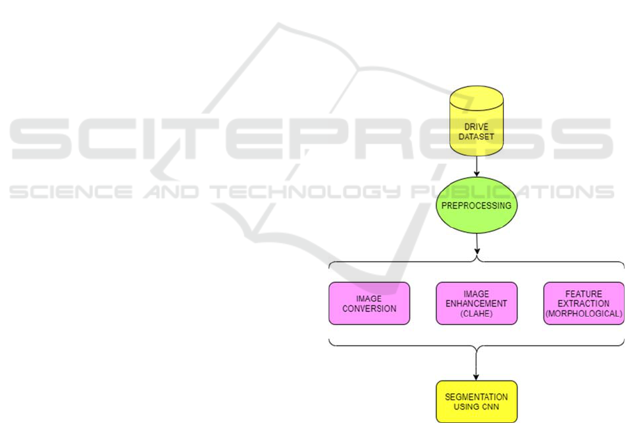
The architecture of the proposed Retinal Blood
Vessel Segmentation method is described in Figure 1.
Figure 1: Basic architecture of proposed work.
The DRIVE dataset is fed into the system which
undergoes Pre-processing, namely- Image
Conversion, Image Enhancement (CLAHE) and
feature extraction (morphological). The image
conversion, converts the dataset images from colour
to grey format, applies the equalizer CLAHE for
converting the image to contrast form i.e.
KDIR 2021 - 13th International Conference on Knowledge Discovery and Information Retrieval
294
Enhancement. The feature extraction phase
undergoes morphological feature, which is followed
by Segmentation using Convolutional Neural
Networks.
3.1 Dataset Discussion
The proposed Retinal Blood Vessel Segmentation
model is executed over the chosen DRIVE dataset
(Staal et al., 2004). The DRIVE dataset contains data
from the diabetic program of Netherlands which is
collected from 400 people within age of 25-90 years.
This wide age group is considered to avoid model
overfitting and is a potential age range for the diabetic
patients. This dataset is also considered by several
researchers (Yao et al., 2016; Albargathe et al., 2021;
Escorcia-Gutierrez et al., 2021) and comprises of 20
training and 20 test images respectively.
3.2 Pre-processing
Once the DRIVE dataset is extracted, the system
undergoes pre-processing phase as follows.
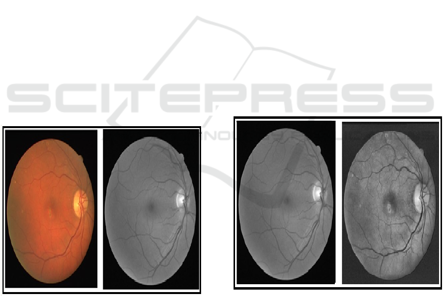
3.2.1 Colour to Grey Conversion
The grey scaling process converts an image from
different colour spaces into the shades of grey, as is
seen in Figure 2.
(a) Colored Image (b) Grey Scale Image
Figure 2: Color to grey scale conversion.
The coloured spaces are such as RGB, CMYK,
and HSV etc. that are converted into the shades of
grey- i.e., in between complete black and complete
white. The python programming based OpenCV
library is used to perform the grey scale conversion.
The primary reason of doing this conversion is to
provide accurate results and faster processing, as
there are only two values- 0 and 1 in the resultant
colour histogram.
3.2.2 Applying Clahe
CLAHE is an adjustable extension of Histogram
Equalization which is followed by the threshold. It
helps in preserving the local contrast characteristics
of an image dynamically.
In the initial steps, stress is on the local contrast
rather than global contrast of the image. The global
histogram balance doesn’t stress on local contrast
enhancements, and subsequently have minor contrast
differences which is extremely common in NPDR
imagery and is totally missed if the quantity of pixels
falling in a specific dark shade is too little. To take
care of this issue, the proposed calculation is
characterized to work adaptively on the image that is
to be improved, not like normal standard histogram
adjustment. It improves the contrast upgrade on local
image data in a divide and overcome way,
subsequently effectively handles the global distortion
of the image.
All in all, the fundamental thought of the
calculation is to separate the image into various little,
non-covering context oriented areas which are called
Tiles (Figure 3).
(a) Original Image (b) Enhanced Image
Figure 3: Image enhancement using CLAHE.
3.2.3 Feature Extraction
In the feature extraction phase, the morphological
features of the images of an eye are fed into the
machine learning model to obtain the relevant results.
The encoder-decoder approach is used to convert an
Retinal Blood Vessel Segmentation using Convolutional Neural Networks
295
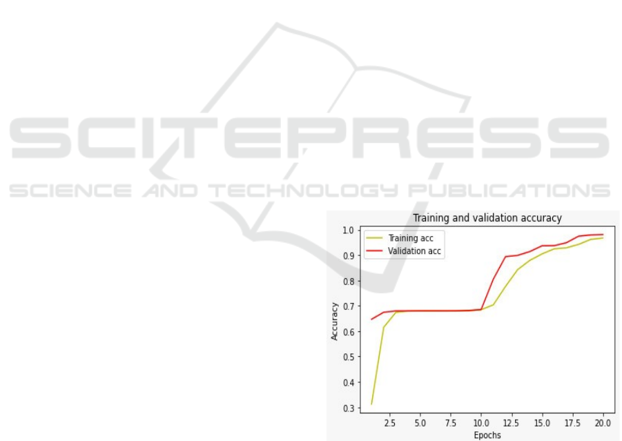
image data into embedding’s which is fed into the
Convolutional Neural Networks.
3.3 Segmentation
Identifying the sets of pixels which can go together is
the problem of image segmentation. Pixel-level
categorization is another name for this method. To put
it in another way, it comprises dividing images into
many segments or objects. One of the most important
applications in the computer vision area is image
segmentation (Moccia et al., 2018). It is used in a
variety of fields, including medicine and intelligent
transportation.
Image segmentation has advanced dramatically
with the deep neural networks (Siddiqui et al., 2020).
Among the deep learning concepts, CNN which is
also known as CovNet is useful for image detection
and recognition (Noh et al., 2019). The CNN inputs
image data, passes it through different layers-
convolutional, pooling, flattened and fully connected
layers and classifies data into appropriate category.
3.3.1 Convolutional Layer
It takes input as an image, extracts features and
preserves relationship among image pixels. It stores
the feature of an image as different sizes of matrix
then reduces the size of the matrix by taking filters of
varied sizes. The obtained reduced matrix is called as
Feature Map.
3.3.2 Non-Linear Activation Function
It uses non-linear activation function i.e., ReLU-
Rectified Linear Unit, mathematical function which
is given in equation (1).
(1)
3.3.3 Pooling Layer
It reduces the dimension of features map while
retaining important features of the image. The
pooling layers contains several variations like max
pooling, average pooling, and min pooling. In this
work, max pooling is used which contains maximum
values of the feature maps.
3.3.4 Flattening Layer
It is used to flatten the pooled matrix to its
corresponding vector which is serving as an input for
fully connected layer.
3.3.5 Fully Connected Layer
It contains several layers with many nodes. It
contains an input layer (first layer), output layer (last
layer), and hidden layers (other layers). In the fully
connection, each node is connected to every node of
its next layer. In the last layer, sigmoid function
categorizes the image to its most accurate category
while within rest of the layers, ReLU function is
applied.
4 RESULTS & ANALYSIS
The results of the proposed method are computed
mainly in terms of the model accuracy.
4.1 Model Accuracy
On training over the DRIVE dataset, using encoder-
decoder of CNN, the model gives an accuracy of
0.9806.
4.2 Training and Validation
The accuracy curve and loss curve are illustrated here.
4.2.1 Accuracy Curve
The training and validation accuracy curve of the
method is described in Figure 4.
Figure 4: Training and validation accuracy curve.
4.2.2 Loss Curve
The training and validation loss curve of the
proposed method is described in Figure 5.
outputnegativeo
outputpositive
xxf
,
,1
),0max()(
KDIR 2021 - 13th International Conference on Knowledge Discovery and Information Retrieval
296
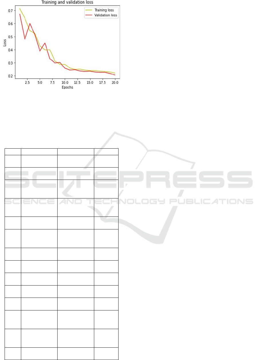
Figure 5: Training and validation loss curve.
4.3 State-of-Art Comparison
The proposed work is compared with respect to the
state-of-art by the other researchers (Table 1).
Table 1: Comparative analysis over DRIVE dataset.
S.No. References Method Accuracy
1 Marın et al.,
2010
Neural
Networks
0.9452
2
Fraz et al.,
2012
Hybrid
Approach
0.9430
3 Sreejini and
Govindan,
2015
Multi-scale
Matched Filter
0.9633
4 Singh and
Srivastava,
2016
SDOG-MF 0.9645
5
Yao et al.,
2016
CNN 0.9360
6
Soomro et al.,
2018
Independent
Component
Analysis
0.9530
7
Soomro et al.,
2019
FCM + CNN 0.9590
8
Wang et al.,
2019
Cascade
Classification
0.9541
9
Jebaseeli et
al., 2019
Tandem Pulse
CNN
0.9897
10 Saroj et al.,
2020
Frechet PDF
0.9544
11 Albargathe et
al., 2021
H-Minima
0.9672
12 Escorcia-
Gutierrez et
al., 2021
Markowitz 0.9556
13 Gegundez-
Arias et al.,
2021
Modified UNet
0.9547
14 Proposed
Work
CLAHE +
CNN
0.9806
5 CONCLUSIONS
In this work, CLAHE + CNN is applied for the retinal
blood vessel segmentation of images over the DRIVE
dataset. The method undergoes pre-processing- grey
scale conversion and CLAHE, feature extraction
using morphological feature, segmentation, training
and prediction using CNN. The results are evaluated
in terms of the model accuracy as 0.9806 which is
quite competitive with respect to the state-of-art work
over the DRIVE. Because of the ease-to-use and good
performance, the proposed method accelerates the
diagnosis of Diabetic Retinopathy. In future, model
accuracy can be enhanced further using more deep
learning strategies.
REFERENCES
Albargathe, S. M. B. K., Kamberli, E., Kandemirli, F., and
Rahebi, J., (2021). Blood vessel segmentation and
extraction using h-minima method based on image
processing techniques. Multimedia Tools and
Applications, 80(2): 2565-2582.
Bell, J. A., Kivimaki, M., and Hamer, M. (2014).
Metabolically healthy obesity and risk of incident type
2 diabetes: A meta‐analysis of prospective cohort
studies. Obesity Reviews, 15(6): 504-515.
Chaudhuri, S., Chatterjee, S., Katz, N., Nelson, M., and
Goldbaum, M. (1989). Detection of blood vessels in
retinal images using two-dimensional matched filters.
IEEE Transactions on Medical Imaging, 8(3): 263-269.
doi: 10.1109/42.34715.
Dabelea, D., Mayer-Davis, E. J., Saydah, S., Imperatore,
G., Linder, B., Divers, B. J., Bell, R., Badaru, A.,
Talton, J. W., Crume, T., and Liese, A. D. (2014).
Prevalence of type 1 and type 2 diabetes among
children and adolescents from 2001 to 2009.
Jama, 311(17): 1778-1786.
Escorcia-Gutierrez, J., Torrents-Barrena, J., Gamarra, M.,
Romero-Aroca, P., Valls, A., and Puig, D. (2021). A
color fusion model based on markowitz portfolio
optimization for optic disc segmentation in retinal
images. Expert Systems with Applications, 174, 14 697.
Fong, D. S., Aiello, L., Gardner, T. W., King, G. L.,
Blankenship, G., Cavallerano, J. D., Ferris, F. L., and
Klein, R. (2004). Retinopathy in diabetes. Diabetes
Care, vol. 27, no. suppl 1, s84–s87.
Fraz, M. M., Barman, S. A., Remagnino, P., Hoppe, A.,
Basit, A., Uyyanonvara, B., Rudnicka, A. R., and
Owen, C. G. (2012). An approach to localize the retinal
blood vessels using bit planes and centerline detection.
Computer Methods and Programs in Biomedicine,
108(2): 600–616.
Gegundez-Arias, M. E., Marin-Santos, D., Perez-Borrero,
I., and Vasallo-Vazquez, M. J. (2021). A new deep
learning method for blood vessel segmentation in
Retinal Blood Vessel Segmentation using Convolutional Neural Networks
297

retinal images based on convolutional kernels and
modified u-net model. Computer Methods and
Programs in Biomedicine, 205, 106 081.
Jain, A., Yadav, D., and Arora, A. (2021). Particle swarm
optimization for Punjabi text summarization.
International Journal of Operations Research and
Information Systems (IJORIS), 12(3): 1-17. IGI Global.
Jebaseeli, T. J., Durai, C. A. D., and Peter, J. D. (2019).
Segmentation of retinal blood vessels from
ophthalmologic diabetic retinopathy images.
Computers & Electrical Engineering, 73: 245-258.
Marın, D., Aquino, A., Gegu´ndez-Arias, M. E., and Bravo,
J. M. (2010). A new supervised method for blood vessel
segmentation in retinal images by using gray-level and
moment invariants-based features. IEEE Transactions
on Medical Imaging, 30(1): 146–158.
Moccia, S., Momi, E. De., Hadji, S. El., and Leonardo, S.
(2018). Mattos Blood vessel segmentation algorithms-
Review of methods, datasets and evaluation metrics.
Computer Methods and Programs in Biomedicine,
158: 71-91.
Noh, K. J., Park, S. J., and Lee, S. (2019). Scale-space
approximated convolutional neural networks for retinal
vessel segmentation. Computer Methods and Programs
in Biomedicine, 178: 237-246.
Odstrcilik, J., Kolar, R., Budai, A., Hornegger, J., Jan, J.
Gazarek, J., Kubena, T., Cernosek, P., Svoboda, O., and
Angelopoulou, E. (2013). Retinal vessel segmentation
by improved matched filtering: Evaluation on a new
high-resolution fundus image database. IET Image
Processing, 7(4): 373–383.
Rajagopalan, N., Venkateswaran, N., Josephraj, A. N., and
Srithaladevi, E. (2021). Diagnosis of retinal disorders
from optical coherence tomography images using
CNN. PLOS One, 16(7), e0254180.
Saroj, S. K., Kumar, R., and Singh, N. P. (2020). Frechet
pdf based matched filter approach for retinal blood
vessels segmentation. Computer Methods and
Programs in Biomedicine, 194, 105 490.
Siddiqui, F., Gupta, S., Dubey, S., Murtuza, S., and Jain, A.
(2020). Classification and diagnosis of invasive ductal
carcinoma using deep learning. In Proceedings of the
10th International Conference on Cloud Computing,
Data Science & Engineering (CONFLUENCE 2020),
242-247. IEEE.
Singh, N. P. and Srivastava, R. (2016). Segmentation of
retinal blood vessels by using a matched filter based on
second derivative of Gaussian. International
Journal of Biomedical Engineering and Technology,
21(3): 229-246.
Soomro, T. A., Khan, T. M., Khan, M. A., Gao, J., Paul, M.,
and Zheng, L. (2018). Impact of ICA-based image
enhancement technique on retinal blood vessels
segmentation. IEEE Access, 6, 3524-3538.
Soomro, T. A., Afifi, A. J., Shah, A. A., Soomro, S., Baloch,
G. A., Zheng, L., Yin, M., and Gao, J. (2019). Impact
of image enhancement technique on CNN model for
retinal blood vessels segmentation. IEEE Access, 7,
158183-158197.
Sreejini, K., and Govindan, V. (2015). Improved multiscale
matched filter for retina vessel segmentation using PSO
algorithm. Egyptian Informatics Journal, 16(3): 253-
260.
Staal, J., Abràmoff, M. D., Niemeijer, M., Viergever, M.
A., and Ginneken, B. Van. (2004). Ridge-based vessel
segmentation in color images of the retina. IEEE
Transactions on Medical Imaging, 23(4): 501-509.
Sun, J., Wan, C., Cheng, J., Yu, F., and Liu, J. (2017).
Retinal image quality classification using fine-tuned
CNN. In Fetal, Infant and Ophthalmic Medical Image
Analysis, pp. 126-133. Springer.
Wang, X., Jiang, X., and Ren, J. (2019). Blood vessel
segmentation from fundus image by a cascade
classification framework. Pattern Recognition,
88: 331-341.
Yao, Z., Zhang, Z., and Xu, L. Q. (2016). Convolutional
neural network for retinal blood vessel segmentation. In
Proceedings of the 9th International Symposium on
Computational Intelligence and Design (ISCID), 1:
406-409. IEEE.
END NOTES
i
Facts & Figures: Accessed on Feb 2021.
https://www.idf.org/aboutdiabetes/what-is-diabetes/facts-
figures.html
KDIR 2021 - 13th International Conference on Knowledge Discovery and Information Retrieval
298
